In this article we will discuss about:- 1. L.S. Anterior End of Balanoglossus 2. T.S. Proboscis of Balanoglosus 3. T. S. Collar Region 4. T.S. Branchiogenital Region.
L.S. Anterior End of Balanoglossus:
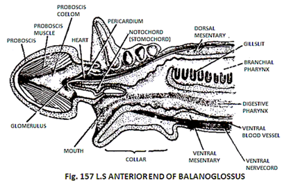
1. It is the longitudinal section of anterior part of Balanoglossus.
2. In it the three fundamental regions of body of the animal are visible i.e., proboscis, collar and trunk.
In the trunk region only branchio-gential region is present which is characterized by the following features:
(a) The pharynx is-divided into an upper respiratory and a lower digestive cavity.
(b) The upper cavity has “U” — shaped gill-slits.
(c) The cavities of pharynx are separated by a longitudinal fold of skin.
3. The collar has a buccal cavity in the centre, the dorsal wall of which is produced into the proboscis as a forwardly directed stomocord or buccal diverticulum.
4. The collar is some what rectangular in out line and is situated just behind probosics.
5. The probosics is the anterior most and some what rounded structure and is characterized by the following features:
(a) Most of its anterior part is filled with muscles.
(b) A little space is left in its posterior part, the proboscis coelom.
(c) In the proboscis coelom project a group of structures the central complex which includes:
(1) A stomocord
(2) A pericardium
(3) A heart and
(4) A glomerulus.
(d) All these organs are connected to the wall of proboscis through dorsal and ventral mesentries.
6. In the region of collar inside the dorsal body wall runs a dorsal blood sinus and e dorsal nerve cord and in the ventral body wall runs a ventral blood sinus and a ventral nerve cord.
7. Between collar and proboscis is present a wide aperture the mouth.
T.S. Proboscis of Balanoglosus:
Fig. 158 T.S PROBOSCIS BLANOGLOSSUS
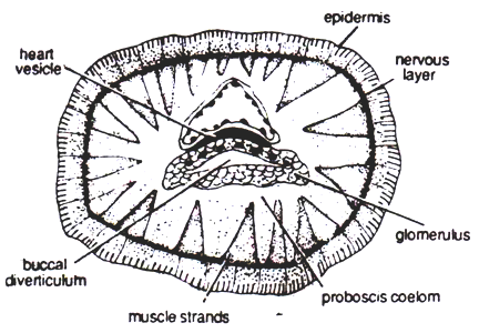
It is the slide of T.S. proboscis of Balanoglossus shows following structures:
i. Body wall represented by epidermis muscle layers and basement membrane.
ii. The epidermis is single layered.
iii. A thin nerve layer lies below the epidermis.
iv. The muscle layer consists of a thick longitudinal muscle layer and thin circular muscle layer.
v. In the centre lies a basal organ or central complex made up of glomerulus, buccal diverticulum and cardiac (heart) vesicle.
vi. Around basal organ the whole space is filled with alternate muscle and connective tissue strands arranged diagonally.
T. S. Collar Region–Balanoglossus:
Fig. 159 T.S COLLAR BALANOGLOSSUS
It is the slide of T.S. of collar region of Balanoglossus and it shows following features:
i. The body wall is represented by single layered epidermis and nervous layer.
ii. Musculature (collar cord) poorly developed. It comprises of circular & longitudinal muscle layers.
iii. Notochord is present but only in anterior part (hemichordata).
iv. A dorsal tubular nerve cord is present (chordate).
v. A distinct pharynx is present perforated by gill-slits (chordate).
vi. No skull, jaws or endo and exoskeleton present.
vii. In addition to dorsal nerve cord a ventral nerve cord is also present.
viii. Body is divisible into proboscis, collar and trunk and Tail is absent.
T.S. Branchiogenital Region of Balanoglossus:
1. It is a transverse section of Balanoglossus passing through the branchio genital region.
2. It is characterised by the following features:
(a) It is some what rounded and bowl-shaped and is made up of a pair of lateral wings, the genital pleurae.
(b) In the centre between the two wings is situated a raise centre – the pharynx.


