List of three common saprophytic fungus: 1. Mucor 2. Yeast 3. Penicillium.
Contents
Saprophytic Fungus # 1. Mucor:
Mucor, also called mould, is a very common saprophytic fungus growing abundantly on decayed organic matters, particularly on those rich in carbohydrates—starch and sugar. Soft white cottony patches of Mucor are frequently found on rotten bread, vegetables and dung.
Plant Body:
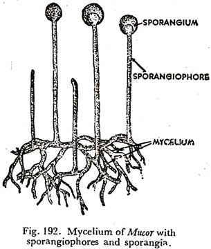
The plant body is a copiously branched mycelium, which is a collection of slender non-septate threads called hyphae (sing, hypha). The wall, made, of fungus-cellulose, enclose cytoplasm with many vacuoles and innumerable nuclei (Fig. 192).
So they are coenocytic. Glycogen and oil globules are present to serve as reserve food. The hyphae become thinner and thinner, the more they penetrate into the substratum for absorbing nourishment. Non-septate mycelium becomes septate on attaining old age and during reproduction.
Reproduction:
Mucor is reproduced by asexual and sexual methods. During asexual reproduction a number of stout aerial hyphae shoot up from the superficial mycelium. After growing to a certain extent, the tip of each of them swells, and some protoplasm with reserve food flows to the enlargement from the adjoining region. Protoplasm collects more densely towards periphery, the central portion remaining comparatively thin and vacuolated.
A good number of vacuoles arrange themselves between the outer denser protoplasm, called sporoplasm, and the central thinner protoplasm, known as columella-plasm.
The flattened vacuoles coalesce, and as a result, a distinct cleft is formed separating the two regions; now a wall is constructed along the cleft delimiting the central sterile dome-shaped region, called columella, which projects into the enlargement. By progressive cleavage or furrowing the sporoplasm now breaks up into a good number of angular masses, each with cytoplasm and many nuclei.
They round off, tough black walls are secreted and finally become spores. Mucor spores are also called gonidia (sing, gonidium). The enlargement containing the spores is the sporangium or gonidangium, and the hypha bearing the sporangium at the tip is called sporangiophore or gonidangiophore (Fig. 193).
The outer wall of the sporangium dissolves in water and the spores are liberated. Often a number of spores remain held in suspension. On suitable medium each spore germinates forming one or more germ tube which gives rise to new mycelium.
Sexual reproduction in Mucor takes place by conjugation. Usually hyphae of two sexually different strains, designated as + strain and — strain, send out club-shaped branches called pro-gametangia, which touch at the tips.
Terminal portion of each branch swells and is cut off by a transverse septum. The compartments thus formed, function as gametangia, and their protoplasmic contents as gametes. The gametes here are multinucleate and hence called coenogametes.
The remaining portion of hyphal branch is known as suspensor. On dissolution of the wall between them, the gametes fuse, cytoplasm with cytoplasm and nuclei with nuclei in pairs. The nuclei which do not fuse in pairs ultimately disintegrate.
The zygote (zygospore) thus formed enlarges and secretes a tough wall around it which is often warty or spiny. After a period of rest it germinates, when the outer wall bursts and the inner wall with the protoplasmic contents comes out as an un-branched tube (Fig. 194). This is called promycelium.
Tip O promycelium enlarges and produces spores, each of which can give rise to a new mycelium. The spores produced in the sporangium resulting from the germination of a zygospore are either all + or all —, never both.
Though Mucor produces isogametes, it shows distinct differentiation of sex. American botanist Blakslee showed in 1904 that zygote formation in Mucor is possible only when gametes produced by two different strains meet.
He termed them as + strain and — strain, rather than male and female for the two mycelial types- Morphologically, there is not much difference between the two, only + mycelium grows a bit more vigorously than its — counterpart.
Such species are called heterothallic. Parthenogenesis is not uncommon in this fungus. One of the gametes may behave like a zygospore without actual pairing. This is called azygospore or parthenospore.
Yeast condition or Torula stage of Mucor:
If a portion of non-septate mycelium is put in sugar solution, it readily develops septa and finally breaks up into many one- celled parts called oidia. Like yeast, oidia multiply by budding and can also excite alcoholic fermentation in sugar solution. This is yeast condition or torula stage of Mucor.
Saprophytic Fungus # 2. Yeast (Saccharomyces):
Yeast is a common saprophytic fungus growing in sugary substances. They are abundantly present in grape juice, vine-yards, nectaries of flowers and sugary exudates of plants like date- palm, juice and palmyra-palm juice.
Plant Body:
The plant body is very simple. Yeasts are unicellular organisms. Each cell is elliptical or round in shape having a distinct cell wall. There is granular cytoplasm, a single nucleus and granules of glycogen and protein and oil globules as reserve food. It was believed that the nucleus is a degenerate one due to occurrence of a vacuole, but now it has been found to be a true nucleus.
Reproduction:
Under favourable conditions, i.e., when there is sufficient food, yeast cells reproduce vegetatively by budding. This is the most common method of reproduction in this fungus. A bud or protuberance arises at one end of the cell and gradually enlarges.
The nucleus divides into two by mitosis. One nucleus with some cytoplasm and food flow from the mother cell to the bud. By constriction the bud is separated from the mother cell. By this process a large number of buds are produced which may often remain in the form of short chains (Fig. 196). The idea that the nuclei divide directly by amitosis during the process of budding has been found to be incorrect.
A few of them are called fission yeasts, in which the protoplast of the mother cell is separated into two parts by the formation of a septum, rather than by constriction as in budding. When food supply is exhausted yeast cells produce spores.
Individual cells enlarge and the nuclei divide once, twice, or thrice, forming 2,4, or 8 nuclei in each cell. Cytoplasm collects round each nucleus and ultimately resistant spores are formed.
The spores are called ascospores and the mother cell (sporangium) is known as ascus. The ascus wall breaks to liberate the ascospores. Each of them goes on reproducing by budding in suitable medium. This process, described as asexual, is really parthenogenetic (Fig. 197).
Sexual reproduction takes place in some species of yeast by conjugation. Two yeast cells approach each other and touch, where short protuberances are formed.
Dissolution of the wall results in the formation of a short conjugation tube connecting the two cells. The two nuclei move to the tube which broadens considerably and, as a result, the whole thing takes up more or less barrel-shaped appearance. The nuclei fuse in the tube forming zygote (diploid) nucleus.
The zygote nucleus usually divides thrice, of which the first division is reduction division. Cytoplasm collects round each nucleus and usually eight spores are delimited. The spores are the ascospores and the barrel-shaped sporangium is the ascus (Fig. 198). Ascospores come out of the ascus and reproduce by budding is suitable medium.
Alcoholic Fermentation:
Yeast has the property of setting up alcoholic fermentation in sugary solution. We know that alcoholic fermentation is an energy- releasing process brought about by micro-organisms under anaerobic conditions. Yeast cells secrete an enzyme, zymase, which decomposes sugar into alcohol and carbon dioxide with liberation of energy. CO2 comes out as bubbles forming froth.
It may be represented thus:
C6H12O6 + Zymase-2C2 H5OH + 2CO2 + Zymase + Energy (25 cal.)
(Sugar) (Enzyme) (Alcohol)
For this particular property yeasts are used commercially in breweries for the manufacture of alcoholic beverages like wines, beers, etc. The same principle applies in the preparation of indigenous liquor, toddy, from sugary exudates. They are also used in bakeries or ‘raising’ of breads.
Yeast cells excite alcoholic fermentation in bread paste (dough), and carbon dioxide bubbles, while escaping on application of heat, raise the bread. Yeasts are rich in vitamin B complex. So they have nutritional value as well.
Saprophytic Fungus # 3. Penicillium:
Penicillium is a common saprophytic fungus growing on decayed organic matters like bread, jam, jelly, vegetables and fruits and even on damp shoes and leather. It is known as green or blue mold. The spores -of this fungus are abundantly present everywhere and often cause considerable damage to fruits and vegetables.
Some species are also used in industries. Sir Alexander Flemming isolated the wonder drug, penicillin from Penicillium notatum in 1929. The antibiotic penicillin had revolutionised medical science and proved to be a real boon to humanity.
The mycelium is composed of much branched septate hyphae occurring in tangled masses. They ramify extensively on the substratum and many of them penetrate into it to serve as rhizoids. The hyphal cells are multi-nucleate.
Reproduction
This fungus reproduces mainly by asexual method, through the spores called conidia, which are formed in very large number. Sexual method of reproduction has also been reported in some species, though the stages are not very clearly known.
Asexual:
Some stout hyphae come out erect from the mycelium and function as conidiophores. Smaller branches develop from the tip of the conidiophore, which again divide to form a row of closely- packed branches called sterigmata. Un-branched chains of asexual spores—conidia, are cut off from the tip of the sterigmata in basipetal order (Fig. 198A).
The terminal portion of conidiophores with branches and chains of conidia together looks like a broom and is called penicillus—meaning broom. The conidia are globose, ovoid or elliptical with smooth or spiny surface and usually green in colour.
They are uninucleate at the early stage but may become multinucleate in some cases. The conidia are easily dispersed by wind, and germinate on a suitable substratum. A germ tube is first formed which ultimately develops into a new mycelium.
Sexual:
Sexual reproduction, though not clearly known, is oogamous. A hypha comes out erect, enlarges, becomes club-shaped and develops into the ascogonium. The nucleus divides again and again to form 32-64 nuclei dispersed in the cytoplasm of the ascogonium. In the meantime another branch comes out of a neighbouring hypha which twines round the ascogonium.
The terminal part of that branch is cut off by a septum, swells and forms the antheridium. It comes in contact with the ascogonium where a pore is formed for movement of the protoplast of the antheridium to the ascogonium. Though gametic union has not been established it may be assumed that the process takes place. By formation of septa the multinucleate ascogonium gives rise to a row of bi-nucleate cells.
Many hyphae with bi-nucleate cells now develop from these cells—which are called ascogeneous hyphae. They become septate, each cell having two nuclei, and the terminal cell develops into an ascus. The two nuclei of the ascus fuse to form the zygote nucleus, which then divides thrice, the first division being reductional, and ultimately produces eight ascospores.
Meanwhile sterile vegetative hyphae send out many branches around the sex organs forming a closed fruit body—the ascocarp. This cover made of hyphal cells is known as cleitothecium, the inner layer of which is nutritive in function. With maturity of the Ascospores the asci dissolve leaving them free and scattered in the cleistothecium. The periderm now decays liberating the ascospores which are easily blown off by wind.


