In this article we will discuss about the life cycle of morchella with the help of suitable diagrams.
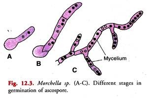
Mycelium of Morchella (Fig. 12.3C):
It is inconspicuous but extensive and subterranean. It grows a few inches deep in the soil and thus hidden from view. The mycelium consists of a mass of loosely interwoven branched hyphae. The hyphae ramify through the interstices of the soil nourishing themselves on the organic materials. They are septate. Each segment or cell contains several nuclei. Under adverse circumstances the mycelia produce sclerotia.
Development of Ascocarp:
It begins as a dense knot of hyphae. Under favourable conditions of food and moisture, the mycelial hyphae grow rapidly and branch repeatedly. They get interwoven to form dense, compact masses.
These masses of hyphae are called the hyphal knots. They are formed just beneath the surface of the soil. Each hyphal knot in the presence of abundant food and moisture (after rain) develops into an aerial stalked fructification called an ascocarp.
It is cup-shaped only when young. During further growth the hymenium becomes convex. The asci thus face outwards. The smooth hymenium later becomes complicated by unequal growth of the hymenial surface which is thrown into folds and presents a honey-combed or sponge-like surface.
This device increases the fertile area and thus the number of asci. Since the stalk and the terminal honey-combed cap of the fructification are delicate structures the morels grow best under moist conditions and shade.
Ascocarp (Fig. 12.1):
The fructification or the ascocarp is a spore producing structure. The mature ascocarp consists of a stalk-like portion, the stipe surmounted by a hollow, conical, cap-like object, the pileus (Fig. 12.1).
The stipe is cream coloured. It is thick, fleshy and hollow with a waxy surface. The fructification ranges from 2.5 cm to 12 or 14 cm in height. The colour varies from a dirty greyish white to a dark brown depending on the species and the age.
The terminal conical cap of the pileus constitutes the fertile portion of the ascocarp. It is hollow and fleshy. The honey combed surface of the pileus gives it the appearance of a sponge.
The pileus is pitted, sometimes ridged and is about the same length as the stipe. When young the surface of the pileus is quite smooth.
Towards maturity there appears a network of ridges and depressions (pits) on its surface due to unequal growth of the hymenial surface.
The ridges are sterile and are usually of the same colour as the stalk. The depressions or pits are the fertile areas and have a brown tint. The ridges unite the fertile areas which form an irregularly distributed hymenium or thecium.
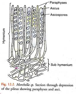
A thin section (Fig. 12.2) through the depression reveals that it is lined by a fertile layer called the hymenium or thecium. The latter consists of numerous asci which are long, cylindrical, operculate cells.
They stand side by side perpendicular to the surface of the depression forming a palisade-like layer. Each ascus usually contains eight hyaline, ascospores arranged in a uniseriate manner.
The asci are positively phototrophic. They bend towards the source of light. Intermingled with the asci are elongated, sterile slender hyphae called the paraphyses. Beneath the hymenium is the sub-hymenium or hypothecium.
A cross-section through the stipe reveals that it is made up of pseudoparenchymatous tissue. This tissue in the region of the depression of the pileus forms the hypothecium.
Dehiscence of Asci:
The tips of mature asci project above the level of the hymenium and turn towards light being positively phototrophic. The residual cytoplasm within each ascus changes into sugars of high osmotic value.
The latter absorb water. As a result a considerable amount of pressure is set up in the asci. The pressure set up within the asci aided by the turgor pressure set up in the paraphyses causes the release of ascospores through a preformed apical pore which opens by a lid hinged at the top of the ascus.
The internal force projects the ascospores high up into the air where they are caught up by the air currents. The explosions of the asci are successive and not simultaneous.
The pileus gradually shrinks as it dries up. Eventually it withers. The ascospores are large colourless, oval and multinucleate when mature.
Dispersal of Ascospores:
It is reported that the catabolic activities in the ripe fruiting body are high during the period of dehiscence. This is indicated by the marked rise in the temperature of the tissue.
Conventional currents are set up in the surrounding air. These are induced by the warm tissue of the ripe fructification.
These conventional currents of air are suggested to play an important role in the dispersal of the liberated ascospores. They are carried by wind to distant places.
Ascospore Germination (Fig. 12.3):
On falling on a suitable soil (moist humus soil) each ascospore germinates (A) to produce a new mycelium soon after release. The ascospores that happen to fall on unsuitable soil perish.
Ascospores do not remain viable after one year near the soil surface. According to the latter, the ascospores germinate and develop extensive mycelium at 15°C under highly nutritious non-competitive conditions.
However, the ascospore germinates at a temperature as low as 2°C but rate of germination and hyphal growth is slower at lower temperature. Above 15°C germination is inhibited.
Sexual Reproduction in Morchella:
The sex organs which constitute the accessory parts of sexual process are completely suppressed in Morchella. Consequently the sexual process is extremely simplified.
It involves two distinct processes, namely, Plasmogamy and Karyogamy. The latter is immediately followed by meiosis.
(a) Plasmogamy:
It consists in the union of cytoplasmic contents of two cells without nuclear fusion which is delayed. This results in a sequence of binucleate cell generations constituting the dikaryophase.
Geis (1940) reported that plasmogamy occurs at a very late stage during the development of the ascocarp in the subhymenial layer. It takes place just before the formation of asci.
Plasmogamy in Morchella takes place by the following two methods:
(i) Somatogamous Copulation (Fig. 12.4):
Two vegetative hyphae of the subhymenium region of the pileus come in contact. The intervening walls between the copulating cells dissolve at the point of contact (A).
The two multinucleate protoplasts intermingle in the fusion cell (B). Two functional nuclei, one from each copulating cell, form a dikaryon. All other nuclei in the fusion cell disappear. Later ascogenous hyphae arise from the fusion cell containing the dikaryon (C).
Each young ascogenous hypha receives a pair of nuclei. These are the derivatives of the parent dikaryon. The ascogenous hyphae afterwards become septate.
The terminal cells of these hyphae function as ascus mother cells (D). They are binucleate. The nuclei are the derivatives of the parent dikaryon.
(ii) Autogamous Pairing:
It has been reported in M. elata. In this case any vegetative cell of the sub-hymenium (hypothecium) may establish a dikaryon.
It is accomplished by pairing of two of its own vegetative nuclei. It is called autogamous pairing. The remaining nuclei in the cell degenerate. The cells with dikaryons (dikaryotic cells) develop ascogenous hyphae as in somatogamous copulation.
(b) Karyogamy:
The two nuclei in the ascus mother cell fuse. The fusion cell with a diploid nucleus is called the young ascus. It represents the transitory diplophase in the life cycle.
The asci in Morchella thus develop from the tip cells of ascogenous hyphae without the formation of croziers.
(c) Meiosis:
The young ascus cell elongates. The synkaryon in the ascus undergoes two successive divisions. These constitute meiosis. During meiosis the number of chromosomes in the resultant four daughter nuclei is reduced to half the diploid number.
The four haploid nuclei undergo the third division. It is mitotic. In this way eight haploid daughter nuclei are formed in each ascus cell. Each of these is fashioned into an ascospore.
With the formation of ascospores the haploid or gametophyte phase is again initiated in the life cycle of Morchella.
It is speculated that the overwintering stage in the life cycle of Morchella is the sclerotium.


