In this article we will discuss about the life cycle of peziza with the help of suitable diagrams.
Mycelium of Peziza:
It is well developed, frequently perennial and consists of a dense network of hyphae. The hyphae are branched and septate. The cells are uninucleate.
The hyphae are hidden from view as they ramify within the substratum. They from a complex system which extracts nourishment from the substratum. The fruiting bodies are above ground.
Reproduction in Peziza:
1. Asexual Reproduction:
It takes place by the formation of conidia and chlamydospores. The conidia are exogenously formed spores. They are abstricted from the tips of conidiophores. Each conidium germinates to form a new mycelium.
The chlamydospores are thick-walled resting cells. They are intercalary in position. They may be formed singly or in series within the cells of the hyphae. Under suitable conditions each chlamy-dospore germinates and gives rise to a new mycelium.
2. Sexual Reproduction:
The sexual apparatus is wholly lacking in Peziza vesiculosa. This does not prevent the development of a fructification or the fig. 12.12 (A-B) which is aerial and relatively a short-lived structure. The sexual process does take place. It is extremely simplified and consists in the association of two purely vegetative nuclei in a pair.
The adult mycelium consists of a tangled mass of hyphae. Certain vegetative cells in the centre of the tangled hyphal mass have been seen to possess nuclei which become associated in pairs.
These pairs of nuclei are called the dikaryons. The dikaryotic condition is brought about either by autogamous pairing or by somatogamous copulation between the vegetative cells of the adjacent hyphae of the tangled hyphal mass.
The cells with the dikaryons give rise to the ascogenous hyphae which become multicellular by cross walls. Their cells are binucleate. The terminal binucleate cell of each ascogenous-hypha functions as an ascus mother cell.
Formation of croziers in the development of asci has not been reported in P. vesiculosa. The ascogenous hyphae and dikaryotic cells from which they are developed together with the ascus mother cells represent the dikaryophase in the life cycle of Peziza.
The two nuclei of the ascus mother cell fuse to form the synkaryon. The young ascus with the synkayon represents the transitory diplophase (Fig. 12.14). The synkaryon undergoes three successive divisions. Of these the first and the second constitute meiosis.
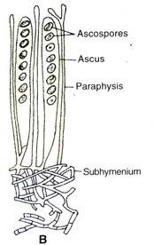
This results in the formation of eight haploid nuclei which become organised into ascospores. The mature ascus is an elongated, cylindrical cell (Fig. 12.13B).
The ascus wall is lined by a thin layer of cytoplasm (epiplasm) which encloses a central vacuole filled with sap. In the vacuole lie the oval ascospores.
The erect asci lie side by side lining the cavity of the cup-shaped apothecium (Fig. 12.13A). The asci near the margin of the cup bend towards the source of light being positively phototropic.
Interspersed between the asci are the Sterile hyphae called paraphyses. The rest of the apothecium consists of densely interwoven, branched hyphae forming a pseudoparenchymatous tissue which supports the hymenium (Fig. 12.13A).
The apothecia (Fig. 12.12A) are sessile or shortly stalked cup-shaped structures regular in form and large in size varying from 2 cm. to several inches in diameter. In P. vesiculosa the apothecium is of pale fawn colour but P. aur antia has brilliant orange apothecium.


