In this article we will discuss about:- 1. Meaning and Definitions of Fungi 2. Characteristics of Fungi 3. Occurrence 4. Vegetative Structure 5. Reproduction.
Contents
Meaning and Definitions of Fungi:
Fungi (singular fungus — mushroom, from Greek) are chlorophyll-less thallophytic plant. Due to absence of chlorophyll, they are heterophytes i.e., depend on others for food. They grow in various habitats and show much diversity in their structure, physiology and reproduction. They developed long back in the geological time scale.
Their existence was found from fossil records of Pre-Cambrian period. Information from ancient literature indicates that the fungi were used as food by human beings. At present, the fungi are used in medicine and also as food in addition to other aspects. The fungi cause diseases of crops (spots, rusts and smuts etc.) and on human beings (Aspergillosis, Blastomycosis etc.).
Some of the plant diseases like late blight of potato (c.o. Phytophthora infestans) and brown spot of rice (c.o. Helminthosporium oryzae) caused famine in Ireland (1845) and in West Bengal (India) (1943), respectively.
More than 5,000 genera and 50,000 species of fungi have been recorded, but their number may be much more than the actual record. The subject which deals with fungi is known as Mycology (mykes — mushroom; logos— study) and the concerned scientist is called mycologist.
The various definitions of fungi as proposed by mycologists are:
1. Alexopoulos (1962):
Alexopoulos (1962) defined fungi as “nucleated, spore-bearing, achlorophyllous organisms which generally reproduce sexually and asexually and whose usually filamentous, branched somatic structures are typically surrounded by cell walls containing cellulose or chitin or both”.
2. Alexopoulos and Mims (1979):
Alexopoulos and Mims (1979) defined fungi as eukaryotic spore bearing, achlorophyllous organisms that generally reproduce sexually and asexually, and whose usually filamentous, branched somatic structures are typically surrounded by cell walls containing chitin or cellulose, or both of these substances, together with many other complex organic molecules”.
Characteristics of Fungi:
1. Fungi are cosmopolitan in distribution i.e., they can grow in any place where life is possible.
2. They are heterotrophic in nature due to the absence of chlorophyll. On the basis of their mode of nutrition, they may be parasite, saprophyte or symbionts.
3. The plant body may be unicellular (Synchytrium, Saccharomyces) or filamentous (Mucor, Aspergillus). The filament is known as hypha (plural, hyphae) and its entangled mass is known as mycelium.
4. The hypha may be aseptate i.e., coenocytic (without septa and containing many nuclei) or septate. The septate mycelium in its cell may contain only one (monokaryotic), two (dikaryotic) or more nuclei.
5. The septa between the cell may have different types of pores: micropore (Geotri- chum), simple pore (most of the Ascomycotina and Deuteromycotina) or dolipore (Basidiomycotina, except rusts and smuts).
6. The cells are surrounded by distinct cell wall (except slime molds), composed of fungal cellulose i.e., chitin; but in some lower fungi (members of Oomycetes), the cell wall is composed of cellulose or glucan.
7. The cells generally contain colourless protoplasm due to absence of chlorophyll, containing nucleus, mitochondria, endoplasmic reticulum, ribosomes, vesicle, microbodies, etc.
8. The cells are haploid, dikaryotic or diploid. The diploid phase is ephemeral (short-lived).
9. In lower fungi like Mastigomycotina, the reproductive cells (zoospores and gametes) may be uni- or biflagellate, having whiplash and/or tinsel type of flagella. But in higher fungi like Zygomycotina, Ascomycotina, Basidiomycotina and Deuteromycotina, motile cells never form at any stage.
10. In response to functional need, the fungal mycelia are modified into different types such as: Plectenchyma, Stroma, Rhizo- morph, Sclerotium, Hyphal trap, Appreso- rium, Haustorium, etc.
11. The unicellular fungi, where entire plant body becomes converted into reproductive unit, are known as holocarpic fungi (e.g., Synchytrium). However, in many others, only a part of the mycelial plant body is converted into reproductive unit, thus they are called eucarpic fungi (e.g., Pythium, Phytophthora).
12. They reproduce by three means: Vegetative, asexual and sexual.
(a) Vegetative reproduction takes place by fragmentation (Mucor, Penicillium, Fusarium), budding (Saccharomyces, Ustilago) and fission (Saccharomyces).
(b) Asexual reproduction takes place by different types of spores. These are zoospores (Synchytrium), conidia (Pythium, Aspergillus), oidia (Rhizo- pus), chlamydospore (Fusarium), etc. The spores may be unicellular (Aspergillus) or multicellular (Alternaria).
(c) With the Exception of Deuteromycotina, Sexual Reproduction takes Place by the following Five Processes:
Gametic copulation (Synchytrium), Gametangial contact (Pythium, Phytophthora), Gametangial copulation (Rhizopus, Mucor), Spermatization (Puccinia, Podospora) and Somatogamy (Polyporus, Agaricus).
General Account:
The fungi are chlorophyll-less thallophytes or achlorophyllous non-vascular plants.
Occurrence of Fungi:
The fungi are cosmopolitan in distribution and occur in all possible habitats. Due to absence of chlorophyll, they depend on other for food, that is why they may be saprophytes, parasites or symbionts.
Most of the fungi are terrestrial which grow in soil, on dead and decaying organic material. Some grow on both plants and animals. They can grow on foods like jam, bread, fruits etc. Some members are also found in water — aquatic fungi. They are also present in the air. Thus the fungi are universal in their distribution.
Vegetative Structure of Fungi:
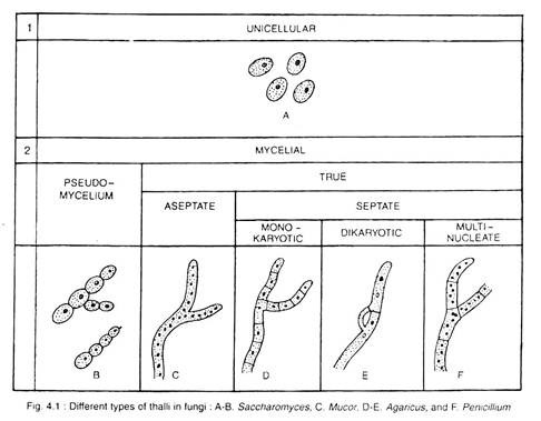
Except certain unicellular forms (Fig. 4.1 A) like Synchytrium, Saccharomyces; the fungi are generally thalloid and filamentous type. The filaments are known as the hyphae (sing, hapha). The tangled mass of these hyphae is the mycelium. The hypha may be aseptate which contains many nuclei and vacuolated cytoplasm. The multinucleate tubular aseptate hypha is called coenocyte (Fig. 4.1 C).
On the other hand, hypha that has partition walls dividing the hypha into many cells is called septate (Fig. 4.1 D-F). The septate mycelium in its cell may contain only one (monokaryotic), two (dikaryotic) or many nuclei (multinucleate) with vacuolated protoplasm.
The septa between the cells may have different types of pores. In Geotrichum, small pores are present on the wall, called micropore (Fig. 4.2A), when only one bigger sized pore is present on the wall, it is called simple pore (Fig. 4.2B), found in most of the Ascomycotina and Deuteromycotina.
In Trichomycetes and Mucorales, the pore is surrounded by an overarching bifurcation of the margin of partition wall which looks like a bordered pit of tracheid. A biumbonate, electron dense plug of material is present between the two bifurcations (Fig. 4.2C).
But in Basidiomycotina, except rust and smuts, the complicated type of pore with single opening is called dolipore (dolium, a large Jar or cash, i.e., barrel) septum (Fig. 4.2D).
Lastly in lower fungi, the older part and the basal region of reproductive part forming partition wall, without any pore, is called solid septum (Fig. 4.2E).
The cells have distinct cell wall (except slime molds), made up of fungal cellulose i.e., chitin along with other substances. Fungal cells have chlorophyll-less vacuolated protoplasm, which contains nuclei/nucleus, mitochondria, endoplasmic reticulum, ribosomes, microbodies, vesicles, etc. (Fig. 4.3). The chief reserve foods are glycogen and oil.
Mycelium that contains only one nucleus (n) in each cell is called monokaryotic mycelium or primary mycelium. Whereas, in many cases, cells of mycelium contain two nuclei i.e., binucleate (n + n), formed due to transfer of nuclei from one monokaryotic mycelium to the other and initially form dikaryotic cell and then to dikaryotic mycelium or secondary mycelium by growth with the help of clamp connection.
The cell may contain diploid nucleus (2n), formed by the fusion of two haploid nuclei (n) of opposite strain. But this stage is ephimeral (Fig. 4.1 E, F).
In lower fungi i.e., members of sub-division Mastigomycotina, the reproductive cells (zoospores or gametes) may be uni- or biflagellate with (9 + 2) organisation having either whiplash (acronematic type) i.e., smooth-walled flagella and/or tinsel (pantonematic type) i.e., flagella covered with many minute hair-like mastigonemes or flimers, those originate from the axile filament (Fig. 4.4, 4.5).
The tinsel type gives forward movement and the whiplash type gives beating movement. But, in all other fungi placed under subdivisions Zygomycotina, Ascomycotina, Basidiomycotina and Deuteromycotina, motile cell does not occur at any stage.
Composition of Cell Wall:
The cells are surrounded by outer rigid structure, the cell wall. Its composition varies in different groups of fungi. According to Aronson (1965) and Bartnicki-Garcia (1970), the cell wall consists of about 80-90% polysaccharides along with proteins (1-15%) and lipids (2-10%).
The most common cell wall material is chitin. But in some other fungi, cellulose or other glucans are present. The chemical structure of cellulose and chitin is given below. Bartnicki-Garcia (1968) reported that the composition of cell wall varies in different groups of fungi.
These are cellulose- glycogen (Acrasiomycetes), cellulose-glucan (Oomycetes), cellulose-chitin (Hyphochytridiomycetes), chitin-chitosan (Zygomycetes), chitin- glucan (Chitridiomycetes, Asco-, Basidio- and Duteromycotina), mannan-glucan (Saccharo- mycetaceae and Cryptococcaceae), mannan- chitin (Sporobolomycetaceae), Polygalacto- samine-galactan (Trichomycetaceae).
Hyphal Forms:
In response to functional need, the fungal mycelia are modified into different types of structure:
1. Plectenchyma:
When the component hyphae completely interwoven to form a compact thick tissue, it is called plectenchyma.
It is of two types:
Prosenchyma or prosoplectenchyma, where the hyphae remain more or less parallel to each other, retain their individuality and do not fuse (Fig. 4.6B) and pseudoparenchyma or para- plectenchyma, where the hyphae are completely fused to each other, form a compact mass and lose their individuality. In cross- section the whole mass looks like parenchyma of angiospermic plant (Fig. 4.6B).
2. Sclerotia (sing. Sclerotium):
It is a compact structure of different shapes and sizes, formed by the aggregation of mycelia. It may be round to elongated pod-like, very minute dots to large ball-like structure weighing approximately 14 kg (30 lbs). It may survive for many years as resting stage (Fig. 4.6A).
3. Rhizomorph:
In this case, hyphae are aggregated longitudinally in varying degree of complexity, where the hyphae lose their individuality and the whole structure behaves as an organised unit. They can withstand adverse environmental conditions and after few years, they can start growing (Fig. 4.6E).
4. Haustoria (sing. Haustorium):
These are intracellular outgrowth of the mycelium that grows intercellularly for absorption of nutrient from host cells. They are of various shapes and sizes, like simple, knob-like, coiled, branched, etc. They penetrate the cell wall and generally do not rupture the cell membrane during absorption (Fig. 4.6F, G).
5. Appresoria (sing. Appresorium):
It is the swollen tip of germ tube or mycelium of plant pathogenic fungi which helps the mycelium to adhere to the surface of the host and also helps in penetration (Fig. 4.6H).
6. Hyphal trap:
Certain fungi develop sticky hypha or hyphal loops to catch predators like nematode, protozoa, small animals etc., known as hyphal trap. The fungi of this kind is known as Predaceous fungi (Fig. 4.61).
7. Stroma:
It is a solid body of various shapes and sizes, formed by the compact aggregation of mycelium. Reproductive structures and fruit bodies are developed inside the stroma (Fig. 4.6D).
Reproduction in Fungi:
In unicellular fungi (Synchitrium, Saccharomyces), entire vegetative cell is transformed into a reproductive unit, called Holocarpic. However, in others (Pythium, Penicillium, Helmintho- sporium), only a part of the vegetative body forms reproductive unit and the rest portion remains as vegetative, called Eucarpic.
The fungi reproduces by all the three means:
Vegetative, Asexual and Sexual.
1. Vegetative Reproduction:
It takes place by the following ways:
(a) Fragmentation:
It is common in filamentous fungi (Rhizopus, Alternaria, Fusarium) where the hyphae break up into two or more fragments due to some external force and each one develops into a new individual (Fig. 4.7A, B).
(b) Budding:
It takes place in unicellular fungi (Saccharomyces, Schizosaccharo- myces). A small outgrowth, the bud emerges out from the parent cell. Nucleus divides into two and one passes to the bud. The bud is then separated by partition wall, but continues its growth.
Later on, it breaks off from the mother and grows individually. Sometimes, the process repeats very fast and the buds remain attached with the mother in chain, that looks like mycelium, called pseudomycelium (Gr. pseu- do, false + mycelium). This process takes place in ascospores and basiclio- spores of some fungi (Fig. 4.7C, 4.1 B).
(c) Fission:
Normally unicellular fungi (Saccharomyces, Schizosaccharomyces) reproduce by this method, where the vegetative cell elongates, and divides into two daughter cells of equal size by simple constriction in the middle with simultaneous nuclear division (Fig. 4.7D).
 2. Asexual Reproduction:
It takes place by means of several types of spore generally form during favourable condition. The spores may be unicellular (Penicillium, Aspergillus) or multicellular (Fusarium, Helminthosporium). They may be exogenous, developed on conidiophore (Penicillium) (Fig. 4.8C) or endogenous, developed in sporangium (Mucor) (Fig. 4.8L) or pycnidium (Ascochyta pisi).
Some of the spores are:
(a) Zoospore:
The zoospores may be uni- or biflagellate, generally pear-shaped, produced in sporangium, e.g., Synchytrium, Phytophthora (Fig. 4.8J).
(b) Conidia:
These are exogenously produced non-motile spores develop by constriction at the end of specialised hyphal branches, called conidiophores. They may produce singly (Phytophthora, Pythium) or in chain (Penicillium, Aspergillus) (Fig. 4.8A, B, C).
(c) Oidia:
In some fungi (Mucor mucedo), the hyphal tips often divide by transverse wall into large number of small segments, may remain in chain or becomes free from each other, these are known as oidia. The oidia on germination develop into new plants (Fig. 4.8D).
(d) Chlamydospore:
The chlamydospores are thick walled round to oval in outline, coloured brown or black. They produce either terminally or in intercalary at some intervals throughout the length of hyphae, e.g., Fusarium (Fig. 4.8E, F, G).
(e) Sporangiospores:
These are globose, multinucleate, non-motile aplanospores, formed inside the sporangium. The sporangiospore germinates by producing germ tube. Later on, it develops profusely branched mycelium (Fig. 4.8K, L).
Sexual Reproduction:
It is the process of union between two compatible nuclei. The nuclei in some members are contributed by two well-organized gametes.
The whole process of sexual reproduction consists of three phases, in the sequence of plasmogamy, karyogany and meosis:
(i) Plasmogamy:
It involves the union of two protoplasts, brings two haploid nuclei close together in the same cell.
(ii) Karyogamy:
It involves the fusion of two haploid nuclei brought together during plasmogamy. This results in the formation of diploid nucleus i.e., zygote, which is ephemeral (short-lived).
(iii) Meiosis:
It follows karyogamy and reduce the number of chromosome from diploid zygote nucleus to original haploid number in the daughter nuclei.
The plasmogamy i.e., the first phase of sexual reproduction, differs in different fungi.
The different methods of plasmogamy are:
(a) Planogametic Copulation:
Planogametes are motile gametes. This process involves the fusion of two gametes, where either one or both are motile.
Depending on the structure and nature of gametes, it is of three types:
Isogamy, Anisogamy and Oogamy:
(i) Isogamy:
The uniting gametes are morphologically similar, but physiologically different. This process is common in primitive unicellular fungi, e.g., Synchytrium (Fig. 4.9A).
(ii) Anisogamy:
Both the uniting gametes are morphologically similar, but different physiologically and in size. The smaller one is more active, considered as male and the larger less active one as female, e.g., Allomyces (Fig. 4.9B).
(iii) Oogamy:
Both the uniting gametes are morphologically and physiologically different. The male gamate is smaller and motile, and the female gamete is larger and non-motile, e.g., Monoblepharis (Fig. 4.9C).
(b) Gametangial Contact:
The uniting gametes are present in different gametangium, thus the male and female gametangia are known as antheridium and Oogonium (Ascogonium in Ascomycotina), respectively. The gametes are never released from gametangium. Both the gametangia come in close contact and transfer male gamete to the egg through fertilization tube. The gametangia do not lose their identity, e.g., Ascobolus, Pythium (Fig. 4.9D, E).
(c) Gametangial Copulation:
The process involves the fusion of the entire content of the uniting gametangia.
Such fusion occurs in the following two ways:
(i) The two gametangia fuse by the dissolution of their common wall resulting into the formation of a single cell in which content of both the gametangia mix with each other and their morphological identity are completely lost, e.g., Rhizopus, Mucor (Fig. 4.9F).
(ii) The entire thallus acts as gametangium. Both the gametangia come in close contact and the male gametangium transfer its entire content to the female gametangium through the pore developed in contact area e.g., Rhizophidium (Fig. 4.9G), Polyphagus.
(d) Spermatisation:
Certain fungi produce many unicellular non-motile, male cells, the spermatia. The spermatia are brought in contact by agents like wind, water and insect either to the trichogyne of the ascogonium or to somatic hyphae or even to special receptive hyphae. The wall at the point of contact dissolves and content of spermatia passes to the female organ, e.g., Puccinia, Podospora (Fig. 4.9H).
(e) Somatogamy:
In many higher fungi belonging to Asomycotina and Basidiomycotina, the development of gametes and gametangia are completely lacking. In such fungi, somatic hyphae anastomose with each other to bring together the compatible nuclei. It is regarded as a reduced and efficient form of sexuality, designated as somatogamy, e.g., Polyporus, Agaricus, Morchella (Fig. 4.9I).


