In this essay we will discuss about Bacteria. After reading this essay you will learn about:- 1. Meaning of Bacteria 2. Salient Features of Bacteria 3. Morphology 4. Cell Wall 5. Size 6. Flagellation 7. Behavioural Responses 8. Endospores 9. Reproduction 10. Some Important Genera.
Contents:
- Essay on the Meaning of Bacteria
- Essay on the Salient Features of Bacteria
- Essay on the Morphology of Bacteria
- Essay on the Cell Wall of Bacteria
- Essay on the Size of Bacteria and the Significance of their Being Small
- Essay on the Flagellation in Bacteria
- Essay on the Behavioural Responses of Bacteria
- Essay on the Endospores in Bacteria
- Essay on the Reproduction in Bacteria
- Essay on the Some Important Genera of Bacteria
Contents
- Essay # 1. Meaning of Bacteria:
- Essay # 2. Salient Features of Bacteria:
- Essay # 3. Morphology of Bacteria:
- Essay # 4. The Cell Wall of Bacteria:
- Essay # 5. Size of Bacteria and the Significance of their Being Small:
- Essay # 6. Flagellation in Bacteria:
- Essay # 7. Behavioural Responses of Bacteria:
- Essay # 8. Endospores in Bacteria:
- Essay # 9. Reproduction in Bacteria:
- Essay # 10. Some Important Genera of Bacteria:
Essay # 1. Meaning of Bacteria:
The bacteria constitute a very wide group of microorganisms that exhibit a fascinating diversity in morphology, habitat, nutrition, metabolism, and reproduction.
Although they are not very complex morphologically, the tiny bacteria nevertheless have highly complex physiological, biochemical, cytological, and genetically characteristics making them a valuable tool for understanding the various intricacies of life.
Due to their extreme simplicity in structure, small size favouring rapid cell division, highly resistant nature and diversified mode of nutrition, bacteria are of universal occurrence. They are present in our mouth and flourish in intestine. They are present in air we breathe and in food we eat. They abundantly occur in fresh and salt water, soil water and even in ice.
Their most favourable habitat is soil, where they occur in abundance mainly in the upper half feet. In a handful of garden soil, the bacterial population may outnumber the human population on the earth. They live in all conditions not fatal to living beings and are among the most numerous of all living beings present in almost every conceivable environment.
Some bacteria are deadly parasites of plants, animals and human beings; some live as mutualists with plants or as commensals in the alimentary canals of animals. Some bacteria may remain viable when cooled up to -190°C, while others may remain viable when boiled up to 78°C.
Essay # 2. Salient Features of Bacteria:
Bacteria (eubacteria) are microscopic and least differentiated living organisms, believed to be the first primitive organisms on our planet. They are the typical prokaryotes and also possess characters resembling both the plants and the animals. Bacteria show considerable variation in characters almost themselves, they possess many characters common to all.
Such common characters of bacteria are the following:
1. They are omnipresent and occur in all possible habitats one can think of.
2. Most of the bacteria have heterotrophic (absorptive) mode of nutrition, i.e., they absorb their food directly from their external environment, and may be saprophytic, mutualistic or parasitic. Some bacteria are autotrophic; they possess bacteriochlorophyll and synthesize their own food.
3. Bacteria are usually single-called and morphologically least complex of all the living organisms.
4. Bacteriochlorophyll pigments, whenever present, are located within involuted cytoplasmic membranes; well organised plastids are absent.
5. Bacterial cell wall is a dense layer surrounding the plasma membrane and functions to give shape and rigidity to the cell. The main constituent or back-bone of bacterial cell wall is peptidoglycan (also known murein, muranic acid or mucopeptide), which is biochemically unique and is absent in cell walls of archea (archaebacteria) or any eukaryote.
6. A well organised nucleus, characteristic of eukaryotes, is lacking and descrete chromosomes are also absent. The nuclear material is not surrounded by nuclear membrane and is usually called nucleoid.
7. Bacterial DNA have no histone proteins and nucleosomes. They also lack ‘introns’, the non-coding sequences.
8. The organelles like mitochondria, endoplasmic reticulum, Golgi apparatus are absent.
9. The function of mitochondria is carried out by complex localized in folding of plasma membrane known as mesosomes.
10. Ribosomes occur abundantly and freely in the cytoplasm of a bacterial cell. Each ribosome has a sedimentation coefficient of 70S and is made up of two subunits of 50S and 30S each consisting of roughly equal amounts of rRNA and protein.
11. Bacteria reproduce asexually and multiply most commonly by binary fission.
12. True sexual reproduction is absent. The sexuality is completed by genetic recombination methods called conjugation, transformation and transduction.
13. The motile bacteria possess one or more flagella. Each flagellum is composed of eight parallel chains of flagellin molecules, a type of protein.
Essay # 3. Morphology of Bacteria:
The following categories of bacteria are recognized on the basis of diversity in their morphological features:
(i) Unicellular Bacteria:
1. Cocus [pI., Cocci]:
The cocci bacteria [Fig. 4.1. and 4.2(A)] are unicellular and spherical varying from 0.5 to 1.25 µ in diameter. They exist either singly (micrococcus), in pairs (diplococcus), in chains (streptococcus), in clusters (staphylococcus), or in cubical masses of 8 or more cells (sarcina).
2. Bacillus [pI., bacilli]:
Bacilli bacteria (Fig. 4.2B) are unicellular and hyphen (-) or small rod-shaped ranging about 1.5 µ in diameter and 10 µ in length. They occur either singly (microbacillus), in pairs (diplobacillus), in chains (streptobacillus), or in palisade arrangement. Bacillus anthracis, B. subtilis, Lactobacillus and Clostridium are the common examples of bacilli bacteria.
3. Vibrio [pI. Vibrios]:
When the bacilli bacteria are so curved that they look like a comma, they are called vibrios Fig. 4.2(C). They seldom exceed 10 µ in length and 1.5 to 1.7 µ in diameter, e.g., Vibrio comma.
4. Spirillum [pl. Spirilla]:
When the bacilli bacteria are coiled like a cork-screw through 1-5 complete turns, they are referred to as Spirilla [Fig. 4.2(D)]. They range from 10-50 µ in length and 0.5 to 3 µ in diameter, e.g., Spirillum undulum and 5. volutans.
5. Stalked Bacteria:
Stalked bacterial [Fig. 4.2(E)] are unicellular bacteria having well defined stalks. In some cases, the stalk is a part of the cell (Caulobacter), in others it is formed as a result of secretion from the cell (Gallionella). Usually these bacteria have sticky, knob-like base that join each other forming a rosette-like structure.
6. Budding Bacteria:
Budding Bacteria [Fig. 4.2 (F)] are unicellular and are globose having a small, thin tube-like structure. The whole call looks like a loot-ball. The tubular structure elongates and swells forming a new cell. This process results in a network of globular cells, e.g., Rhodomicrobium.
(ii) Filamentous Bacteria:
Some bacteria have filamentous and branched mycelial body and for a long time they were considered to be fungi. It was the prokaryotic nature of their cells that enabled the microbiologists to put them with bacteria. These filamentous bacteria (Fig. 4.2G) are called ‘Actinomycetes’ and vary from 1-5 µ in diameter.
Some actinomycetes are pathogenic to man, while others cause important plant diseases. Mostly these are present in soil. Actinomycetes are very important bacteria as they are one of the most important sources of antibiotics, e.g., Mycobacterium, Actinomyces, Streptomyces, Actinoplanes, etc.
(iii) Myxobacteria [Gliding bacteria]:
The myxobacteria (Fig. 4.2H) love mostly soil though they are also present in dung and water. The cells of myxobacteria are cigar-shaped and usually form a colony in a common slimy mass. They are peculiar for their ‘gliding movement’ as they lack flagella and hence also called gliding bacteria. Though some of the gliding bacteria do not form any fruiting body, others form.
At the time of fruetification the cells come closer and heap-up. The mucilaginous covering around them becomes hard and the whole structure looks a tree with the branches hearing brightly coloured oval or spherical cysts. In each cyst hundreds of bacterial cells are present which glide out when the cyst-wall ruptures. Examples of myxobacteria are Beggiatoa, Chondromyces, etc.
(iv) Spirochaete [pI. Spirochaetes]:
Spirochaetes (Fig. 4.2. I and J) are the bacteria having spiral-shaped body but lacking a rigid cell wall. They measure 3-495 µ in length; are flexuous and motile, lacking flagella and the movement is spinning or whirling brought about by flexions of the body. The flexion is caused by contraction of fibrils called crista.
Each end of crista is anchored in the cytoplasm. Spirochaetes are found in fresh, sea and polluted waters. They divide by binary fission and do not produce resting spores. Examples of spirochaetes are Cristispira, Treponema, Spirochaeta etc.
Essay # 4. The Cell Wall of Bacteria:
The study of the bacterial cell wall dates back to more than five decades when Salton and Home (1951) described the structure of cell wall for the first time; this was later confirmed by electron microscopic studies. Cell wall is a dense layer surrounding the plasma membrane and functions to give shape and rigidity to the cell.
Concentration of dissolved solutes inside a bacterial cell like that of E. coli develops turgor pressure estimated at 2 atmosphere which is roughly the same as the pressure in an automobile tier. This amount of pressure is counterbalanced by the cell wall.
Bacteria can be divided into two major groups, namely, gram-positive and gram-negative on the basis of differences in their cell-wall structure. The main constituent or back-bone of bacterial cell wall is peptidoglycan (also known murein, muramic acid or mucopeptide) which is biochemically unique and is absent in the cell walls of Archaea (archaebacteria) or any eukaryotc.
Though the peptidoglycan is present in both gram-positive and gram-negative bacteria cell walls, it alone does not make the whole structure of the complex cell wall and is accompanied with other biochemical components.
Peptidoglycan:
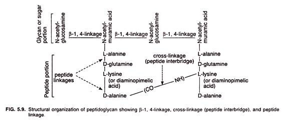
Peptidoglycan, the main constituent or back-bone of bacterial cell wall, consists of two parts: a glycan or sugar portion and a peptide portion.
The glycan portion is made up of alternating units of N-acetylglucosamine and N-acetyImuramic acid bonded with each other by β-1, 4-linkages. The peptide portion is a short-chain composed of four amino acids (L-alanine, D-glutamine, either L-lysine or diaminopimelic acid, and D-alanine) connected with each other by piptide-linkages and hence is called tetrapeptide chain.
The two adjacent of different tetrapeptide chains are interlinked by a cross-linkage (peptide interbridge). The type and extent of cross-linkages may vary among different species. In some species, the cross-linkage forms between the carboxyl group (-CO-) of an amino acid in one tetrapeptide chain and amino group (-NH-) of an amino acid in other tetrapeptide chain (Fig. 5.9).
In others, a pentaglycine chain is used to link two tetrapeptide side chains (Fig. 5.10). It is these cross-linkages that provide rigidity to the peptidoglycan which helps protecting the cell against osmotic socks exerted on it.
However, the diaminopimelic acid (DAP) does not occur in the peptidoglycan of all bacteria; only all gram-negative bacteria and some gram-positive bacteria possess it.
Most of the gram-positive bacteria have amino acid lysine instead of DAP. Another unusual feature of the peptidoglycan (i.e. the bacterial cell wall) is the presence of two amino acids that have the D-configuration, D-alanine and D-glutamine. It is because in proteins amino acids are always of L-configuration.
The chemical structure of one of the repeating units of peptidoglycan is given in Fig. 5.11 and an overall structure of peptidoglycan in Fig. 5.12.
Gram-Positive Cell Walls:
The cell wall of gram-positive bacteria (Bacillus, Streptococcus, etc.) appears as a thick homogenous layer (Fig. 5.13A), and mainly consists of peptidoglycan (up to 90%). The remainder being made up of proteins, polysaccharides, and teichoic acid. Teichoic acids are acidic polysaccharides, which lie on the outer surface of the peptidoglycan, and are covalently bonded with it.
Their functions are not known with certainty; they are considered to affect the passage of ions, thereby help maintain the cell wall at a relatively low pH so that self- produced enzymes (autolysins) do not degrade the cell wall. Other functions are also attributed to teichoic acid such as binding metals and acting as receptor sites for some viruses.
Gram-Negative Cell Walls:
The wall of gram-negative bacteria (Rhizobium, Escherichia, Salmonella, etc.) is biochemically far more complex than of gram-positive bacteria and appears usually trilayered (Fig. 5.13B). The innermost layer is the plasma membrane of the cell made up of phospholipid bilayer; the middle layer is the peptidoglycan (10% or less), and the outer most layer represents the outer membrane.
The region between the inner plasma membrane and the outer membrane is called periplasmic space. The inner half of the outer membrane is similar to the plasma membrane, but the outer half contains lipopolysaccharides (‘fat-carbohydrates’) in place of phospholipids.
The outer membrane is present outside the thin peptidoglycan layer (Fig. 5.14). Braun’s lipoprotein is the most abundant protein occurring in the outer membrane. It is a small lipoprotein covalently joined to the underlying peptidoglycan and embedded in the outer membrane by its hydrophobic end.
Braun’s lipoprotein joins the outer-membrane and peptidoglycan so firmly that both can be isolated as a single unit. There are, however, a special type of porin proteins present in the outer membrane. Three porin molecules cluster together and span the outer membrane to form a narrow channel through which molecules smaller than about 600-700 daltons can pass.
The most unusual constituents of the outer membrane are its lipopolysaccharides (LPSs). The latter are large, complex molecules consisting of three parts: lipid A, the core polysaccharide, and the ‘O’ side chain or ‘O’ antigen. Lipid A is hurried in the outer membrane while the remaining core polysaccharide and ‘O’ side chain project from the surface.
Lipid A is a major constituent of lipopolysaccharide and helps stabilize the outer membrane. Lipid A often is toxic and functions as an endotoxin. The core polysaccharide usually contains charged sugars and phosphate and contributes to the negative charge on the bacterial surface.
‘O’ side chain or ‘O’ antigen is a polysaccharide consisting of several peculiar sugars and varies in composition between bacterial strains. ‘O’ side chains rapidly change their nature to avoid detection and thus help bacteria to thwart host defences.
Difference in Cell Walls of Gram-positive and Gram-negative Bacteria:
Gram –positive:
1. Cell wall appears thick and homogenous.
2. Peptidoglycan comprises upto 90% of the cell wall hence more rigid.
3. Besides peptidoglycan, there are teichoic acids, other polysaccharides and proteins in the cell wall.
4. Teichoic acids are the main surface antigens.
5. More sensitive to wall attacking antibiotics like penicillin.
Gram-negative:
1. Cell wall appears thin and usually tri-layered.
2. Peptidoglycan comprises only 10% or less of the cell wall hence less rigid.
3. Besides peptidoglycan, there are phospho-lipids, proteins and lipopolysaccharides in the cell wall. Teichoic acids are absent.
4. Lipopolysacchrides are the main surface antigens.
5. Less sensitive to wall attacking antibiotics like penicillin.
Outer membrane serves as protective barrier. Despite its permeability to small molecules due to porin proteins, the outer membrane prevents or slows the entry of bile salts, antibiotics, lysozymes and other toxic substances which might kill or injure the bacterium.
As a result, infections with gram- negative bacteria are often more difficult to treat. Since teichoic acid is absent and the peptidoglycan is less in amount, the wall of gram-negative bacterium is less rigid as compared to that of gram-positive one.
Essay # 5. Size of Bacteria and the Significance of their Being Small:
Bacteria vary in size from cells as small as 0.1-0.2 µm in diameter to those more than 50 µm in diameter. The dimensions of an average rod-shaped bacterium, Escherichia coli, for example, are about 1 x 3 µm. For comparison, typical eukaryotic cells may be 2 µm to more than 200 µm in diameter. Bacteria are thus extremely small in comparison to eukaryotes.
Small size of bacteria (and almost all other prokaryotes) affects a number of their biological properties. For convenience, the rate at which the nutrients are taken in and wastes are passed out of a cell is in general inversely proportional to cell size. This is because transport rates are to some degree a function of the membrane surface area available; small cells have more surface area available than do large cells.
As a cell increases in size, its surface area-to-volume (S/V) ratio decreases. The surface area-to- volume (S/V) ratio of a sphere (cell) is expressed as 3/r. A small cell having a smaller r (radius) value has a higher S/V ratio than a larger cell (Fig. 4.3A) and, therefore, can enjoy more efficient exchange of nutrients with its surroundings than can a large cell, show more rapid growth rates and the formation of larger cell populations.
The parameters of rapid growth and larger cell populations greatly affect microbial ecology. It is so because high numbers of rapidly metabolizing cells can cause major physio-chemical changes in an ecosystem even over very short periods of time.
Essay # 6. Flagellation in Bacteria:
The pattern of flagellar arrangement (flagellation) is a good identification mark in bacteria (Fig. 4.3B). Flagella are either confined to the pole or poles or it may be present all-round the body of the bacterium. However, bacteria can be grouped as under on the basis of flagellation.
Atrichous — Bacteria that lack flagella.
Monotrichous — Single flagellum on either of the poles of the bacterial cell.
Amphitrichous — One flagellum or more on each pole of the bacterial cell.
Lophotrichous — Flagella in groups present on one pole of the bacterium.
Pcritrichous — Flagella present allround the body of the bacterial cell.
Essay # 7. Behavioural Responses of Bacteria:
Flagellar machinery of a bacterial (prokaryotic) cell has evolved responses to the gradients of physical and chemical agents in nature to which they often encounter. These responses to gradients may be positive or negative by directing movements of the body either toward or away respectively. Such gradient-directed movements are called taxes (sing, taxis).
A variety of taxes are found taking place in bacteria depending upon which type of gradient governs the movement. However, the different types of taxes (behavioral responses) are: chemotaxis, phototaxis, aerotaxis and magnetotaxis.
(i) Chemotaxis:
When motile bacteria react to chemical stimuli, they accumulate in some areas or retreat from others. Such stimulus-response behaviour is called chemotaxis. The attraction of organisms by chemical stimuli is brought about in the following manner (Fig. 4.4). Peritrichously flagellated bacteria (e.g., Escherichia coli) have two kinds of motile behaviour: straight-line swimming (called ‘runs’) and tumbling.
The latter interrupts the straight movement and causes reorientation. If the bacteria are placed in a concentration gradient of an attractant, the linear swimming movement will last many seconds if it is in the direction of the optimal concentration of the attractant, but in the opposite direction it is interrupted after only a few seconds.
Whilst the tumbling movement leads to a completely random selection of the new swimming direction, the direction- dependent duration of the linear swimming motion results in accumulation of the organisms in the region of optimal substrate concentration. The sensing of, and response to, stimuli is due to specific chemosensors, the binding proteins present in the cell envelop.
Bacteria possess a memory system that allows them to compare the concentrations of chemicals as they swim along, so that they effectively detect chemical concentrations over distances many times the length of a cell. In some cases, these are independent of substrate assimilation. Thus, some mutants show unchanged chemotactic reactions to certain nutrients, though they have lost the capacity to utilise them.
(ii) Phototaxis:
Many phototrophic microorganisms move towards light, a process called phototaxis. The phototrophic purple bacteria depend on light for their energy supply. Therefore, it is not surprising that they possess a phototactic mechanism and accumulate in illuminated areas. The advantage of phototaxis is that it allows a phototrophic microbe to orient itself most efficiently for photosynthesis.
If one projects a small spot of light onto a thick suspension of Chromatium on a slide previously kept in the dark, the organisms can be seen to collect in the illuminated area. Moreover, it appears that the organisms cannot leave the illuminated area, once they have entered it, in the course of their random movements. On entering the dark zone, abrupt reversal of flagellar motion propels them back into the light zone.
This reversal is so sudden that this response has been called ‘scotophobotaxis’ (shock reaction). Even slight differences in light intensity between areas of illumination can evoke this reaction. Some Chromatium species accumulate in areas that receive only 0.7% more light than the environment. This contrast sensitivity (with regard to illumination) is similar to that of the human eye (0.4%).
In some species, such as the highly motive phototrophic bacteria (e.g.. Rhodospirillum centenum), the entire colonies of cells show phototaxis and move in unison toward the light.
(iii) Aerotaxis:
Bacterial movements towards or away from oxygen or air is called aerotaxis. Some motile bacteria reveal their metabolic capacities relative to oxygen or air by their aerotactic movements.
When bacterial suspensions are placed between slide and coverslip, aerophilic bacteria accumulate near the edge of the coverslip and in the vicinity of air bubbles, demonstrating their requirement for aerobic conditions and their dependence on aerobic respiration for energy.
Strictly anaerobic bacteria, on the other hand, tend to collect in the centre, whilst microaerophilic bacteria, such as some pseudomonads and spirilla, keep a certain distance from the air interface.
(iv) Magnetotaxis:
Some bacteria contain inclusion of iron granules (called magnetosomes) which permit them to orient their movement in response to magnetic fields. This phenomenon is called magnetotaxis. A number of bacteria (rods, spirilla, cocci) has been isolated recently from the surface layers of sediments in freshwater ponds and the sea.
These have been found to orientate themselves in a magnetic field and swim in the direction of the field lines. They possess unusual amounts of iron (0.4% of their dry weight) as ferromagnetic iron oxide (magnetite) accumulated in the form of magnetosomes which are localised close to the area of flagellar insertion.
Bacteria isolated in the northern hemisphere seek the north; the field lines are directed downwards with a gradient of about 70°. Magnetotactic behaviour thus enables these bacteria to migrate downwards into the oxygen-poor or oxygen-free sediments.
Since the magnetotactic bacteria are anaerobic or microaerophilic, this mechanism and behaviour finds a ready ecological explanation. When such organisms are transferred to the southern hemisphere, they cannot survive; only a few ‘wrongly polarised’ cells, however, can grow and multiply. It indicates that the polarity is not genetically governed process.
Essay # 8. Endospores in Bacteria:
Some bacteria produce ‘endospores’ within their cell by a process called ‘sporulation’. Endospore-producing bacteria occur most commonly in the soil and the genera Bacillus and Clostridium are the best studied of endospore-producing bacteria.
These spores are extraordinarily resistant to environmental stresses such as heat, ultraviolet radiation, gamma radiation, chemical disinfectants, and desiccation and can remain dormant for extremely long periods of time.
Endospores are of great practical significance in food, industrial and medical microbiology due to their resistance and dangerous pathogenic nature of several species of endospore- producing bacteria. This is because it is essential to develop adequate methods to sterilize solutions and solid objects.
1. Structure:
The endospore (so named because of its formation within the cell), which is readily seen under the light microscope as strongly refractile bodies (Fig. 4.5A), due to being very impermeable to usual dyes (e.g., methylene blue), is structurally much more complex in that it possesses many layers that are absent in vegetative cells (Fig. 4.5B).
The outermost layer is exosporium, a thin delicate covering made of protein. Beneath the exosporium, there is a thick spore-coat consisting of several protein layers which are spore- specific. The spore-coat is impermeable and responsible for the spore’s resistance to chemicals.
Below the spore-coat is the cortex which may occupy as much as half the spore volume. Cortex consists of loosely cross- linked peptidoglycan. Inside the cortex, there is the core-wall which surrounds the core membrane and the core or spore protoplast. The latter possesses cytoplasm, nucleoid, ribosomes etc. but is metabolically inactive.
The core or spore protoplast of a mature endospore contains abundant dipicolinic acid and calcium ions normally existing in the form of calcium-dipicolinate complex (Fig. 4.6) and is in a partially dehydrated state as it contains only 10-30% of the water content of the vegetative cell. Because of it, the consistency of the core cytoplasm is that of a thick gel.
In addition to low water content, the pH of the core cytoplasm is about one unit lower than that of the vegetative cell and contains high levels of core-specific proteins, namely, small acid- soluble spore proteins (SASPs).
SASPs are considered to perform at least two important functions:
(i) They bind tightly to DNA in the core and protect it from ultraviolet radiation, dessication and dry heat and
(ii) They function as a carbon and energy source at the time of endospore germination to give rise to new vegetative cell.
2. Differences between Endospore and Vegetative Cell:
A mature endospore differs greatly from the vegetative cell from which it was formed. These differences are given in Table 4.1.
3. Formation of Endospore:
Endospore formation (called sporulation or sporogenesis) involves a very complex series of events in cellular differentiation. Endospore formation takes place only in such a vegetative cell which ceases growth due to lack of nutrients. The complex process of sporulation can be divided into seven stages (I to VII) which are shown in Fig. 4.7.
4. Endospore Resistance:
Bacterial endospores can retain viability for many years. A few viable endospores of Bacillus subtilis and B. licheniformis were found in the soil attached to plants that had been stored under dry conditions at the Kew Gardens Herbarium for 200-300 years.
Endospores can even retain viability for millennia, and viable endospores have been found in geological deposits where they must have been dormant for thousands of years.
What factors are responsible for such prolonged viability of endospores? It has long been thought that dipicolinic acid was directly involved in heat-resistance of endospore, but heat- resistant mutants now have been isolated in which dipicolinic acid is absent. Then, if not DPA, what factors make endospore so resistant to heat and other lethal agents.
It is now being believed, however, that there are several factors probably involved in endospore resistance. These are calcium-dipicolinate and acid-soluble protein stabilization of DNA, protoplast dehydration, the spore-coat, DNA-repair, and the greater stability of cell proteins in bacteria adopted to growth at high temperatures.
5. Germination:
The conversion of endospore into active vegetative cell appears a complex process and involves three steps: activation, germination and outgrowth. Activation is the process that prepares endospore for germination. It is most easily accomplished by heating at sub-lethal but elevated temperature.
An activated endospore undergoes germination which is characterized by swelling and rupture or absorption of spore-coat, loss of dipicolinic acid, degradation of small acid-soluble spore proteins (SASPs), loss of resistance to heat and other stresses, loss of refractility, and enhancement in metabolic activity.
The final stage is the outgrowth which involves visible swelling due to water uptake and synthesis of new RNA, DNA and proteins. The spore protoplast emerges from the broken spore-coat, develops into an active bacterial cell and begins to divide.
Essay # 9. Reproduction in Bacteria:
Reproduction in bacteria (almost in all monerans or prokaryotes) is asexual, taking place by means of binary fission, arthrospore formation, conidia formation and budding. Some of the bacteria produce dormant structures (endospores, cysts) to survive adverse conditions; these structures germinate producing vegetative cells when favourable conditions return.
(i) Binary Fission:
Binary fission is the most common method of reproduction in bacteria. It is a process is which a parent cell divides to produce two equal-sized daughter (progeny) cells. In binary fission (Fig. 4.8), the parent cell first enlarge and then its cell wall and plasma membrane begin to grow inward in the middle region.
Simultaneously, the circular bacterial chromosome replicates in characteristic prokaryotic manner resulting in two daughter bacterial chromosomes (Fig. 4.9).
The inward growth of the cell wall and plasma membrane proceeds forming a transverse septum in the middle in such a way that it divides the parent cell into two equal-sized daughter (progeny) cells each with a complete bacterial chromosome. Finally, the daughter cells separate from each other and become independent. Repeating the process results in multiplication of the bacterial population.
Binary fission is a quick process. Although the time required to complete a binary fission (called generation time or doubling time) varies considerably among different bacteria, some like E. coli needing only 20 mm under optimal conditions. At this rate, one cell of E. coli will multiply by binary fission to yield 512 cells in three hours, 3, 2768 cells in five hours, and so on.
(ii) Budding:
Budding (Fig. 4.10), a type of division characterized by an unequal division of cellular material, takes place in some rod-shaped bacteria which develop small outgrowths generally at one end. These outgrowths, finally, bud off from the parent cells as daughter cells and mature into bacterial cells. Rhodomicrobium is a budding bacterium.
(iii) Sexual Reproduction:
No true sexual reproduction is knows to occur in bacteria (almost in all monerans or prokaryotes). As normally defined, a true sexual reproduction represents a method of propagation of new individuals involving union of two compatible nuclei accompanied with plasmogamy, karyogamy, and meiosis occurring in a regular sequence at specified points in the life of an organism.
The two compatible nuclei contain a complete set of genetic material. Contrary to it, only incomplete sets of genetic material are transferred from one prokaryotic cell to the other, no karyogamy and meiosis is reported. Thus the bacteria in particular and all prokaryotes in general lack true sexual reproduction, and their requirements of sexuality are met by different processes of genetic recombination.
Although sexual reproduction with the formation of a zygote (karyogamy) and subsequent meiosis does not occur in bacteria (in all prokaryotes), genetic recombination takes place following horizontal gene transfer. In horizontal gene transfer, genes are transferred from one independent, mature bacterium (donor) to another (recipient).
This process is quite different from transfer of genes from parents to offspring (vertical gene transfer, in sexual reproduction) in eukaryotes. In eukaryotes, the recombination is reciprocal, i.e., all of the DNA is conserved in gametes that eventually arise from meiosis and recombination. Recombination in bacteria is a one-way DNA transfer from donor to recipient.
Transfer of DNA from a donor bacterium to the recipient bacterium takes place in three different ways: direct transfer between two bacteria temporarily in physical contact (conjugation), transfer of a naked DNA fragment (transformation), and bacteriophage-mediated transfer of DNA (transduction).
Essay # 10. Some Important Genera of Bacteria:
(i) Agrobacterium:
Agrobacterium tumefaciens, the causative agent of ‘crown gall’ disease of fruit trees (Fig. 4 11), is one of extensively studied phytopathogenic bacteria. In plants, crown gall is initiated during the first few days following infection by the bacterium, but once initiated, continued presence of the inciting bacterial cells is no longer necessary to maintain the gall state (tumerous state) in the plant.
A. tumefaciens, attacks the susceptible plants in the wound regions, transfers its ‘tumorigenic DNA’ or Ti-plasmid’, which is responsible for uncontrolled multiplication of cells leading to the formation of gall. This characteristic of A. tumefaciens has made it of immense significance in genetic engineering of plants.
As we know, for efficient integration of foreign gene into a host, a cloning vector is required. Several such vectors are available, but the most important of these for crop plants is natural Ti-plasmid (pTi); Agrobacterium tumefaciens carries Ti-plasmid and can transfer a small DNA fragment of this plasmid to host cells.
The technique of using this bacterium as a vector has been relatively most successful in dicotyledonous crops belonging to the genera Solanum, Nicotiana, Petunia and Brassica, rather than monocotyledonous crops.
(ii) Escherichia Coli:
E. coli is a straight rod of 0.4-0.7 x 2.0-4.0 µm, commonly found in the intestinal tract of mammals, usually single cell but may form short chains, does not produce spores or capsules, stains readily, gram-negative, motile with peritrichous flagella or nonmotile, grows readily on simple nutrient media, aerobic but in presence of carbohydrates becomes facultatively anaerobic, generally nonpathogenic in nature but some strains have been found to produce disease under exceptional circumstances.
About 90% or more strains of E. coli ferment lactose with production of acid and gas. One of the characteristics of E. coli is the production of colicins, a specific type of metabolite which is lethal to its own members as well as to related ones; the colicins, in this way, probably play a major role in maintaining the normal populations of E. coli.
E. coli are one of the leading species of ‘coliform bacteria’; the common test to determine the contamination of water by faecal matter is coliform test. Therefore, by testing their presence one can determine the potability of drinking water.
Besides, this bacterium has frequently been used by microbiologists in studying various processes of microorganisms such as physiology, biochemistry, cytology and genetics. They are also being used frequently as cloning organisms in newly born discipline of genetic engineering.
(iii) Lactobacillus:
The lactobacilli are rods, varying from long and slender to short coccobacilli; they commonly form chains, may rarely show motility by peritrichous flagella (when present), are nonsporing, and gram-positive becoming gram-negative with increasing age and acidity.
They are found in dairy products and effluents, grain and meat products, water, sewage, beer, wine, fruits and fruit juices, and pickled vegetables. They are also parasitic occurring in mouth, intestinal tract, and vagina of many homothermic animals, including humans.
The features that make lactobacilli microbiologically significant are:
(1) Their ability to ferment sugars resulting in the production of lactic acid, making it possible to use them in the production of fermented plant and dairy products or the manufacture of industrial lactic acid,
(2) Production of gas and other volatile products and,
(3) The heat resistance of most of the high-temperature lactobacilli, which enables them to survive pasteurization or other heating devices, like that given to the curd in the manufacture of swiss and similar cheeses. Contrary to it, some lactobacilli, e.g., L. viridescens and L. salinmandus have been found growing in refrigerated meats and are exceptional because of their ability to grow at low temperatures.
(iv) Rhizobium:
Rhizobia are rod-shaped (0.5-0.9 x 1.2-3.0 µm) (Fig. 4.12), gram-negative, nonsporing, and generally pleomorphic under adverse growth conditions. These bacteria are usually nonmotile or motile; motile, forms have two to six peritrichous flagella or a single polar or sub-polar flagellum. They are aerobic chemoheterotrophs requiring optimum temperatures of 25-30°C for their growth.
Rhizobia are symbiotic nitrogen-fixing bacteria and are the most important contributors of fixed nitrogen to soil. Nitrogen fixation by these bacteria can take place only when they grow in association with the host plant; they fail in fixing nitrogen when living free of the host.
However, rhizobial species invade root hairs of leguminous plants and incite production of root nodules, wherein they live endosymbiotically.
(v) Corynebacterium:
Corynebacterium (G. koryne = club) are gram-positive, aerobic; nonmotile, rod-shaped organisms frequently having a swollen end giving club-shaped appearance.
They consist of an extremely diverse group of bacteria including animal and plant pathogens as well as saprophytes. Some species such as C. diphtheriae are pathogenic causing diphtheria. However, some Corynebacteria are pleomorphic and form coccoid elements during growth.
(vi) Mycobacterium:
Mycobacterium are rod-shaped, pleomorphic, and may undergo branching or filamentous growth. The filaments however, become fragmented into rods or coccoid elements on slight disturbance. Mycobacterium possesses a distinctive straining property called acid-fastness which is due to the presence of unique lipid component called mycotic acids only found in the genus Mycobacterium.
M. tuberculosis is the causal agent of ‘tuberculosis’ in humans. When grown on solid media, these bacteria generally form tight, compact and often wrinkled colonies. M. leprae is another species that causes leprosy in humans.


