In this essay we will discuss about Protozoa:- 1. Definition of Protozoa 2. General Characters of Protozoa 3. General Organisation 4. Cytoplasm 5. Nucleus 6. Locomotor Organelles 7. Modes of Locomotion 8. Behaviour 9. Contractile Vacuole and Osmoregulation 10. Reproduction 11. Economic Importance.
Contents:
- Essay on the Definition of Protozoa
- Essay on the General Characters of Protozoa
- Essay on the General Organisation of Protozoa
- Essay on the Cytoplasm of Protozoa
- Essay on the Nucleus of Protozoa
- Essay on the Locomotor Organelles of Protozoa
- Essay on the Modes of Locomotion of Protozoa
- Essay on the Behaviour in Protozoa
- Essay on the Contractile Vacuole and Osmoregulation in Protozoa
- Essay on the Reproduction in Protozoa
- Essay on the Economic Importance of Protozoa
Contents
- Essay # 1. Definition of Protozoa:
- Essay # 2. General Characters of Protozoa:
- Essay # 3. General Organisation of Protozoa:
- Essay # 4. Cytoplasm of Protozoa:
- Essay # 5. Nucleus of Protozoa:
- Essay # 6. Locomotor Organelles of Protozoa:
- Essay # 7. Modes of Locomotion of Protozoa:
- Essay # 8. Behaviour in Protozoa:
- Essay # 9. Contractile Vacuole and Osmoregulation in Protozoa:
- Essay # 10. Reproduction in Protozoa:
- Essay # 11. Economic Importance of Protozoa:
Essay # 1. Definition of Protozoa:
The Protozoa may be defined as ‘microscopic, acellular animalcules existing singly or in colonies, without tissues and organs, having one or more nuclei. When in colonies, they differ from Metazoa in having all the individuals alike except those engaged in reproductive activities’.
Essay # 2. General Characters of Protozoa:
1. The protozoans are small, generally microscopic animalcules.
2. Simplest and primitive of all animals with very simple body organisation, i.e., protoplasmic grade of organisation.
3. Acellular animals, without tissues and organs.
4. Body naked or covered by pellicle but in some forms body is covered with shells and often provided with internal skeleton.
5. Protozoans are solitary or colonial; in colonial forms the individuals are alike and independent.
6. Body shape variable; it may be spherical, oval, elongated or flattened.
7. Body protoplasm is differentiated into an outer ectoplasm and inner endoplasm.
8. Protozoans may have one or more nuclei; nuclei may be monomorphic or dimorphic, vesicular or massive. Vesicular nuclei are commonly spherical, oval or biconvex, consist of a central body, the endosome (nucleolus) encircled by a zone of nuclear sap.
9. Locomotory organelles are pseudopodia, flagella, cilia or none.
10. Nutrition may be holozoic (animal-like), holophytic (plant-like), saprozoic or parasitic. Digestion intracellular, takes place inside the food vacuoles.
11. Respiration occurs by diffusion through general body surface.
12. Excretion also occurs through general body surface but in some forms through a temporary opening in the ectoplasm or through a permanent pore, the cytopyge.
13. Contractile vacuoles perform osmoregulation in freshwater forms and also help in removing excretory products.
14. Reproduction asexual or sexual; asexual reproduction occurs by binary fission, multiple fission, budding or sporulation and sexual reproduction is performed by gamete formation or conjugation.
15. Life cycle often exhibits alternation of generation, i.e., it includes asexual and sexual phases.
16. Encystment usually occurs to tide over the un-favourable conditions and it also helps in dispersal.
17. The single celled body of Protozoa performs all the vital activities of life and, therefore, no physiological division of labour is exhibited by them.
18. The protozoans exhibit mainly two modes of life, free-living inhabiting freshwater, saltwater and damp places, and parasitic living as ecto- and endoparasites. They are also commensal in habit.
Essay # 3. General Organisation of Protozoa:
(i) Size:
The Protozoa are usually microscopic and not visible to the naked eyes. Their size varies from 2 microns to 250 microns (one micron (m) is equal to 1/1000 mm or 0.001 mm). Babesia, Leishmania and Plasmodium are the smallest protozoans known so far. Some of the larger Protozoa, like Amoeba and Paramecium, can be seen with naked eyes.
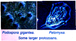
Spirostomum, a ciliate, grows to 3 mm long. Pelomyxa, a giant amoeba, attains a diameter of about 1 to 5 mm. Porospora gigantea, a Sporozoa, grows to about 16 mm long. Cycloclypeus, a Foraminifera, exceeds a diameter of about 50 mm and some shelled marine protozoans have diameters of about 63 mm.
(ii) Shape or Form of Protozoa:
Protozoa, the most primitive of all organisms, exhibit nearly all types of body shape. The body shape of a Protozoa is definite but it may usually vary within narrow limits. The body shape is usually determined by the consistency of cytoplasm, limiting membranes of the body, shells and skeleton. Amoeba has an irregular, asymmetrical body shape because of the absence of rigid body envelope.
However, the floating forms usually possess spherical body shape like Noctiluca the body shape is usually elongated in active swimmers like Euglena and Paramecium, the body shape may be flattened in creeping forms like Oxytricha. Shells and tests like those of Difflugia and Arcella determine the shape of these species.
It may be funnel-shaped like Stentor, bell-shaped like Vorticella and so on. Some of the protozoans like Radiolarians exhibit spherical symmetry and attached species like Vorticella exhibit more or less radial symmetry.
(iii) Body Envelope and Skeleton of Protozoa:
In Protozoa, the body envelope and external skeletal layers mark the boundary line between the body protoplasm and external environment. These protect them from external environmental hazards. The body envelope, being selective in nature, allows exchange of substances across it and helps in perceiving various types of stimuli.
However, the body envelope, in protozoans, may be either plasma lemma or pellicle. In some species like Amoeba proteus, the body envelope is a thin plasma membrane or plasma lemma which is mucopolysaccharide in nature.
It helps in adhesion to the substratum and in the exchange of various materials. The pellicle is comparatively thicker, tough, elastic and proteinous in nature; it helps in maintaining the general shape of the protozoans and performs the usual functions as referred earlier.
The skeletal layers are secreted in still other protozoans in which their protoptasmic body remains protected. These include cyst, theca, lorica and test or shell. The cyst is an external temporary sheath formed both by free-living and parasitic individuals. It is primarily secreted during un-favourable conditions.
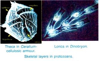
The encysted individuals comfortably tide over the environmental hazards. The theca is another skeletal layer found in many dinoflagellates like Ceratium and Glenodinium. It is a coat of closely-fitted armour of cellulose, comparable to the thick cell walls of higher plants.
The lorica, in majority of dinoflagellates, is differentiated into a number of plates arranged into a definite pattern; but in some forms, it may be formed of two valves.
The lorica is still another skeletal layer found in certain protozoans like Salpingoeca, Monosiga, Dinobryon, Synura splendida and Poteriodendron, etc. In fact, it is a coat of less-closely fitted armour of protozoans than the theca. The lorica is usually vase-shaped or tubular having an opening for the emergence of the anterior part of the animal or its appendages.
The base of the lorica is either attached directly to the substratum (in sessile, individuals like Salpingoeca) or it may terminate in a stalk like Monosiga. In colonial loricated protozoans, one lorica may be attached to another lorica directly as in Dinobryon or one lorica may be attached to another lorica by a stalk as in Poteriodendron and Synura splendida.
The shells or tests are still other skeletal layer of protozoans; these are of common occurrence. There are loose armour with one or more openings over the body of protozoans like Arcella, Difflugia, Euglypha, etc. In Arcella the shell is thin and composed of pseudochitin (proteins plus carbohydrates) and ventrally it has an aperture from which 3 or 4 pseudopodia project out.
In Difflugia the shell is made of sand and other foreign substances like fragments of foraminiferan’s shell and sponge spicules. These foreign substances get embedded in a secreted matrix by the animal, working like cement, to form the shell. The foraminiferan’s shells are made of calcium carbonate, while shells found in some rhizopods like Euglypha are siliceous being made of silica.
The radiolarian’s shells are internal skeletal layer lying between ectoplasm and endoplasm. It forms a central capsule, which is composed of pseudo-chitin or silica or strontium sulphate and secreted by the cytoplasm. The central capsule is perforated by one to many pores through which the extra- capsular cytoplasm extends out as fine pseudopodia.
Essay # 4. Cytoplasm of Protozoa:
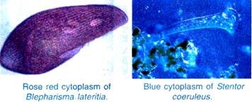
The cytoplasm of Protozoa is generally colourless but certain coloured species are also found; Blephcirisma lateritia is rose-red and Stentor coeruleus is blue. The cytoplasm is commonly divided into peripheral clear ectoplasm and inner granular endoplasm. These two may change from one to the other as is reported in Amoeba proteus.
The cytoplasm contains various organelles like mitochondria, Golgi apparatuses, endoplasmic reticulum, ribosomes, lysosomes, centrioles, microtubules, plastids, etc. The organelles listed above are typically the same as found in a typical Metazoa cell. The other structures exclusively found in Protozoa are various types of vacuoles, stigma, trichocysts, etc.
Essay # 5. Nucleus of Protozoa:
The nuclei of Protozoa exhibit a greater variety of size, shape and structure than the nuclei of Metazoa. The nucleus of Protozoa has a nuclear membrane, nucleoplasm, oxychromatin, basichromatin, and there may be a nucleolus. The nuclear membrane remains intact even in cell division.
There are various kinds of nuclei in Protozoa:
1. Vesicular nucleus has a large amount of nucleoplasm, the chromatin is small in quantity and it forms small granules, the achromatin (oxychromatin) is more fluid and its network, if present, is coarse, there is a round endosome of basichromatin or oxychromatin, or of both, e.g., Euglena, Arcella, Entamoeba.
2. Massive or compact nucleus has a small amount of nucleoplasm, there is a large amount of chromatin forming evenly scattered small granules, the achromatin is viscid forming a fine network, e.g., Amoeba. In the majority of Protozoa the nuclei show a structure intermediate between the vesicular and massive nuclei.
3. Polyenergid nucleus has several sets of chromosomes, instead of one set inside the nuclear membrane, this is due to mitosis occurring repeatedly inside the nuclear membrane. But the sets of chromosomes are finally liberated and each forms a new nucleus. The polyenergid condition is a provision for spore formation, e.g., Radiolaria.
Usually Protozoa (Mastigophora, Sarcodina and Sporozoa) have a single nucleus, but some (Ciliophora and Opalinata) have more than one. When the nuclei are more than one, they may be alike or different. Some Sarcodina may have many similar nuclei, e.g., two in Arcella and hundreds in Pelomyxa.
In Trypanosoma (a flagellate) there are two dissimilar nuclei, the principal one is trophonucleus which regulates metabolism and trophic activities; the second one is a kinetonucleus which controls the locomotory organelles, the former is vesicular and the latter of massive type.
4. Chromosome nucleus, in this case the nucleus retains chromosome’s in interphase, e.g. Amoeba sphaeronucleus. In Opalina many monomorphic nuclei are found.
5. Dimorphic nuclei are found in Ciliophora, the larger one is macronucleus containing trophochromatin, it controls vegetative functions, it divides amitotically, and in conjugation it disappears and is replaced by material from the synkaryon. The shape of the macronucleus is much varied.
The second nucleus is a small round micronucleus, there may be one or more micronuclei; it contains idiochromatin and controls reproduction. It divides mitotically in binary fission and conjugation; it gives rise to the macronucleus when the latter becomes effete and disintegrates.
The shape of nuclei also varies; it may be spherical, e.g., Entamoeba, kidney-shaped, e.g., macronucleus of Paramecium, horse-shoe-shaped, e.g., macronucleus of Vorticella and beaded , e.g., macronucleus of Stentor.
Essay # 6. Locomotor Organelles of Protozoa:
The Protozoa perform locomotion or movement by various organelles; pseudopodia characteristic of Sarcodina, flagella characteristic of Flagellata (Mastigophora), cilia characteristic of Ciliata and other contractile structures of pellicle, myonemes, characteristic of Sporozoa and few others. The seat of locomotion lies in the ectoplasm, since locomotor organelles either arise from it or are present in it.
(i) Pseudopodia:
Pseudopodia are generally temporary outgrowths of protoplasm from any part of the body, they are found in those Protozoa which are “naked” or have a very thin pellicle. Pseudopodia may be of ectoplasm or they may also have a core of endoplasm. Following kinds of pseudopodia are met with.
(a) Lobo-podia are blunt, short or finger-like, they are made of ectoplasm with a core of fluid endoplasm, e.g., Arcella and Amoeba.
(b) Filo-podia are fine, long threads, often with rounded ends, at times they may branch; they are made of only hyaline ectoplasm, e.g., A. radiosa-Radiolaria.
(c) Rhizopodia or reticulopodia are thin, long and branching, the branches of adjacent pseudopodia may anastomose to form a network which also serves as a trap for capturing food, e.g., Elphidium.
(d) Axopodia are long, stiff threads made of ectoplasm, with a hard central axial filament of endoplasm, unlike others they are semi-permanent, e.g., Actinophrys. Axopodia are not organelles of locomotion but are only for capturing food.
(ii) Flagella:
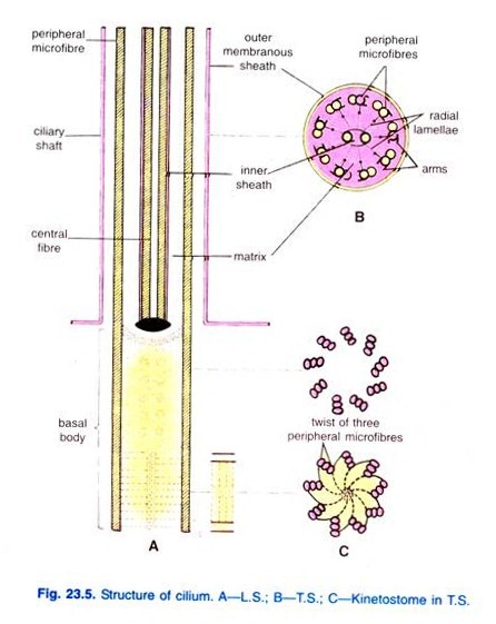
Flagella are extremely fine fibres having a central axoneme made of two longitudinal fibrils, and an enveloping protoplasmic sheath having nine double longitudinal fibrils forming a ring. All 20 fibrils lie in a matrix of dense cytoplasm and they fuse at the base to join a basal granule or kinetosome. The kinetosome may be joined to the nucleus by a rhizoplast.
The not act as a centriole then it is connected by a rhizoplast to a centriole or to the nucleus. At the tip of the main flagellum may be a very fine end piece or mastigoneme, or the main axis of the flagellum may bear fine, flexible lateral processes or mastigonemes on one side or on both sides. Mastigonemes constitute the so-called flimmer or ciliary flagellum.
Flagella are recognised to be of the following types depending upon the arrangement of mastigonemes in them:
(i) Stichonematic:
When the mastigonemes are present on one side of the flagellum, it is called stichonematic flagellum, e.g., flagellum of Euglena.
(ii) Pantonematic:
When the mastigonemes are present in two or more rows arranged on both the sides of the flagellum, it is called pantonematic, e.g., flagellum of Paranema.
(iii) Acronematic:
When the mastigonemes are absent and the distal end of the flagellum ends in a terminal filament, e.g., flagellum of Chlamydomonas and Volvox.
(iv) Pentachronematic:
When the mastigonemes are present in two rows on the lateral sides of the flagellum but the flagellum ends in terminal filament, e.g., flagellum of Urcoelus.
(v) In some cases, the flagellum is simple without mastigonemes and/or terminal filament, Chilomonas, Cryptomonas.
The number and arrangement of flagella varies greatly in Mastigophora. They may be one to eight in number. The free-living forms are usually with one to two flagella but the parasitic forms may have up to eight.
Usually flagella originate from the anterior end of body but in some cases like Trypanosoma, it originates from the posterior end and travels along the margin of undulating membrane and becomes free anteriorly. Flagella are primarily organelles of locomotion and secondarily for capturing food.
(iii) Cilia:
Cilia are exactly like flagella in structure and there is no real distinction between them, except in the method of working. In primitive forms cilia cover the entire body, but in more specialised forms cilia are restricted to certain regions only.
Cilia arise from kinetosomes, from each kinetosome arises a rhizoplast which does not join the nucleus, nor do cilia bear any mastigonemes. Running slightly to the right of a longitudinal row of kinetosomes is a delicate thread called kinetodesma.
A row of kinetosomes with its kinetodesmata forms a longitudinal unit called kinety, all kinetia of an animal constitue its infraciliary system. Infraciliary system is characteristic of all ciliates, even in those forms in which cilia are lost in the adults the infraciliary system is retained.
The infra-ciliary system of ciliates differs from that of flagellates in the following respects:
(a) The cilia are generally shorter and more numerous than flagella.
(b) In ciliates the infraciliature is not joined to the nucleus, not are kinetia inter-connected; in flagellates rhizoplasts join the kinetosomes to the nucleus, and the kinetia may be interconnected
(c) In cell division of ciliates the cleavage is perkinetal because it cuts across all kinetia, the upper halves go to one daughter cell and the lower halves to the other. This type of division is called homothetigenic in which the daughter cells are duplicates of each other.
In cell division of flagellates the cleavage is interkinetal because it is longitudinal and parallel to kinetia, so that kinetia are not cut but are shared by daughter cells. This type of division is called symmetrigenic in which the daughter cells are not duplicates but mirror images of each other. The normal number of kinetia of an animal is restored by division of kinetosomes.
(d) Cilia have no mastigonemes as in flagella.
The cilia may form the following composite motile organelles:
(a) Membranelles are membranes formed by fusion of two or more adjacent transverse rows of short cilia, they are found in the peristome making powerful sweeps.
(b) Undulating membranes are made of one or more longitudinal rows of cilia fusing together, they are found in the peristome or cytopharynx and are used for food collection, e.g., Vorticella. The undulating membrane of Trypanosoma is only a web of ectoplasm, it is not made of cilia and it is locomotory.
(c) Cirri are formed by fusion of two or three rows of cilia on the ventral side of some ciliates, they are locomotory and may also be tactile, e.g., Stylonychia.
(iv) Myonemes:
Myonemes are contractile fibrils in the ectoplasm; they may be surrounded by a canal; they may be straight or may form a network. Myonemes have alternate rows of light and dark substance, e.g., Stentor. They are found in flagellates, ciliates and sporozoans. They are primarily organelles for metaboly, e.g., Paramecium, and secondarily for locomotion by muscle like contraction in Monocystis and sporozoan like Plasmodium.
Essay # 7. Modes of Locomotion of Protozoa:
Different modes of locomotion are reported in Protozoa due to the presence of different types of locomotor organells in them.
Thus, the various modes of locomotion found in Protozoa are as follows:
1. Amoeboid movement performed by pseudopodia and characteristic of Amoeba.
2. Flagellar locomotion performed by flagella and characteristic of flagellates like Euglena.
3. Ciliary locomotion performed by cilia and characteristic of ciliates like Paramecium.
4. Gliding or metaboly performed by myoneme fibrils and characteristic of Sporozoa like Monocystis, Plasmodium.
Some ciliates and flagellates also exhibit this type of locomotion:
(i) Amoeboid Movement:
Amoeba moves from one place to other by pseudopodia. The pseudopodia are finger-like temporary processes given out from any part of the body and withdrawn. The pseudopodium is formed by the projection of ectoplasm in which endoplasm flows. Thus, the cytoplasm which enters into pseudopodia is naturally drawn from the other parts of the body.
Amoeba moves by the formation of pseudopodia, characteristic of this animal, is known as amoeboid movement. Various theories have been put forth to explain the way of formation of pseudopodia and amoeboid movement.
However, change of viscosity theory or sol-gel theory proposed by Hyman (1917), later supported by Pantin (1923-26) and Mast (1925), explains well the way of the formation of pseudopodia and amoeboid movement. According to them the amoeboid movement is the result of changes within the colloidal protoplasm from fluid plasmasol to more solid plasmagel and vice- versa.
Accordingly, the amoeboid movement is the net result of the following four steps occurring simultaneously in the body of Amoeba:
(i) Attachment of the body of Amoeba to the substratum.
(ii) Conversion of plasmasol into plasmagel, i.e., gelation at the anterior advancing tip.
(iii) Conversion of plasmagel into plasmasol, i.e., solation at the posterior opposite end of the body.
(iv) Contraction of the plasmagel at the posterior end of the body to push the plasmasol forward.
These processes are repeated again and again and, thus, Amoeba moves ahead. The speed of amoeboid movement varies from 2 microns to 3 microns per second. This is supposed to be the most primitive type of animal movement.
Recently, a number of proto-zoologists have worked out the process of amoeboid movement variously. But nearly all of them conclude that the theory given by Hyman and supported by Pantin and Mast is essentially correct.
(ii) Flagellar Movement:
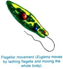
The free-living flagellates like Euglena moves by lashing the flagellum and by the movement of the whole body. Flagella performs lashing movements with a rowing action or undulating motion. In rowing the flagellum is held rigid but slightly concave in the direction of the stroke, in recovery it bends and is drawn back.
In undulating motion it beats obliquely and undulations pass along the flagellum from the base to the tip causing the animal to rotate. The movement of flagellum results in a forward propulsion of the animal in a spiral fashion. The Euglena locomotes about 15 microns to 30 microns in a second.
(iii) Ciliary Movement:
The ciliates are characterised by the possession of numerous, small, fine, thread-like ectoplasmic processes, the cilia. The ciliary movement is like that of flagellar movement. The cilia are generally directed backward, whose constant beating pulls the animal forward. But if the cilia are directed forward, their constant beating pulls the animal backward.
However, the ciliary movements result in a forward propulsion of the animal in a spiral path. Paramecium is a typical example exhibiting ciliary locomotion. The speed of ciliary locomotion varies from 400 microns to 2,000 microns per second.
(iv) Gliding Movement:
The sporozoans usually exhibit a characteristic contractile movement due to the presence of myoneme fibrils. The myoneme fibrils are highly contractile and elastic in nature. They contract and expand bringing about a change in the shape of the body. Thus, the animal moves or glides from its original place. This type of movement is usually exhibited by parasitic forms like Monocystis.
The myoneme fibrils, found in certain free living forms like Euglena, cause waves of contraction to pass from anterior to the posterior end of the body. Thus, there occurs a series of changes in the shape of the body which help in crawling worm-like movement.
Essay # 8. Behaviour in Protozoa:
The movements or reactions performed by an animal due to its sensitivity, in response to its environmental changes constitute the behaviour of the animal. The responses the animal makes for environmental changes follow a constant pattern for that animal, thus, each animal has its own characteristic behaviour. Any environmental change to which an animal responds is called a stimulus.
Movements made by an animal in response to stimuli may be positive or negative. If the animal is attracted by a stimulus and turns towards it, then the response is positive; but if the animal is repelled by stimulus and moves away from it, then the response is negative. Reactions to stimuli are classified into kinesis and taxes. Kinesis is that reaction in which the stimulus leads to an increase in the movements of the animal.
Taxes are those reactions which are directed to the stimulus, they depend on the possession of some sense organ. An animal may react to light, contact, temperature, gravity, chemicals or electric current. Positive reactions are rare exceptions in Protozoa, majority of their reactions are negative.
The response of an animal to a stimulus depends not only on the nature of the stimulus, but also on the condition of the animal at the moment.
(i) Amoeba:
Amoeba has no structures for reception of stimuli but its responses to stimuli are due to the irritability of its protoplasm. To gentle contact an Amoeba reacts positively and forms a food cup, but to forceful contact it reacts negatively, it first stop then forms new pseudopodia and moves away either at right angle or in the reverse direction.
If its surrounding water is changed to distilled water, or sugar or salt or weak acid is added to its surrounding water, then pseudopodia are withdrawn and body becomes rounded, this is followed by formation of a single pseudopodium by which the animal moves away.
If a chemical is applied to a small area of the body, the stimulated region contracts away from the stimulus, and the animal moves away in the opposite direction. If the temperature of its environment is raised between 30° and 35°C, then all movements cease, death results at 40°C, but Amoeba becomes most active between 20° and 25°C.
Strong light causes gelation of plasmasol in the illuminated side, but the opposite side forms pseudopodia and the animal moves away.
Amoeba avoids darkness, medium light is most favourable which is called optimum. Sudden and intense increase of light causes stopping of all movement and rounding off of the body, but if the intense light is maintained then after some time movement is resumed, this shows that Amoeba can adapt itself to new conditions. The response of Amoeba to different stimuli varies with the strength of the stimulus.
(ii) Euglena:
If a drop of water containing Euglenas is placed in direct Sunlight and half of the drop is shaded, then it will be seen that animals avoid both the lighted and shaded parts and collect in the middle area of medium light which is the optimum. Like most organisms containing chlorophyll, Euglena reacts positively towards light, it orientates itself parallel to the light rays and moves towards the sources of the illumination.
But very strong light makes Euglena stop, then move back and pivot on its posterior end, while the anterior end traces a wide circle; this behaviour is avoiding reaction.
The anterior end of Euglena is sensitive to light and it rotates as it moves forwards but when the light falls upon it from one side only, it reacts by swerving violently, and it orientates itself parallel to the rays of light whenever its photoreceptor is shaded by the stigma. The stimuli other than light, Euglena behaves in much the same way as other Protozoa, giving a negative reaction to unpleasant changes of environment.
(iii) Paramecium:
The behaviour of Paramecium is stereotyped, but it is well adapted to preserve it and to keep it within the bonds of a normally favourable environment. Special organelles for perception of stimuli are not seen, but the anterior end seems to be more sensitive than other parts of the body.
When Paramecium comes in forceful contact with a solid object, it reacts negatively, the ciliary beat is reversed by which it moves backwards and pivots on its posterior end, while the anterior end revolves in a circle; this behaviour is an avoiding reaction by which different samples of surrounding medium are brought into its peristome, if a sample is favourable the animal moves forwards in the direction of the sample but if the sample is un-favourable it moves away at an angle.
Detection of the external environment is probably by means of cilia, perhaps all the cilia are sensory receptors, but there are some long, stiff cilia which play no role in locomotion and are probably entirely sensory. Jarring of the surround-medium causes the animal to swim downwards.
To all contact it gives the avoiding reaction, but when it is stimulated violently by contact or chemicals, it responds by discharging its trichocysts. If a small drop of 1/50% acetic acid is placed in a large drop of water containing Paramecia then they collect in the acid drop; but if the strength of acid is increased to 1/10%, then they move away from the acid drop.
Thus, they swim into weak chemicals but give the avoiding reaction when they come in contact with boundary of the drop, this produces their aggregation. On the other hand, in a stronger chemical they give the avoiding reaction when they meet the outer boundary of the drop of acid so that they are unable to enter it. Light has no effect on Paramecium, but it gives an avoiding reaction to strong light and ultra-violet rays.
The optimum temperature for Paramecium is between 24° and 28° C., and up to a point it becomes more active at higher temperature and less active at lower temperature, but to sudden temperature changes it gives an avoiding reaction. Paramecium is weakly negative to gravity in its reaction.
If a weak electric current is passed in the medium containing Paramecia, it will be seen that the animals swim towards the cathode because the current causes a partial reversal of ciliary beat. With stronger current more cilia beat backward than forwards, so that the animal swims towards the anode.
The reactions of Protozoa show similarities in many details with those of higher animals, and their reactions are quite adequate to maintain them within their normal environment.
The behaviour of Protozoa is explained by certain theories:
1. Tropism theory of Loeb says that Protozoa respond to stimuli in a forced compulsory way, not because of choice, but because they cannot behave in any other way; they act like automatous being driven entirely by external stimuli.
2. Trial and error theory of Jennings maintains that Protozoa escape un-favourable conditions and find more favourable ones by repeated avoiding reactions amounting to trial and error. The animals are not forced into or away from un-favourable regions like automatons, but they are in control of their activities and direct their own course. The expression klinokinesis is now substituted for trial and error behaviour and avoiding reaction.
Essay # 9. Contractile Vacuole and Osmoregulation in Protozoa:
(i) Shape, Size and Occurrence:
The contractile vacuoles are usually large, colourless, pulsatile fluid-filled organelles found in majority of protozoans. These vacuoles are nearly always found in freshwater Flagellata, Sarcodina and Ciliata. The contractile vacuoles are also found in some marine ciliates but these are not at all found in parasitic protozoans.
The contractile vacuoles are found in their simplest form in Sarcodina like Amoeba. In this case, these are usually spherical vesicles or sometimes irregular and bounded by a limiting membrane. These vacuoles are found surrounded by a circlet of mitochondria which provide energy for their pulsating activity.
In Flagellate like Euglena, the contractile vacuole is somewhat complicated and surrounded by a large number of accessory contractile vacuoles. In Ciliata like Paramecium, the contractile vacuoles are much complicated and found surrounded by 5 to 12 radiating canals or feeding canals which collect water from the various parts of the body.
The number of radiating canals varies in different ciliates. The radiating canals discharge their contents in the main contractile vacuole, thus, serving as feeders.
(ii) Situation and Number:
The position of contractile vacuole is not definite in Sarcodina and, therefore, can be found anywhere in the endoplasm. In Flagellata, e.g., Euglena the contractile vacuole is found situated near the anterior end at the side of reservoir. In Ciliata like Paramecium, the contractile vacuoles are usually two in number, situated one at each end of the body.
The number of contractile vacuoles varies in the different groups of Protozoa but its number remains constant, in the same species. However, it is single in Amoeba, single in Euglena, two in Paramecium but these are many in Radiolaria and Heliozoa.
(iii) Structure:
Contractile vacuole is an empty space filled with fluid. Electron microscopic studies have revealed that its limiting membrane is lipoprotein in nature, like that of the plasma membrane.
(iv) Mode of Working:
The mode of working of contractile vacuole includes two steps, the diastole and the systole. The diastole is the phase of enlargement of the contractile vacuole to its maximum size and systole is the phase of its contraction to expel its contents.
A contractile vacuole is usually formed by the fusion of a large number of very small droplets in the area where contractile vacuole is to be formed (Amoeba) or around the mitochondria contractile vacuole (Euglena).
The systole occurs by the sudden burst of the contractile vacuole in Sarcodina but in Flagellata like Euglena, it empties in reservoir and so is the case with other forms. As referred, the energy required for the working of contractile vacuole is furnished by the mitochondria surrounding the vacuole.
However, the exact mechanism of working of contractile vacuole is not yet understood, even then the following theories have been put forth to explain its working:
1. Osmotic theory:
This theory explains that the water from the surrounding cytoplasm enters into the contractile vacuole by osmosis.
2. Filtration theory:
This theory explains that the water from the cytoplasm is forced into the contractile vacuole through its membrane due Fig- to internal hydrostatic pressure. Kitching has, however, contradicted this theory.
3. Secretion theory:
This theory states that the water is actively secreted into the vacuole during diastole through the vacuole wall. This theory, too, is not widely accepted.
(v) Function and Significance:
Contractile vacuole performs the function of osmoregulation by removing excess of water content from the body. In addition to its water regulatory function, the contractile vacuole is also believed to be excretory in function.
It has been observed that the water from the surrounding media continuously enters in the body of freshwater protozoans, therefore, water content of the protoplasm increases, i.e., there is an increase in the internal hydrostatic pressure.
This increased water content or hydrostatic pressure of the protoplasm inhibits the normal functioning of the body and if it continues to increase then a time may come when the body of the individual may burst.
Thus, the contractile vacuole helps in removing the excess water content of the protoplasm, i.e., it helps in maintaining the internal hydrostatic pressure.
Actually, the body fluid is hypertonic to the surrounding medium in freshwater forms. In case of marine and parasitic protozoans the surrounding media is nearly isotonic and, therefore, no excess water enters in the body. Hence, contractile vacuole is usually absent in these forms.
Essay # 10. Reproduction in Protozoa:
The process of reproduction is to continue one’s kind in nature. The mode of reproduction in Protozoa is highly variable among different groups although it is primarily a cell division. Protozoa reproduce both asexually and sexually.
(i) Asexual Reproduction:
In asexual mode of reproduction no special sex cells are involved but there is always some form of fission present. An essential part of the process is the partition of some parent chromatin substance among daughter individuals. Hence, the fission of the cell body is preceded by the division of the nucleus.
Asexual reproduction occurs by the following methods:
1. Equal or Binary Fission:
Equal or binary fission takes place for reproducing and also for gamete formation. Usually there is a centriole within the nucleus, but unlike Metazoa no asters are formed, moreover the nuclear membrane persists intact during division in most Protozoa.
The nucleus elongates and divides amitotically into two parts which travel apart, then the cell constricts in the middle to form two daughter cells. Macronuclei of ciliates divide amitotically.
Binary fission is simple in Sarcodina like Amoeba where the plane of division is not definite and it is usually transverse in Ciliates like Paramecium but in most flagellates like Euglena it is longitudinal in which the nucleus elongates transversely, but the cell divides lengthwise, while binary fission is oblique in certain cases like Ceratium.
In binary fission (in flagellates) a single flagellum is retained usually by one daughter cell, and the basal granule divided into two, the new basal granule forms a flagellum in the other daughter cell.
When there are many flagella they are distributed among the daughter cells which grow new flagella to complete the number. Cilia are shared by daughter cells and new cilia are formed by kinetosomes to complete the number. Chromatophores usually divide but contractile vacuoles rarely divide, they are generally shared or are made anew. Complex organelles are destroyed and then re-made in the daughter cells.
2. Multiple Fission:
The nucleus divides repeatedly without division of the cytoplasm, later the cytoplasm separates into as many parts as there are nuclei, usually some residual cytoplasm is left un-segmented. If multiple fission produces four or more young ones by equal cell division, and the young ones do not separate till the process is completed, then such cell division is spoken of as repeated fission, e.g., Vorticella.
Multiple fission produces small cells which may grow into adults or they may become gametes which require fertilisation to form sporozoites (Plasmodium). The multiple fission may lead either to asexual or sexual reproduction.
Products of multiple fission of a zygote generally form spores, sometimes the products of any multiple fission are called spores. A spore may be enclosed in a spore case (Monocystis) or they may be naked. The naked spores may be amoeboid (Entamoeba) or flagellated (Chlamydomonas) or ciliated (Suctoria). Spores may be gametes or serve for the distribution of the species.
All types of fission occur within a cyst or without encystment. Cyst formation is common in freshwater and parasitic Protozoa, though all Protozoa do not form cysts. In cyst formation the animal becomes rounded, loses its organelles of locomotion, it ejects the food vacuoles and contractile vacuoles disappear.
The animal then secretes a.gelatinous covering which hardens into a chitinous epicyst, inside this a membranous endocyst is secreted; the cyst may have more than two layers. The function of the cyst is protection of the animal against un-favourable conditions of environment, or it may be reproduction. The cysts can be carried by wind or some other agent and are, thus, important in dispersal.
Protozoa have the following kinds of cysts:
(i) Resting cysts enable an organism to proceed undisturbed in its normal activities (Euglena);
(ii) Resistance cysts are formed against un-favourable conditions of environment (Amoeba);
(iii) Gamocysts are those in which union of gametes takes place for reproduction (Gregarina);
(iv) Oocysts contain a zygote (Plasmodium);
(v) sporocysts are those in which multiple fission occurs to form sporozoites (Monocystis).
Finally excystment takes place on return of favourable conditions, but the individual leaving the cyst is never the same as the one that underwent encystment, it has a complement of new organelles and renewed vigour. The excystment may be through a minute pore in the cyst, but more usually it is due to the protozoan secreting some enzymes which rupture the cyst wall.
3. Plasmotomy:
An asexual division of a multinucleate animal in which the cytoplasm divides but the nuclei do not is called plasmotomy (Opalina, Pelomyxa). Later each daughter cell regains the normal number of nuclei by nuclear division.
4. Budding or Gemmation:
An unequal division of the parent body produces one or more buds which may separate from the parent, the nucleus of the bud is a part of the parent nucleus, e.g., Arcella. The bud is smaller than the parent; the buds may grow into adults or may become gametes.
When buds are formed on the surface of the parent, then this is known as exogenous budding, e.g., Noctiluca produces hundreds of buds on its surface as small protuberances.
When the buds are formed inside the cytoplasm and may remain within the parent, then the process is called endogenous budding, e.g., Arcella. Endogenous budding may be a method of asexual reproduction or it may bring about formation of gametes, e.g., Arcella becomes multinucleate, protoplasm collects around the nuclei to form many amoebulae which escape from the parent and grow into adults.
5. Parthenogenesis:
Parthenogenesis is the ability of the gametes to develop into adults without fertilisation by gametes of the opposite sex, the gamete possessing this power is almost always the female one, e.g., in Actinophrys two individuals get enclosed in a cyst, each divides to form two gametes, one gamete of an individual conjugates with a gamete of the other individual, the remaining gamete of each individual develops parthenogenetically into an adult.
Thus, gametes which have been unable to undergo cross fertilisation develop by parthenogenesis. Potential gametes of Chi amy domonas will grow and divide to become adults when they have missed syngamy. Endomixis of ciliates is also a parthenogenetic phenomenon. The chromosome condition in parthenogenesis may be expected to be haploid since no fertilisation occurs, but it is generally diploid.
6. Regeneration:
Regeneration is the capacity to form new tissues to replace a lost part, this capacity varies inversely with the complexity of an organism.
In Protozoa any nucleated portion is capable of regeneration, while non-nucleated portions are not, e.g., Stentor has a long chain-like nucleus, if the animal is cut transversely into say three parts, then each piece having a portion of the nucleus will regenerate the missing portions and three Stentors will be formed.
(ii) Sexual Reproduction:
Sexual reproduction occurs by the following methods in Protozoa:
1. Syngamy or Copulation:
Syngamy is the union and complete fusion of two gametes of the same species. If the two gametes are identical morphologically, though they may be different physiologically, then they are isogametes and their syngamy is isogamy (Monocystis). If the gametes differ in size and morphology, then they are anisogametes and their syngamy is anisogamy (Plasmodium).
The smaller, usually numerous and motile gametes are male or microgametes; the larger, generally few and inactive gametes are female or macrogametes. Meiosis or reduction division occurs generally in the formation of gametes, but in many flagellates meiosis is post-zygotic, that is, it occurs after the formation of the zygote.
The fusion of two gametes produces a zygote, its nucleus is formed by the fusion of nuclei of gametes, and it is called a synkaryon.
The zygote may develop directly into an adult, or it may encyst and undergo multiple fission. Syngamy whether isogamous or anisogamous is always exogamous, that is, the fusing gametes come from different parents, hence, sex distinction may be said to exist in Protozoa, though sexes may not be distinguished.
2. Conjugation:
Conjugation is a temporary union of two Protozoa of the same species for an exchange of nuclear material without the fusion of their cytoplasm, e.g., in Paramecium caudatum. In ciliates there is no formation of distinct gametes.
A sexual process somewhat intermediate between syngamy and conjugation occurs in Vorticella in which one individual forms one to four microgametes by repeated fission, and the other individual forms a macrogamete by nuclear modification, the macrogamete is a hologamete because it is not formed by fission.
Thus, Vorticella shows sexual dimorphism in its gametes, a microgamete fuses with a macrogamete to form a zygote. The zygote by three divisions produces seven cells which grow into adults.
In both syngamy and conjugation there is a rejuvenation of the animal by replacement of the macronucleus with material from the synkaryon, both processes produce new types of individuals by combination of genes, hence, they give the race a better chance of survival.
3. Automixis:
In some Protozoa the nucleus divides into two, the two nuclei fuse together, this is called automixis. If the two nuclei which fuse are present in a single cell, then the process is called autogamy, but if the two fusing nuclei are present in two different cells, then the process is known as paedogamy.
Autogamy occurs in a single Paramecium aurelia which provides both the fusing nuclei to form a synkaryon. Paedogamy occurs in Actinosphaerium and Actinophrys in which two cells of a secondary cyst and their two remaining nuclei fuse to form a zygote which reproduces by binary fission.
Sexual reproduction of Protozoa differs from the sexual reproduction of Metazoa in that the protozoan is both somatic and gametic.
For many generations there is a somatic phase in which binary fission occurs, then one generation is gametic in which syngamy or conjugation takes place. The function of binary fission is reproduction or increasing the number of individuals; and the function of syngamy or conjugation is rejuvenation, but not reproduction although it is called “sexual reproduction”.
In the life cycle of some Protozoa binary fission alternates with syngamy, this alternation may have regular sexual and asexual generations (Elphidium), but more usually binary fission is repeated for many generations continuously, and it is broken only occasionally by syngamy or conjugation.
Probably the occasional conjugation occurs only when the physiological condition of the animal becomes different from normal (Paramecium).
4. Endomixis:
It is a type of nuclear re-organisation which usually occurs when conjugation is prevented. In this case fusion of pro-nuclei does not take place. But the macronucleus is re-organised from micro-nuclear material. The re-organised macronucleus accelerates the metabolic activities of the individual and helps in the renewal of the vigour as is reported in Paramecium aurelia.
5. Hemixis:
It has been reported in the various species of Paramecium like P. caudatum, P. aurelia and P. multimicronucleatum. In this case, the macronucleus throws away its many fragments of different sizes in the cytoplasm which are absorbed in it. The left out part of macronucleus, then starts behaving in a normal way and becomes the fresh macronucleus.
The micronucleus, however, plays no part in hemixis and remains inactive and unchanged during this process.
Essay # 11. Economic Importance of Protozoa:
Protozoa is a group of acellular microscopic animals which have occupied almost all possible ecological habitats on the earth. For instance, they live in water, in moist surface of soil, in air and even as commensals and parasites in animals and plants. Man being a biological species is invariably effected by these organisms.
Some of them are beneficial in the sense that they are helpful in sanitation, provide food, make oceanic ooze and help in the study of various biological phenomena, while a large number of them are harmful to man because they cause serious diseases in man and domestic animals such as cattle, poultry and fishes.
A. Beneficial Protozoa:
Some Protozoa are of great importance for mankind and other animals.
According to their utility they can be classified into following categories:
1. Helpful in Sanitation:
Some protozoans play a vital role in the sanitary betterment and improvement of the modern civilisation in keeping water safe for drinking purpose. The protozoans living in polluted water feed upon waste organic substances and, thus, purify it. However, some protozoans feed on the bacteria holozoically and purify the water indirectly.
2. Protozoa as Food:
Although Protozoa have microscopic body but provide directly or indirectly the sources of food supplies to man, fish and other animals. The larvae of most aquatic insects feed on aquatic protozoans. The insect larvae are taken as food by clams, prawn and young fishes which are the ultimate source of food of man.
Pelagic protozoans such as Foraminifera and Radiolaria sink after death to the bottom of ocean and forming the fundamental source of food supply along with organic debris for the deep sea fauna. Few Protozoa have chlorophyll and are capable of synthesizing food by photosynthetic activity.
3. Commensal Protozoa:
The commensal protozoans are those which live on or in body of other animals (hosts) and derive some benefit from the relationship, however, the other partner is neither benefited nor injured.
They may be of following two types:
(i) Ectocommensal Protozoa:
The ectocommensal Protozoa live on the surface of the body of host. Various ciliates and suctorians lead an ectocommensal life on molluscs, arthropods, fishes and frogs, etc. The bodies of such hosts simply serve as substratum for these ectocommensals.
(ii) Endocommensal Protozoa:
The endocommensal protozoans live inside the body of hosts. For example, Trichomonas, Giardia, etc., (Mastigophora), Entamoeba coli (Rhizopoda), and Nyctotherus, Balantidium and Opalina (Ciliata), live as endocommesals within the alimentary canal of man, frogs, cockroaches and others.
They feed on bacteria and so have beneficial effect. In man, Balantidium coli (an endocommensal) feed upon harmful bacteria in the colon.
4. Symbiotic Protozoa:
The symbiotic Protozoa are those protozoans which live in symbiotic relationship with other animals. In symbiotic association, the two partners become so dependent on each otheij that one cannot get along without the other and their separation results in the death of both.
The most important symbiotic protozoans (symbionts) are some intestinal flagellates such as Trichonympha, Colonympha, etc., of termites and wood roaches. These symbionts help in the digestion of cellulose which is converted into glycogen. The glycogen is utilised by the symbionts and the hosts.
5. Commercial Uses of Protozoan Skeletons:
The skeletons of dead Foraminifera and Radiolaria sink to the sea bottom and form the oceanic ooze. This solidifies and convert into rock strata. Such sort of strata of oceanic ooze are white chalk cliffs of Dover and England, and stone beds of Paris, Cairo and North America.
A large number of Paris buildings are built of the limestone which is exclusively composed of the shells of genus Hiliolina. Similarly pyramids of Egypt are constructed by lime-stone deposits of Nummulite shells. Sometimes, the skeletal deposits are used as filtering agents and as abrasives.
6. Zoological Importance of Protozoa:
The protozoans have been found as an ideal material for cytological, cytochemical, physiological, biochemical, and genetical studies, because of their small size, simple organisation, quick reproduction and easy availability.
B. Harmful Protozoa:
Almost all harmful protozoans lead a parasitic mode of life. They parasitize almost every species of plants and animals including the man and cause various fatal diseases.
1. Parasitic Protozoa:
The parasitic protozoans live on or within the body of other organisms (known as host) for the sake of food, shelter and continuance of their races. The parasitic Protozoa belong to different groups of phylum Protozoa and members of class Sporozoa are exclusively parasitic.
Such sort of taxonomic distribution of parasitic protozoans helps in drawing the conclusion that the parasitic mode of life is secondary state and the parasitic protozoans have evolved frequently and independently from different groups of free-living ancestors.
Kinds of parasitic Protozoa:
The parasitic Protozoa can be classified into following two categories according to their occurrence on or within the body of host.
1. Ectoparasitic Protozoa:
The Protozoa, which live on the external body surface of the host plant or animal, are known as ectoparasitic Protozoa. The ectoparasitic protozoans are less common and they occur in the epidermal layer of some animals.
For example:
1. Hydramoeba hydroxena is the ectoparasite of Hydra and it feeds on the ectodermal cells.
2. Ichthyophthirius multifiliis is an ectoparasite of freshwater fishes and it occurs in epidermal cells. It causes integumentary blisters.
3. Leishmania tropica is an ectoparasite of man which lives in skin and causes oriental sores.
2. Endoparasitic Protozoa:
The protozoans which occur inside the body of the host are known as endoparasitic protozoans and because generally they cause diseases in their hosts, therefore, they are also known as pathogenic Protozoa.
In general, the pathogenic endoparasitic protozoans can be classified in following groups according to their location inside the host body.
(i) Coelozoic Protozoa:
The endoparasitic protozoans living in the body cavity or coelom are known as coelozoic protozoans, e.g., Entamoeba, Trichomonas and Balantidium.
(ii) Histozoic Protozoa:
The endoparasitic protozoans living in between the cells of tissues and body organs are known as intercellular or histozoic protozoans, e.g., Entamoeba, Giardia, Eimeria, Balantidium, Trypanosoma and many other endoparasitic protozoans.
(iii) Cytozoic Protozoa:
The endoparasitic protozoans living inside the host’s body are termed as intracellular or cytozoic protozoans, e.g., Plasmodium, Leishmania, Babesia, Haemoproteus, Sarcocystis, etc.
The endoparasitic pathogenic protozoans have also been classified into different groups according to their exact location inside the body of host.
1. Endoparasites of mouth:
Some endoparasitic protozoans dwell in mouth or buccal cavity and cause various diseases.
A few most common endoparasites of mouth of man and other animals are as follows:
(i) Entamoeba gingivalis:
E. gingivalis belongs to class Sarcodina and dwells in tartar of teeth and abscesses of gums of man, dogs and cats. It causes pyorrhoea disease and its infection is caused by kissing or feeding in the same bowl.
(ii) Leishmania brassiliensis:
L. brassiliensis is a member of class Mastigophora and lives in the oro-nasal mucous membrane of man. It causes espundia or mucoutaneous leishmaniasis. The man is infected by the biting of sandfly.
(iii) Trichomonas tenax:
T. tenax is a member of class Mastigophora which lives in mouth, in tartar and unhealthy gums of man. It causes the disease pyorrhoea and its infection occurs by kissing.
2. Endoparasites of digestive tract:
Various species of pathogenic protozoans live in the digestive tract of man and other animals and cause severe diseases.
Some important pathogenic endoparasites of digestive tract are as follows:
(i) Entamoeba Histolytica:
E. histolytica inhabits the large intestine of man and causes several diseases such as amoebic dysentery and ulceration of colon. In advanced stages of its infection it invades the tissues of liver, spleen, heart, and lungs, etc., and causes amoebic abscesses in them. Its infection occurs due to ingestion of its quadrinucleate cysts along with contaminated food or drinks.
(ii) Giardia intestinalis:
G. intestinalis belongs to class Mastigophora and dwells in large intestine of man. It causes a disease, diarrhoea. Its transmission to man takes place by ingestion of quadrinucleate cysts with contaminated food and drinks.
(iii) Chilomonas:
It is a member of class Mastigophora and lives in large intestine of man. It causes various gastric disorders.
(iv) Trichomonas hominis:
T. hominis is a Mastigophora which dwells in the ileocaecal region of man, monkeys, cats and dogs and is supposed to cause diarrhoea and dysentery. Its infection occurs due to ingestion of contaminated food and drinks which may contain its cysts.
(v) Trichomonas gallinae:
T. gallinae lives in the epithelial lining of the oesophagus and crop of pigeons, turkeys, fowls, etc., and sometimes also lives in liver and lungs. It causes necrotic nodules in these organs.
(vi) Trichomonas gallinarum:
T. gallinarum dwells in the epithelium of lower intestine and liver of turkeys and fowls. It causes disease similar to black head disease.
(vii) Histomonas meleagridis:
H. meleagridis is a Mastigophora which lives in the caecum and liver of turkeys and fowls and causes black head disease in them.
(viii) Nosema apis:
N. apis is a sporozoan which dwells in the intestinal epithelium and Malpighian tubules of honey bees. It causes the noscular disease in them and its infection is caused due to cytoplasmic inheritance.
(ix) Sarcocystis linedemanni:
S. linedemanni is a sporozoan which lives in the striped muscle fibres of sheep’s oesophagus. It causes the degeneration of muscles.
(x) Isospora hominis:
I. hominis belongs to class Sporozoa and is a cytozoic parasite of epithelial cells of intestine of man. Its infection is caused due to ingestion of contaminated food which may carry its oocysts. It causes diarrhoea in man.
(xi) Eimeria sps:
Eimeria belongs to class Sporozoa and its many species dwell in various animals such as sheep, birds and cattle. It causes dysentery and diarrhoea in them.
(xii) Balantidium coli:
B. coli is a member of class Ciliata and it lives in the large intestine of man. It causes a chronic disease known as balantidiosis in man.
3. Endoparasites of blood:
Many species of endoparasitic protozoans dwell in the blood of vertebrates and cause various fatal diseases.
Few important endoparasitic protozoans are described below:
(i) Trypanosoma sps:
Trypanosoma belongs to the class Mastigophora and its various species cause disease in vertebrates.
(ii) Leishmania sps:
Leishmania is a member of class Mastigophora and parasitises man and various domestic animals.
Following species of Leishmania are common in man and other mammals:
(a) L. donovani:
It is a cytozoic endoparasiitic pathogen which lives in the leucocytes and in the endothelial cells of lymph glands, blood capillaries, spleen, liver, bone marrow, etc. It causes the disease visceral leishmaniasis or kala azar in which the man is affected by fever, anaemia and enlargement of liver and spleen. Its infection occurs through the blood-sucking fly, Phlebotomus.
(b) L. infantum:
It is a cytozoic parasite of cells of liver and spleen of children. It causes the enlargement of spleen.
(c) L. brassiliensis:
It is an endoparasite of man, dogs, cats, etc.
(iii) Plasmodium:
It is a sporozoan and its many species lead a cytozoic mode of life and live in red blood cells of various vertebrates.
Few important species are as follows:
P. vivax:
It is intracellular endoparasite of red blood cells and liver cells of man. It causes the malaria disease in man and its infection is caused by biting of blood-sucking female mosquito, Anopheles.
P. falciparum, P. malariae, and P. ovale also cause malaria and have similar nature to P. vivax.
(iv) Babesia:
Babesia belongs to class Sporozoa and its various species are cytozoic of red blood cells of many mammals.
Few important cytozoic endoparasitic species of Babesia are as follows:
(a) B. bigemina:
It is cytozoic parasite of red blood corpuscles of cattle and causes the diseases known as Texas fever or Red water fever with anaemia and diarrhoea. Its infection takes place through bite of vector female tick, Boophilus annulatus.
(b) B. canis:
It is cytozoic pathogen of red blood corpuscles of dog and causes anaemia, malignant jaundice, fever in dogs. It is transmitted to the dogs by biting of ticks.
(c) B. equi:
It is cytozoic pathogen of red blood corpuscles of horses and causes anaemia, jaundice and paralysis of hind limbs. Its transmission to host takes place by biting of ticks.
(v) Haemoproteus:
It belongs to class Sporozoa and is an intracellular endoparasite of red blood corpuscles and endothelial cells of blood vessels of birds.
4. Endoparasites of urinogenital tract:
Various endoparasitic Protozoa dwell in urinogenital organs and cause various pathogenic diseases.
Some important endoparasitic protozoans of urinogenital tract are as follows:
(i) Trichomonas vaginalis:
It occurs in the urinogenital system of women and men. It causes annoying itch, abnormal discharges and various ailments. Infection occurs directly in coitus.
(ii) Trichomonas foetus:
It occurs in the urinogenital system of cattle, horses and sheep. It causes abortions, delayed conceptions and inflammation of perputial sacs. Transmission takes place by direct method in coitus.
5. Other Harmful Protozoa:
Some protozoans affect the mankind indirectly. For example, several species of Protozoa live in soil. They devour the nitrifying bacteria (which produce nitrogen and increase the fertility of soil) and make the soil unfertile, due to which the yield of crop is invariably effected.
Moreover, various species of Protozoa make drinking water polluted and unpalatable. For example, the protozoan Bursaria gives salt marshy smell to water, Ceratium imparts foul smell to water and Eudorina, Pandorina and Volvox, etc., give odour like the ripe cucumber to the drinking water.

