
In this essay we will essay about Cockroach. After reading this essay you will learn about:- 1. Habit and Habitat of Cockroach 2. Geographical Distribution of Cockroach 3. External Structures 4. Integumentary System 5. Body Cavity 6. Alimentary System 7. Respiratory System 8. Circulatory System 9. Excretory System 10. Nervous System 11. Endocrine System 12. Reproductive System 13. Development.
Essay Contents:
- Essay on the Habit and Habitat of Cockroach
- Essay on the Geographical Distribution of Cockroach
- Essay on the External Structures of Cockroach
- Essay on the Integumentary System of Cockroach
- Essay on the Body Cavity of Cockroach
- Essay on the Alimentary System of Cockroach
- Essay on the Respiratory System of Cockroach
- Essay on the Circulatory System of Cockroach
- Essay on the Excretory System of Cockroach
- Essay on the Nervous System of Cockroach
- Essay on the Endocrine System of Cockroach
- Essay on the Reproductive System of Cockroach
- Essay on the Development of Cockroach
1. Essay on the Habit and Habitat of Cockroach:
The cockroaches belong to the class Insecta or Hexapoda and under the order Dictyoptera. The word cockroach has probably originated from the name of a Spanish fruit, “cucaracha”, having disagreeable taste. There are several types of cockroaches. The one which is described here is known as Periplaneta americana.
The word Periplaneta (Gk. Peri = about; planeta = wandering star or planet) indicates the worldwide distribution of this insect. This was possible through its travelling by ships. The other cockroaches (Fig. 18.49) are Blatella germanica, Leucophaea maderae, Blaberus craniifer, Ectobius pallidus and Ectobius panzeri.
Different species of cockroaches differ in size and other characteristics but all of them exhibit certain common features. Well-developed wings are present in both sexes. In males, the wings extend beyond the abdomen. In females, keel-like ovipositor valves are present.

The cockroach, Periplaneta americana, is a common nocturnal, omnivorous household pest which also acts as a scavenger. It prefers dark warm corners of kitchens, godowns, underground drains and places where food and humid atmosphere are available.
Cannibalism is often seen amongst cockroaches. The ability to walk rapidly and the production of a pungent secretion from the abdominal glands are regarded as the defensive mechanisms of this insect.
2. Essay on the Geographical Distribution of Cockroach:
The cockroaches are now distributed throughout the world. The cockroaches probably originated in Southern Asia or Africa and became the dominant fossils of the Carboniferous age (some authors have called it as “The age of cockroaches”).
3. Essay on the External Structures of Cockroach:
The body is dorso-ventrally flattened, elongated and reddish-brown in colour. The cockroach can easily creep inside the small cracks and crevices. The males are usually 35-40 mm in length and females are 29-37 mm.
The body is segmented (Fig. 18.50) and the segments are organised to form three distinct specialised parts, each of which is called a tagma. The three tagmata are— Head, Thorax and Abdomen. All the three tagmata are enclosed by exoskeletal coverings. Several structures occur in each tagma.

1. Head:
It is the anterior most part of the body and is more or less triangular in shape. It is formed by the fusion of six segments which have lost their external demarcations. The exoskeletal covering of the head is called the head capsule and the top of the head capsule is called vertex.
The head capsule includes:
(1) A pair of epicranial plates or occipital sclerites, covering dorsal and posterior parts;
(2) A single piece formed by the fusion of two exoskeletons, frons (which lies below the vertex), clypeus (ventral to the frons) and labrum, hangs in front of head and
(3) Two exoskeletal pieces, the genae, cover each side of the head. On each side, the gena is separated from the frons by a fronto-genal suture and the labrum is attached with the distal border of the clypeus.
Following structures are present on the head region:
(a) Eye:
One pair of prominent sessile eyes are present, one on each side of the head and the two eyes occupy much of the anterolateral wall. Each eye is bean-shaped and compound in nature. The external surface is marked with several polygonal facets, each of which denotes a single visual unit—ommatidium. The eyes are larger in males than in females.
(b) Antenna:
A pair of thread-like elongated antennae is present in the antero-medial indentation of the eyes. Each antenna articulates on a ring-like sclerite at two points—one, which is rigid, is known as antennifer and the other is flexible, called surantennifer. The antennae can be moved freely in all directions.
(c) Ocellus:
The ocelli are paired circular areas one on each side of the eye, near the base of the antennae. Each area acts as a simple eye and is responsible for detecting light.
(d) Mouth:
It is placed at the anterior end of the head. It is provided with several appendages and other associated structures, which are collectively known as mouth parts or trophi.
2. Mouth parts or trophi:
The mouth parts (Fig. 18.51) are composed of paired appendages like mandibles, maxillae and the labium. The other structures which participate in the formation of mouth parts are the hypopharynx and labrum.

Each part of the trophi is discussed below:
(i) Mandibles:
These paired appendages are present one on each side of the mouth. There is a soft cuticular area in the base of the mandible, called the sub-molar region. The remaining part of the mandible is hard. Each mandible bears two surfaces, one on each outer basal angle. These are called condyles.
One condyle articulates with the clypeus of the head and the other one articulates with the gena. The inner border of the mandible is sharply serrated. The teeth of the left mandible lie dorsal to the teeth of the right mandible and both the mandibles work like a saw to cut the food into pieces.
(ii) Maxillae:
These paired appendages are present one on each side of the mandible. Each maxilla is a many-jointed structure and contains following pieces from proximal to distal end—cardo, stipes, lacinia and galea. When the mouth parts are not working, the two galea completely enclose the laciniae.
The basal tip of cardo bears a condyle for articulating with the exoskeleton of the head. An inner groove separates the cardo from stipes. A membrane from the middle of Ate stipes extends up to the head. From the distal end of each stipe arise a many-jointed lateral maxillary palps. This appendage is used for holding the food and thus assists during ingestion.
(iii) Labium:
This median single structure is, in fact, formed by the fusion of two appendages. It is divisible into a proximal part, called submentum and a distal part, mentum.
The submentum articulates with the head immediately near the articulation of maxillae. The free end of mentum carries following paired parts from outer to inner side—many-jointed labial palps, short paraglossae, and small glossae with curved claw at the tip.
(iv) Hypopharynx:
This fleshy central part is bounded dorsally by mandibles, ventrally by the labium and laterally by maxillae. A membrane from the inner border of the labium is continuous with its ventral side. The salivary duct opens in the middle of the ventral side and near the base. The hypopharynx is also termed as tongue or lingua.
(v) Labrum:
It articulates with the distal end of clypeus in the head region. Its dorsal side is hard but ventral side is soft and is known as the epipharynx.
3. Thorax:
The head is connected with the next tagma, the thorax, by a short neck or cervicum. A large membrane connects the head with the thorax. The thorax is divisible into three segments—prothorax, mesothorax and metathorax. The neck is the forward extension of the prothorax.
The dorsal exoskeletal plate or each segment of insects is called tergum and ventral exoskeletal plate of each segment called sternum. The lateral plate of each segment which joins tergum and sternum respectively is called pleuron (p1. pleura). Each plate is subdivided into separate plates and these subdivided plates are called tergites, sternites and pleurites.
A large sclerite covers the prothorax dorsally. It extends anteriorly to cover the head and posteriorly to shield the mesothorax. This sclerite also extends laterally. The central part of this sclerite is lighter in colour than its periphery. A slender line runs along the middle and bifurcates posteriorly. The mesothorax is covered by a round sclerite having a central triangular marking, called the scutellum.
A transverse line above the pointed apex of the scutellum divides the sclerite into an anterior and a posterior part. The anterior part is called prescutum or prealar sclerite, which looks like an independent sclerite but actually it is a part of the mesothoracic sclerite. Posterior to the prescutum lies the scutum which on each side bears one anterior and one posterior tergal processes for the articulation of the fore wing.
The exoskeleton of metathorax resembles that of the mesothorax. But here the prescutum is not so well-marked. The anterior and posterior tergal processes are present on the lateral side of the scutum for the articulation of the hind wings. Each thoracic segment carries a pair of walking legs. All the legs are of similar shape and structure. The first leg is the smallest and the third leg is the largest.
Each thoracic leg (Fig. 18.52) consists of following parts:

(1) Coxa:
Proximal part of the leg which is broad and flat.
(2) Trochanter:
Small part which serves as a joint between the coxa and the next part, femur.
(3) Femur:
Long and flat portion with outer spiny border.
(4) Tibia:
Narrow and long portions with numerous spiny projections called tibial spurs.
(5) Tarsus:
This is the distal part of the leg. It is five-jointed and each part is called a podornere. Beneath the joints there are soft pads, called plantulae. The terminal podomere is known as pretarsus, which bears two claws and a thin hairy pad, called arolium or pulvillus. The mesothorax and metathorax of an adult cockroach carry a pair of wings on the dorsal side of each segment.
During rest the dark coloured leathery mesothoracic wings, called wing cover or tegmina or elytra, cover the thin and membraneous metathoracic wings which are used for flight. The wings are exoskeletal modifications. Each wing is composed of double layers of chitin with branched tracheoles in between.
4. Abdomen:
The abdomen is the longest tagma of the body, which is divisible into ten segments. The entire abdomen is dorso-ventrally flattened. Last few segments are short and closely set. The last part of the abdomen in both the sexes is modified to take part in the formation of an area where reproductive ducts open and the area is provided with certain genital appendages (Fig. 18.53).

A female cockroach bears following structures in the last part of the abdomen:
(1) Hypogynum:
It is formed by the close apposition of the sternites of both the sides of seventh abdominal segment towards the posterior direction. In between these two sternites, a fleshy lobe is placed with one small anterior and a large posterior folds. Dorsally the two folds bear a structure, called paraprocts, within which opens the anus.
(2) Epiprocts:
The tergite belonging to the tenth abdominal segment extends posteriorly as incompletely bifurcated flexible projection, carrying three gonapophyses or ovipositors having sclerites, called valvifers.
(3) Cerci:
These paired structures are borne by the lateral side of the paraproct and articulate distally with the anterior end of the epiproct and the tergites of the tenth abdominal segment.
In addition to the above mentioned structures, the eighth abdominal segment bears three sclerites—two laterals (also called basisternite) and a single median. The spermathecal apertures open within the posterior projection of median sclerite and vulva or female gonopore is present on the ventral side of the two basisternites.
The posterior end of the abdomen in a male cockroach also exhibits specialisation. Here the sternum of the ninth abdominal segment is drawn anteriorly up to seventh abdominal segment and it bears following structures—cerci, epiprocts, styles and gonapophyses. The first two structures correspond to the identical structures of females.
A pair of rudimentary undivided stylets is present on the ninth segment. Three gonapophyses, two laterals and one ventral are placed within a chamber formed dorsally by paraprocts and ventrally by the sternum of ninth abdominal segment. In between lobes of the gonapophyses opens the duct from conglobate gland and the left gonapophysis bears a slender pseudopenis.
4. Essay on the Integumentary System of Cockroach:
The body of cockroach is covered by a cuticle which is impermeable to water. Numerous fine tubules originating from the lower epidermal cells traverse the cuticle.
The cuticle is divisible into:
(a) Inner thick precuticle and
(b) Outer thick epicuticle.
The precuticle is divided into two sub-layers:
(i) The outer pigmented exocuticle and
(ii) And inner endocuticle.
The innermost part of the epicuticle is formed by a substance, called arthroidin and its outer is a thin polymer layer.
This polymer layer is coated externally by a substance, called amphion, which is formed by a combination of wax and cement. The amphion makes the cuticle impervious to water. The most important integumentary glands are cervical glands and abdominal glands. The cervical glands are present within the membranes which cover the neck and its product is called ‘Periplanetin’.
The abdominal glands include dorsal abdominal gland in between the terga of 5th and 6th abdominal segments and ventral abdominal gland in between the sterna of 6th and 7th abdominal segments. These abdominal glands produce a substance having pungent smell which is used for defence.
Muscular system:
The muscles of cockroach may be classified into two broad groups—skeletal muscles and visceral muscles. There are nearly 370 pairs of skeletal muscles, of which 51 pairs are present in the head.
The skeletal muscles supply the mouth parts, thoracic legs, wings and genital appendages. In males, the wing muscles are opaque and pink but in females these muscles are hyaline and white.
When compared with the histology of muscles of vertebrates, the skeletal muscle fibrils of insect show that Z-membrane, I-band, and A-band are prominent but M-line is absent and H-band is inconspicuous. The mitochondira are arranged on the opposite sides of the I-bands.
The visceral muscles include gut and heart muscles. The wall of the gut contains an outer coat of circular muscles and an inner coat of longitudinal muscles.
In the posterior region of crop and in the mid gut the longitudinal muscles are narrow but strongly developed in the anterior region of crop, colon and rectum. The most important heart muscles are fan-shaped alary muscles. The other visceral muscle of heart is a thin circular layer around heart with distinct nuclei.
The histology of heart muscles exhibits the presence of intercalated discs in between the muscle cells, a plasma-lemma having intimate connection with endoplasmic reticulum and complex mitochondria between the myofilaments.
5. Essay on the Body Cavity of Cockroach:
The body cavity in the form of coelom is present only in the embryonic condition. In adult the body cavity is formed by the fusion of embryonic blastocoel with the embryonic coelomic space and is called mixocoel. The wall of the embryonic coelom is used in the formation of different organs. The mixocoel in cockroach is obliterated by a loose tissue called fat bodies.
The rest of the space is occupied by the digestive, excretory and reproductive organs. The blood flows through the mixocoel and for this reason the mixocoel is also called haemocoel and the circulating fluid is called haemolymph.
Two types of cells are present in the fat bodies—one type is binucleated and the other type is with an elongated nucleus. These cells act as storehouse of reserve food which remain in the form of glycogen and are used during starvation.
6. Essay on the Alimentary System of Cockroach:
The alimentary system which is responsible for nutrition includes alimentary canal and digestive glands (Fig. 18.54).

Alimentary Canal:
The alimentary canal is about 6.7 cm in length.
It is divisible into three distinct regions:
(1) Fore gut,
(2) Mid gut and
(3) Hind gut.
1. Fore gut:
It is also known as stomodaeum. It is lined internally by cuticle and includes the mouth, pharynx, oesophagus, crop and gizzard. The mouth denotes the beginning of the alimentary canal. This aperture leads to a small chamber, called the buccal cavity, between the mandibles and maxillae on either side.
The labrum serves as upper lip and the labium acts as lower lip. A short tongue or hypopharynx is present on the floor of the buccal cavity. The buccal cavity opens into a short pharynx which is a small tube. The salivary duct opens within the pharynx near the base of hypopharynx.
The pharynx leads into the next part of the fore gut, which is called the oesophagus and the opening between the two is thick, muscular and guarded by a sphincter. The short and narrow oesophagus is lined externally by a layer of circular muscles and the inner wall contains cuboidal epithelial cells, muscle cells and tracheae.
The oesophagus extends up to the prothorax and is followed by the crop. The dilated sac-like crop constitutes the largest part of the fore gut. The wall T5f the crop is composed of epithelial layer, circular and longitudial muscle layers. The crop extends within the abdominal cavity and acts as a temporary reservoir of food, where ingested food may be retained for two months.
The crop leads into a short thick- walled gizzard or proventriculus, which denotes the last part of the fore gut. It is a bulblike structure and divided into an anterior and a posterior parts.
The wall of the gizzard is highly muscular and contains a thick outer circular muscles and its anterior part contains in its inner wall six chitinous teeth extending towards the cavity of the gizzard. The posterior part of the gizzard possesses two circular hairy cushions. The teeth are used for crushing the food and the hairy cushions work as sieve to permit only the finer particles of food to go inside the midgut.
2. Mid gut:
This undivided part of alimentary canal is also known as mesenteron. It is a slender tube having an internal lining of columnar epithelium. Near the junction of the fore and the midguts, there are eight hollow slender tubes, called hepatic caeca or digestive diverticula. The diameter of each hepatic caecum is nearly 1/3rd of the midgut and histologically it resembles the midgut.
All caeca open within the midgut and are believed to produce digestive juices. A loose network of muscle cells is present on the outer wall of the midgut. In the inner wall, the epithelial cells throw fine filaments within the lumen of midgut. The junction of the midgut and the hindguts is marked externally by the presence of numerous threads called Malpighian tubules which are excretory organs.
3. Hind gut or Proctodaeum:
It is divisible into following parts—Ileum, Colon, Rectum and Anus. The ileum is the first part of the hindgut and has small narrow lumen having epithelial lining. Its outer coat is composed of scattered muscle fibres. The ileum leads to colon, which is broad and slightly coiled.
The inner lining of colon is thrown into irregular folds and is formed by slender epithelial cells having a chitinous covering. The colon continues into a small sac-like rectum. The inner wall of the rectum is raised in the form of papillae.
A special kind of glands, called rectal glands, is present in the rectal wall for absorbing water. Thus the rectum not only stores the residual parts of the food but also helps in osmoregulation. The rectum opens to the exterior through an opening, called the anus. The anus is present in between the two podical plates and is provided with a sphincter muscle.
Digestive glands:
The salivary glands, the inner lining of mid gut and hepatic caeca are the digestive glands of cockroach. A pair of salivary glands lie one on each side of the thoracic cavity. Each gland consists of two leaf-like diffused lobes and a reservoir. The secretory lobes, reservoirs and their ducts together constitute the salivary apparatus (Fig. 18.55).

The lobes of the salivary gland open within the reservoir. From each reservoir a salivary duct runs anteriorly. The salivary ducts of two sides unite to form a common duct which runs along the oesophagus to open into the pharynx and near the base of hypopharynx.
Each lobe of the salivary gland is made up of secretory acini, which are made up of two types of cells:
(a) Cells which are packed with secretory granules. Under electron microscope, the endoplasmic reticulum of the cells appears to be distinct at the time of granule formation,
(b) Cells with an intracellular duct (lined by chitin) and with numerous microvilli.
These cells have very little secretory granules but abundant mitochondria, coarse endoplasmic reticulum and vesicular bodies. The internal lining of the midgut and the hepatic caeca also produce digestive juices.
Feeding and Digestion:
Food:
The cockroach is a macrophagous and omnivorous creature. It feeds on various kinds of organic matter like sugar, cereals, fruits, flesh and plant materials.
Feeding:
The food is detected by sight, smell and touch. The touch receptors include the antennae and maxillary palps. The food is procured by the maxillae and cut into pieces by the mandibles and is passed into the mouth cavity.
Digestion:
Within the buccal cavity, the food comes in contact with saliva and passes through the oesophagus into the crop. Both peristalsis and antiperistalsis take place in the crop. Such activity of the crop is more intense in males than in females. The passage of food from the crop to the gizzard depends upon the ingested fluid.
From the crop, the food passes to the gizzard, where the cuticular teeth crush the food and the hairy cushion permits only finer particles to enter the mid gut. The lining of midgut and hepatic caeca act both as secretory and absorbtive areas. Following enzymes are present in the secretion of these regions— amylase, maltase, invertase, lactase, β-glucosidase, protease and lipase.
The cellulase obtained in the midgut is synthesised by the microorganisms residing there. Most of the digested foods are absorbed only in the midgut. Glucose is absorbed by the caeca.
After the absorption of digested food, the rest passes within the hindgut, where water and salts are absorbed. Residual matter is temporarily stored in the rectum and are periodically rejected through the anus. Food requires nearly 33 hours traveling the entire length of the alimentary canal.
It may be mentioned here that from the junction of gizzard and midgut the epithelial cells constantly throw membranous structures, called peritrophic membrane of uncertain function. These membranous structures are torned up in the anterior region of the hindgut by the internal spines.
Electron microscopic studies have revealed that the peritrophic membranes are made up of several layers and resemble the structures present in saliva. Such similarity in structures indicates that probably peritrophic membranes from midgut mix up with saliva at the time of regurgitation.
7. Essay on the Respiratory System of Cockroach:
The respiration in cockroach is aerial. Our knowledge about the respiratory structures in cockroach is based primarily on the findings from Blatella, because little is known about the same in Periplaneta americana.
The respiratory system (Fig. 18.56) includes:

(a) Ten pairs of spiracles or stigmata,
(b) Three pairs of longitudinal trunks,
(c) Several segmental tracheae and
(d) Branched tracheoles.
Spiracles:
Ten pairs of spiracles (or called stigmata) are present on the lateral sides of the body. Each spiracle is bounded by an annular sclerite, called peritreme, having a filtering apparatus formed by the bristles to eliminate dust particles from the inflowing air.
The first pair of spiracles is the largest and is present on the mesothorax. The second pair is on the metathorax and the remaining eight pairs are on the first eight abdominal segments.
The mesothoracic spiracle has two lips—the anterior lip is rigid and the posterior lip is movable. The two metathoracic lips are united ventrally. No lip is associated with the abdominal spiracles. The thoracic spiracles open directly within the segmental trachea, but the abdominal spiracless open first within a chamber, called atrium, and from this chamber segmental tracheae originate.
Longitudinal tracheal trunks:
Three longitudinal tracheal trunks are present on each side of the abdominal cavity. The dorsal and ventral longitudinal trunks are present near the middle line and the lateral longitudinal trunk is present on the lateral side of the abdominal cavity.
Each lateral longitudinal trunk is divisible into two parts—the anterior part is present between mesothoracic, metathoracic and first abdominal spiracle, and the posterior part extends from second abdominal spiracle to eighth abdominal spiracle.
Each dorsal and ventral tracheal trunk originates from a trachea given by first abdominal spiracle and extends up to a segmental branch from eighth abdominal spiracle. Six tracheae originate from each mesothoracic spiracle which supplies head, prothorax and mesothorax.
From the remaining spiracles on each side three segmental tracheae are given. The longitudinal trunks and the segmental tracheae are swollen at several places and are known as air sacs. The tracheae branch and rebranch to form a network of fine cuticular tubules, called tracheoles, which distribute over the tissue cells.
Taenidia:
Large tracheae are internally supported by a spiral ring of chitin, called taenidia or intima. The taenidia prevent from collapsing of the tracheae. In addition, chitinous fibrils of 10 to 30 mm thickness and an epicuticle of lipoprotein nature lines the lumen of the trachea.
The lumen of trachea is often seen to be filled up with a substance of unknown nature. Smaller tracheae and tracheoles are devoid of taenidia and other chitinous structures. The opening of each tracheole within the tissue is immersed within the body fluid which conveys respiratory gases to and from the cells.
Mechanism of respiration:
During intake of air (inspiration) the abdominal muscles relax to open the anterior four pairs of spiracles, through which air rushes in. The air reaches up to the intercellular spaces through the tracheoles. During expiration, the abdominal muscles contract to drive the air out of tracheal spaces through the last six pairs of spiracles.
According to another view, air flows in and out through all the spiracles and probably there is no direct circulation of air along the longitudinal tracheal trunks. The working of spiracles is under the control of central nervous system. The cockroaches can close all the spiracles and may suspend its respiratory activity for a considerable period of time.
The opening and closure of the spiracles depend upon the carbon dioxide concentration. Usually the exhaled air contains 4% of carbon dioxide and if there is slight increase in its concentration rapid ventilation movement starts. The width of the spiracular opening increases with the rise of temperature from 20 °-33 °C.
8. Essay on the Circulatory System of Cockroach:
The circulatory system of Periplaneta is open or lacunar type as blood opens into spaces, called lacuna, among viscera. The blood or haemolymph flows freely with the haemocoel. It has heart and aorta but no capillaries and the blood bathes tissues directly. The circulatory system consists of blood or haemolymph, heart and aorta.
Blood or haemolymph:
The circulating fluid is called blood or haemolymph. It contains a colourless fluid, called plasma, in which are suspended many haemocytes.
Following particulars are known about the blood of cockroach:
Sp. gravity—1.029 (in mymph)
Total volume—19% of the body weight (in 24 hours old nymph)
Salts present:
Sodium—246 mg per 100 g
Potassium—67 mg per 100 g
Calcium—17 mg per 100 g
Citrate—0.73 mM
Lipoids present:
Phospholipids, Sterols and Triglycerids
Carbohydrates present:
145 mg per 100 g blood (before flight)
250 mg per 100 g blood (after flight)
Proteins and amino acids:
Total protein—740 mg nitrogen per 100 ml of blood
Amino acids—Alanine, Cystine, Glutamic acid, Glutamine, Glycine, Leucine, Methionine, Proline, Serine, Tyrosine and Valine
Others substances:
Uric acid—14.3 mg per 100 g of blood (before flight)
22 mg per 100 g of blood (after flight)
Chitinase—Fairly strong
Glucosidase—traces
The total number of haemocytes in a 24 hours old adult is 9 million. The haemocytes are usually uninucleated but in pathological condition they may be multinucleated. Three types of haemocytes are known in cockroach— Prohaemocytes, Transitional haemocytes and Large haemocytes.
(1) Prohaemocytes:
They are small (6-9 µm in diameter) actively dividing cells. The population of these cells is 23% of the total haemocytes. The nucleus is fairly large in comparison to the basophilic cytoplasm. The nucleus contains much chromatin materials and divides by mitosis. These cells are phagocytic in nature.
(2) Transitional haemocytes:
They constitute 68% of the total haemocytes. Each cell is 9-18 µm in diameter and is phagocytic in nature. These cells divide mitotically only under special condition.
(3) Large haemocytes:
These cells are 18-23 (am in diameter. Each cell has a nucleus with distinct nucleolus and a network of chromatin material. The cytoplasm shows very weak basophilic stain.
According to the work of Jones, there are only two types of haemocytes in Periplaneta americana. They are plasmatocytes and coagulocytes or cystocytes.
The plasmatocytes constitute 60- 95% of total haemocytes. These cells are polymorphic, amoeboid and have spindle-shaped inclusions. The number of coagulocytes varies and the nuclei are large and round. The intranuclear material is bar-shaped.
The blood of cockroach, which is free from the burden of carrying oxygen, serves following functions:
(1) Transportation of dissolved substances.
(2) Transmission of hydrostatic pressure from one end of the body to the other.
(3) Acts as a reservoir of water.
The haemocytes which are present in the blood are responsible for phagocytosis, coagulation and wound healing. During phagocytosis, the haemocytes engulf invading micro-organisms or decaying tissues. After phagocytosis, certain haemocytes fully laden with ingested materials aggregate together.
A few haemocytes form a capsule around them. The coagulation is the result of dual role of haemocytes and plasma. The process begins with one of the proteins in plasma and probably it is the lipoprotein which reacts to form a network.
According to one view, the coagulocytes, being disfigured, break open to release certain granules in the plasma around it. The plasma precipitates around the haemocytes to form islands of coagulated bodies.
Another view about the process of coagulation explains that in the beginning some haemocytes round up and throw thread-like pseduopodia which become sticky and thus lead the cells to agglutinate. The plasma material around them precipitates and the entire island dries up to become hard and ultimately black.
At the time of wound healing, the haemocytes move towards the injured site and form a clot. After 8-10 days the adjoining epithelial cells start to enlarge and invade the haemocyte mass. The mass splits into an inner and an outer layers. The inner layer continues as a connective tissue layer and after a year becomes fibrous. The outer layer disintegrates and becomes melanised. The epithelial cells form the epidermis.
Heart and Pericardial cavity:
The heart of cockroach is an elongated, muscular and contractile tube, placed along the mid-dorsal line of thorax and abdomen (Fig. 18.57). The wall of the heart is composed of outer connective tissue and median muscle cells.
The cavity of the heart is lined by the sarcolemma of median muscle cells. The heart is enclosed with a pericardial sinus, the wall of which has segmented, triangular wing-like bundles of muscles, called alary muscles (Fig. 18.57).


The narrow ends of the alary muscles are inserted into the terga. The heart of cockroaches is composed of thirteen funnel- shaped segmentally arranged chambers lying one above the other. The extended parts of all alary muscles form dorsal perforated diaphragm which divides the perivisceral cavity into the dorsal pericardial sinus and ventral perivisceral sinus or haemocoel surrounding the gut.
Anterior aorta:
At its anterior, the heart continues as a narrow tube, called anterior aorta which branches within the head region and opens into haemocoel on the head. In each segment heart sends a pair of segmental excurrent arteries which open into the haemocoelomic spaces.
The chambers of the heart communicate with the pericardial sinus by valvular openings, called ostia. These openings are present one pair in each segment on the ventro-lateral sides of the heart. The ostia allow the blood to enter into the lumen of the heart from pericardial cavity.
Blood circulation:
Due to the contraction of the alary muscles, the pericardial sinus enlarges, so that the blood of the perivisceral haemocoel flows into the pericardial sinus. The relaxation of these muscles pushes the blood into the tubular heart through the ostia.
The heart and the aorta contract in a metachronous rhythm from behind forwards, pushing the blood anteriorly into the head and then passes into body cavity. The heart beats at the rate of 100-120 per minute at 27 °C. A complete circulation of blood through the body takes 30-60 minutes.

9. Essay on the Excretory System of Cockroach:
Near the junction of the mesenteron (midgut) and proctodaeum (hindgut), there are numerous (70-120) thread-like, blind tubes with lumen and made up of ciliated and cuboidal epithelial cells, called Malpighian tubules (Fig. 18.58A+C)
Origin:
The tubules develop from the undifferentiated region between the midgut and hindgut (Tirelli, 1929).
Location:
The proximal end of each tubule opens within the lumen of the gut and the distal blind end extends within the haemocoel where they are freely bathed by the blood (Fig. 18.58A).

Description:
The tubules are arranged in six bundles. Each bundle contains 15-20 tubules and each tubule has a surface area of 2200 sq. mm.
Histology:
In cross section it appears a ring-shaped structure and is made up of 3-8 ciliated or cubical epithelial large cells containing prominent nuclei (18.58B). These cells are bounded externally by a coat which contains longitudinal muscle fibres and internally by a basement membrane.
The cells towards the lumen of the tube contain de`nse mass of fine processes, called brush border. The inner cell lining of the distal region of the tubule has well developed brush border while in the proximal region they are less differentiated.
Types of Malpighian tubules:
The tubules are of two kinds—one kind takes darker silver nitrate stain than the others. The metabolic wastes which are collected from the haemocoelomic fluid are finally drained within the cavity of the hind-gut. Opinions differ regarding the nature of excretory substances eliminated by the tubules.
According to one view, various nitrogenous substances like urates, uric acid are removed by the tubules along with excess of water. But another view holds that the tubules are only osmo-regulatory in function and thus do not remove nitrogenous substances.
In addition to the Malpighian tubules, the lining of the hindgut also has excretory function and the rectal papillae in the wall of the rectum regulate the exit of water. The urine is finally ejected with the faeces.
Mechanism of excretion:
Functionally the Malpighian tubules are concerned with the removal of waste products of metabolism from the haemolymph. Distal parts of tubules are secretory, that pours nitrogenous urates of K, Na in solution into the lumen of tubules through osmosis where uric acid precipitates as crystals.
Proximal part of tubules are absorptive that takes out water and inorganic base as bicarbonate which in turn returns to the haemolymph (Fig. 18.59). In this way water and bicarbonates are used again and again. This is a mechanism which maintains the osmoregulation of haemolymph. The urine which is produced in the tubules passes into the hindgut where the composition of urine is modified.

10. Essay on the Nervous System of Cockroach:
The nervous system of cockroach includes:
(a) Central nervous system,
(b) Peripheral nervous system,
(c) Sympathetic nervous system and
(d) Sense organs.
(a) Central nervous system:
The central nervous system (Fig.18.60) consists of several specialised parts which control the working of the different structures of the body through the peripheral nervous system.

Each of the specialised parts of the central nervous system is discussed below:
1. Brain or Supra-oesophageal ganglia:
These are paired and large ganglia, present in the posterior side of the head region and on the dorsal side of the oesophagus. Each ganglion consists of a bilobed protocerebrum, a deutocerebrum and tritocerebrum.
2. Sub-oesophageal ganglion:
This ganglion is present in the mid-ventral region of the head and just ventral to the oesophagus. It is formed by the fusion of two ganglia.
3. Circum-oesophageal connectives:
These are short and broad, paired nerves which originate one from each supra- oesophageal ganglion and encircle the oesophagus to unite with the sub-oesophageal ganglion. The two connectives are connected by one large and one small transverse commissures.
4. Ventral nerve cord:
It is formed by two solid nerves, which begin from the posterior end of the sub-oesophageal ganglion and run posteriorly along the mid-ventral line of the body cavity. In each thoracic segment it bears a prominent ganglion and in the abdomen there are six abdominal ganglia. First four abdominal ganglia are present one in each of the first four abdominal segments.
The fifth abdominal ganglion is present near the junction of fifth and sixth segments. The sixth abdominal ganglion denotes the termination of ventral nerve cord. It is twice the size of other abdominal ganglia. This last abdominal ganglion is more or less present on the eighth segment.
(b) Peripheral nervous system:
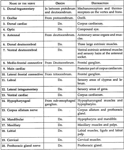
These are nerves which are given out from the ganglia of the central nervous system. The name and distribution of different peripheral nerves which are given from the brain and sub-oesophageal ganglia are shown in the enclosed table (Table 18.2—Arthropoda).
Particulars about the Peripheral Nerves from Brain and Sub-Oesophageal ganglia of Cockroach:
Table 18.2: Arthropoda:
Each thoracic ganglion has eight pairs of nerve roots. With the first pair the ventral nerve cord communicates. The remaining seven pairs of thoracic nerves supply the muscles of the thoracic segment, legs and wings. The metathoracic ganglion in addition sends peripheral nerves to the first abdominal segment.
Each of the first five abdominal ganglia sends out pair of stout nerves to supply different parts of the abdominal segment posterior to it. The sixth abdominal ganglion sends out seven to eight pairs of nerves to the different structures present in the seventh, eighth, ninth and tenth abdominal segments.
(c) Sympathetic nervous system:
It is represented by a slender ganglionated sympathetic nerve, which begins from circumoesophageal connectives to innervate the involuntary muscles of the alimentary canal.
(d) Sense organs:
The sense organ of cockroach may be grouped according to their functions as mechanoreceptors, photoreceptors, thermo-receptors and chemoreceptors. In addition, another kinds of receptors, for responding to humidity changes, are recorded in the cockroach, Blatella. These are called hygroreceptors.
Mechanoreceptors:
Following sensory structures are considered as mechanoreceptors because they are concerned with the reception of stimuli in the form of touch, pressure, vibration and air current.
(a) Cuticular hairs:
These are present in the cuticle either in the form of bristles, spines, plates or collection of hairs. Tactile bristles are present in the cercus and are extremely sensitive to touch. Large spines on the femur and tibia of leg perform similar function.
A collection of hairs, known as hair plates, are present on certain parts of the body for receiving stimuli in the form of touch. A group of specialised hairs which are lodged in sockets occur in the cercus for receiving air movements and low frequency sounds. These are called auditory hairs.
(b) Campaniform stress receptors:
They are present as thick ridges on the segments of the leg, on the labial palps and its adjoining areas. These receptors are believed to respond to different pressure on the cuticular surface.
(c) Chordotonal organs:
These specialised sense organs meant for receiving vibrations are distributed on the legs. These are aggregates of specialised cells which remain arranged either parallelly or in the shape of a fan.
Photoreceptors:
The eyes and ocelli are the two important photoreceptors of cockroach, in addition to the general surface of the body which can also detect light stimuli through cuticular receptors.
(a) Compound eyes:
The position and appearance of the compound eye have already been stated. The external surface of the eye is covered by a transparent cuticle. Each eye is composed nearly of 20,000 visual units, each one of which is called ommatidium (Fig. 18.61A).

Each ommatidium consists of usual structures, like lens, crystalline cone, rhabdome and retinular cells (Fig. 18.61B). The cone is well developed and the rhabdomes extend up to the borders of the cone.
The retinular cells are partially stratified and contain rhabdomes in their inner border. These cells are separated by a slender axial canal. The pigment sheaths which separate the ommatidium are non-retractile. Thus only mosaic type of image formation occurs.
(b) Ocellus:
This is also known as simple eye. It is present near the base of antenna as a white area. It consists of outer flattened corneal cells, 3-5 retinular cells and one rhabdome formed by the inner margins of the retinular cells. It was formerly believed that these ocelli are of uncertain functions.
Recently the light detecting ability of these structures has been proved. It has been demonstrated that cockroaches can respond to the change of light even when the compound eyes are painted. But it fails to do so after the covering of ocelli.
Temperature receptors:
The temperature receptive sense organs are present in pads between the first four tarsal segments of the lge.
Chemoreceptors:
These receptors are responsible for detecting chemical stimuli in the form of smell and taste. The long and annulated antennae are beset with two types of sensory structures—thick-walled bristles and thin-walled hairs.
These bristles and hairs are responsible for the perception of smell. The tips of the maxillary and labial palps, inner surfaces of the mouth parts and the inner border of the mouth and pharynx possess sensory structures for contact, chemo- reception and gustatory (taste) responses.
11. Essay on the Endocrine System of Cockroach:
The body of cockroach contains following endocrine organs corpora cardiaca, corpora allata, prothoracic gland and cervical glands.
These glands work together with five groups of specialised cells in the brain. Of these five groups, three groups of cells are placed anteriorly and send the first nerve to the corpora cardiaca. The remaining two groups are present at the posterior end and they send the second nerve to the corpora Cardiaca.
Corpora cardiac:
It is a pair of small, elongated and irregular glands. At the anterior end it encloses the aorta. At the dorsal side it bears a transverse commissure which extends posteriorly as a process to give rise to the aortic nerves. Ventrally, the corpora cardiaca are connected with a rudimentary ganglion and are drawn posteriorly to connect the corpora allata.
Each corpora cardiacum receives three nerves—two from the neurosecretory regions of the brain and one from unknown origin. The electron microscopic studies have revealed that the cells of corpora cardiaca have profuse endoplasmic reticulum and prominent secretory granules of 600 nm in diameter.
The secretion of corpora cardiaca affects the contractility of muscles lining the gut, the Malpighian tubules and heart. It is believed that the products released by the corpora cardiacum increase the effects of prothoracic glands.
Corpora allata:
This is a pair of small glands present posterior to the corpora cardiaca. Each gland is or less oval. A transverse commissure connects it with the oesophageal nerve. It has nerve connections with sub-oesophageal ganglion, corpora cardiaca and prothoracic glands. The histology of corpora allata shows the presence of profuse mitochondria within the cells but secretory granules are present in limited areas.
The secretion is responsible for following functions:
1. It maintains the juvenile features in the larval stage.
2. It helps in the oocyte formation in adult females.
3. It influences the secretions of secondary sexual organs in both the sexes.
Prothoracic glands:
At the anterior end of the prothoracic ganglion, lies a pair of rope-like prothoracic glands. A tracheal branch remains associated with the gland. The histology of this gland shows the presence of a central part having 6-8 muscle fibres, which remain enclosed by an outer glandular covering of 4-12 cells deep. The secretory granules are of various sizes.
The secretions of prothoracic gland facilitate moulting and after the last moult the prothoracic glands degenerate. It has been experimentally demonstrated that the implantation of two pairs of prothoracic glands in an adult induces it to moult and results into the formation of a giant cockroach.
Cervical glands:
This is a pair of small oval glands, present in the neck region near the posterior opening of the head. This gland is richly supplied with trachea. The outer part of this gland is composed of large glandular cells and the inner part is made up of small cells with unusually large nuclei.
The central part of the gland is occupied by a cavity which traverses within the inner wall. The endocrine nature of this gland has not yet been proved in cockroach but in other insects it is believed to be responsible for producing a hormone to induce moulting. The product which is secreted is called Periplanetin.
12. Essay on the Reproductive System of Cockroach:
Sexes are separate. The members of two sexes may be identified on the basis of their morphological features (Table 18.3).
A. Male reproductive system:
The male reproductive system includes the following organs:
1. Testes
2. Vas deferentia
3. Mushroom gland
4. Conglobate gland or phallic.
5. Male gonapophyses or phallomers and
6. Male gonopore.
1. Testes:
A pair of three lobed small, white structures situated in the lateral side of the abdominal cavity beneath the terga of fourth and fifth abdominal segments. In a cockroach 4.4 cm long, each testis is nearly 1 cm in length.
The testes are longer in the young than in order age. The testes are embedded in the fat body. Each testis is formed of 30-40 transparent testis follicles. In the young, full of sperms grow in the testis (Fig. 18.62).

2. Vas deferentia:
From the posterior end of each testis arises a delicate duct, called vas deferens. The two vas deferentia run posteriorly in the abdominal cavity and then curve towards the centre and open with a sac, called seminal vesicle or vesicula seminales, where sperm are stored. The two seminal vesicles open within a common duct, called the ejaculatory duct.
3. Mushroom gland:
Tufts of numerous blind, slender and thread-like tubules are situated at the junction of vas deferentia and ejaculatory duct. These tubules give a mushroom appearance and form the mushroom gland or utricular gland. The tubules are arranged in two groups.
The long tubules are situated at the periphery of the mushroom gland, called utriculi majores, which secret the inner layer of spermatophores. Short tubules of the mushroom gland, called utriculi brevirostris, forming the bulk of gland which secret a nourishing fluid for the sperm.
4. Conglobate gland or phallic:
A long, flat multi-lobed sac-like structure is situated beneath the ejaculatory duct, called conglobate gland or phallic. Its anterior end is broader and taper posteriorly. The posterior end opens into the genital pouch near the male genital aperture between the ninth and tenth sterna. The function is still not known.
5. Male gonapophyses or phallomers:
A reproductive (genital) pouch is formed near the ventral of the ninth and dorsal side of the tenth segments. The pouch is surrounded by irregular chitinous hooks and plates, called gonapophyses or phallomeres. These help in copulation.
On the left side, the left gonapophysis contains a slender arm with a curved hook, called titillator and in between lobes of the left gonapophyses there is a shorter arm with a hammer-like head, called peudopenis.
6. Male gonopore:
The ejaculatory duct which is unpaired and muscular, runs up to the reproductive pouch and opens through an aperture, called the male gonopore.
B. Female reproductive system:
The female reproductive system consists of a pair of ovaries, oviducts, colleterial glands, spermathecae, gonapophyses and female gonopore.
1. Ovaries:
A pair of large ovaries is placed on the lateral and posterior ends of the abdominal cavity extending 4th to 6th segments. Each ovary is made up of eight beaded tube-like ovarioles. Each ovariole is distaloproximally divisible into six zones (Fig. 18.63).

Zone 1:
The distal-most ligament-like part which connects the ovarioles with the body cavity.
Zone 2:
This is the budding zone, or zone of germarium where future oocytes are produced.
Zone 3:
Here the oocytes grow, but are not arranged in a single file.
Zone 4:
This is the longest region where oocytes are arranged in a single line. The distal end has smallest oocyte and the largest one remains at the proximal end.
Zone 5:
Broad region of the ovariole which contains oocytes rich in yolk. This region is called the region of vitellarium.
Zone 6:
This is the proximal part which opens within the oviduct.
2. Oviduct:
From each ovary originates one short and wide oviduct which is formed by the fusion of ovarioles at the posterior end.
3. Vagina:
The two oviducts unite to form a common chamber, called vagina, which is placed in the median position.
4. Spermathecae:
In between the oviducts, a pair of club-shaped, unequal organs, called spermathecae, is present. The two spermathecae open within the genital pouch on a small spermathecal papilla through an independent median aperture on the ninth segment.
5. Colleterial glands:
A pair of branched tubular glands, called colleterial glands, opens on the dorsal side of the female gonopore. The left gland is larger and opaque while the right one is smaller and transparent. The colleterial glands lie behind and above the ovaries. The secretion of colleterial glands forms the egg-case or ootheca.
6. Genital pouch:
The genital pouch is formed by the modification of the seventh, eighth and ninth abdominal segments. It is bounded ventrally by the sternum of seventh segment, dorsally by the sternum of eighth segment.
The genital pouch can be distinguished into two portions. The anterior smaller portion is called genital chamber into which female gonopore and spermathecal pore open. The posterior larger portion is called oothecal chamber or vestibulum within which oothecae are formed.
7. Female gonapophyses:
The female gonopore opens within genital pouch at the eighth segment. The opening of the pouch is provided with three pairs of chitinous rods, called the gonapophyses. These help in copulation and in depositing the eggs.
13. Essay on the Development of Cockroach:
The female cockroach liberates a smell to attract the male. The male cockroach on receiving the odour, raises its wings and searches the female. During pairing the male protrudes its abdominal segments to fix under the female, thus moving end to end with the female. The pairing continues for an hour. The males which are at least 2-4 days old and females of 4-5 days old are capable of pairing.
The pairing of the two sexes results into the transfer of sperm cells from males within the female as small pinhead sized packets, called spermatophores.
Within the body of the female the sperms remain temporarily stored within spermathecae. Sixteen eggs one from each ovariole travel towards the genital pouch through oviduct. Each egg is centrolecithal, i.e., bulk of yolk is confined to the central part of the egg. Thus the cytoplasm with nucleus is restricted to the periphery.
After fertilization, the fertilized eggs are enclosed in double row within a case, called ootheca which is formed by the secretion of colleterial gland. The ootheca is composed of a protein and contains water.
The female after laying the egg, fixes it on some suitable object with the help of an oral secretion. The development of the embryo continues within the ootheca and after certain period the young hatches out of the case (Fig. 18.64).
 and nymph (late stage) of cockroach")
The development requires 34-99 days and it depends upon temperature. The rate of hatching is 63% and the young takes five minutes time to come out of the inflated ootheca. After coming out, the young eats the egg membrane. The young’s resemble the adults but are soft, white and without gonads and wings.
They are called nymphs. The nymphs undergo several moultings. The stage in between two moultings is called instar. Wings appear at the end of the last moult. The transformation from nymph to adult takes six to eight months and requires 11 moults in males and 12 moults in females.