
In this essay we will discuss about Bacteria. After reading this essay you will learn about: 1. General Characters of Bacteria 2. Occurrence of Bacteria 3. Cell Structure 4. Cytoplasmic Contents 5. Structure (Gram-Positive and Gram-Negative Bacteria) 6. Classification 7. Harmful Activities 8. Useful Activities.
Contents:
- Essay on the General Characters of Bacteria
- Essay on the Occurrence of Bacteria
- Essay on the Cell Structure of Bacteria
- Essay on the Cytoplasmic Contents of Bacteria
- Essay on the Structure (Gram-Positive and Gram-Negative Bacteria)
- Essay on the Classification of Bacteria
- Essay on the Harmful Activities of Bacteria
- Essay on the Useful Activities of Bacteria
Contents
- Essay # 1. General Characters of Bacteria:
- Essay # 2. Occurrence of Bacteria:
- Essay # 3. Cell Structure of Bacteria:
- Essay # 4. Cytoplasmic Contents of Bacteria:
- Essay # 5. Structure (Gram Positive and Gram Negative Bacteria):
- Essay # 6. Classification of Bacteria (Based On Cell Structure):
- Essay # 7. Harmful Activities of Bacteria:
- Essay # 8. Useful Activities of Bacteria:
Essay # 1. General Characters of Bacteria:
1. Bacteria are simple most primitive organisms. They are very minute in size ranging from 0.5 to 2 µ in size.
2. They show prokaryotic type of cell organization.
3. Majority of bacteria are simple unicellular organisms.
4. Because of the presence of rigid and definite cell wall, they are considered as plants. The cell wall is composed of mucopolymers viz. muramic acid, diamino pimelic acid. Cellulose is absent.
5. True nucleus is absent i.e., chromosomes, nucleolus, nuclear membrane etc. are lacking. The nuclear material is in the form of nucleoid (DNA).
6. Membrane bound cell organelle like mitochondria, Golgi bodies, endoplasmic reticulum and plastids etc., are not found in bacterial cells. The function of mitochondria is carried by in-folding (mesosomes) formed by the plasma-membrane.
7. The chlorophyll is absent hence bacteria show heterotrophic mode of nutrition. In photosynthetic bacteria special pigments Bacteriochlorophyll (C55H74O6N4Mg) and bacterioviridin (C55H72O6N4Mg) are found in intra-cytoplasmic membranous structures.
8. Ribosomes are found distributed in cytoplasm and are of 70S type. Sometimes they occur in clusters known as polyribosomes.
9. Motile bacterial cells have one to many flagella which are cytoplasmic in origin and are composed of flagellin a special type of protein and do not show 9 + 2 organization, like other flagella.
10. Reproduction takes place commonly by binary fission. Sexual reproduction was previously considered to be absent but now it has been well established that exchange of genetic material takes place by conjugation, transformation and transduction.
11. Bacterial cells are sensitive to antibiotics.
12. Bacteria show sensitivity to phages.
Essay # 2. Occurrence of Bacteria:
Bacteria are omnipresent. More than 2500 bacterial species are known. Excepting pits of volcanoes, deep strata of rocks or blood of normal animals, they are present all around us. They can withstand extremes of temperature and some of them can be cooled up to -190°C, while a few can be boiled up to 78°C without killing them.
Extremely simple structure, small size, formation of highly resistant endospores, diversity of their mode of nutrition and resistance to un-favourable conditions are some of the important features which contribute to their universal distribution.
Essay # 3. Cell Structure of Bacteria:
The ultrastructure of bacteria can be examined with the aid of electron microscopy and by micro chemical methods which have been highly developed during the past few years. Like other living plant cells, the bacterial cell comprises (1) the outer covering of the cell, (2) the inner cell body or the protoplasm (Fig 1, 2).


1. The Outer Covering of the Cell:
It appears to be made up of three layers:
(a) Capsule or slime layer
(b) Cell wall and
(c) Cell membrane.
(a) Capsule or Slime Layer:
It is the outermost protective layer of the cell wall and is not the constant feature. Its presence or absence depends on the environmental conditions. It is gelatinous or slimy in nature. Under certain conditions of growth the slime accumulates to form a thick conspicuous layer around the cell wall. It is called the capsule or sheath.
Capsule and slime layer of certain bacteria are made up of polysaccharides, a natural substance composed of dextran’s, dextrin, levans etc., and appears, like plant gum. However, in some other types of bacteria it is made up of polypeptides and peptides linked together with glutamic acid molecules.
The layer accumulates the excretory substances and protects the bacteria from un-favourable conditions. Capsule layer formation is also responsible for diseases in human and animals because white blood corpuscles are unable to kill the bacteria due to the presence of capsule.
(b) Cell Well:
The slime layer or capsule is followed by the cell wall. It is the rigid part of the outer covering of the cell. It is 50-100 A thick and consists about 20% of the total cell volume. It is granular, porous, permeable and shows no micro-fibrils.
The purified cell wall material contains lipids, carbohydrates, proteins, phosphorous and other inorganic substances. Rigidity is due to a unique polymer, the mucopeptide, which is based on a back-bone structure of alternating molecules of N-acetyl glucosamine and N-acetyl muramic acid molecules in adjacent chains. (NAG-NAM-NAG-NAM-NAG-). These chains are cross linked by peptide chains (fig. 3).
The cross linking enables the complex to form a rigid network. The muramic acid has so far been found only in the bacteria and Actinomycetes. With this, the amino acids present in the cell wall are glutamic alanine, glycine and either lysine or diamino pimelic acid. Teichoic acid (a polymer of glucose, alanine, glycerol or rabitol) is also found in some bacteria. The Gram-positive and Gram-negative bacteria differ in their cell wall structure.

The cell wall provides definite shape and rigidity, protects the cytoplasm, plays an important part in cell division and regulates the passage of various materials between the external and internal environment of the organisms.
(c) Cytoplasmic Membrane:
The cytoplasmic membrane lies immediately beneath the inner surface of the cell wall and varies from 50-100 Å in thickness. It is a complex highly organised and highly specialized structure and consists molecules of lipids and proteins which are arranged in a fluid mosaic model. It is composed of double layer of phospholipid molecules.
Phospholipids are of two types: Hydrophilic and Hydrophobic. Hydrophilic molecules are towards outer side and hydrophobic molecules are on the inner side. The protein molecules are embedded within this lipid layer. The plasma membrane has several infolding’s in the form of mesosomes
(Fig. 4). The function is a matter of conjecture. They are often called as chondrioids because of their analogy with mitochondria of eukaryotic cells.

Cytoplasmic membrane has some important functions such as:
1. It controls the osmotic behaviour of the cell.
2. The proteins present in the cell membrane act as carriers or permease (to carry selective transport of nutrients and electrons in respiration and photosynthesis).
3. Provides specific site at which DNA remains attached. It is the point from where DNA replication starts.
4. Enzymes of various metabolic path ways (e.g., synthesis of phospholipids, teichoic acid) are located in the cytoplasmic membrane.
2. Protoplasm:
Within the cell wall is the living stuff of the bacterial cell. It is called the protoplasm. In general, the structure and composition of the protoplasm of the bacterial cells is similar to those of other living beings with a few differences. The moisture content is about 70-85% and it is colloidal in nature.
It consists of:
(i) Cytoplasm and
(ii) Nucleoplasm (karyoplasm)
(i) Cytoplasm:
The space between the nuclear region and the plasma membrane is filled with a uniform, granular cytoplasm. It is a dispersed colloidal mixture of water, proteins, lipids, mineral compounds and other substances. The cytoplasm contains organelles (living structures) and inclusions (non-living matter). The non-living inclusions are the storage granules of volutin, glycogen, lipids, globules or protein crystals.
Sulphur and iron are also found in some bacteria (e.g., Beggiatoa). These inclusions serve as store of energy. The organelles present in the cytoplasm are mesosomes, chromatophores, ribosomes, polyribosomes, ribonucleic acid (RNA), nucleoplasm, flagella and pili. However, mitochondria, plastids, golgi bodies and endoplasmic reticulum etc. are absent.
(ii) Nucleoid (Nucleoplasm or Karyoplasm):
The existence of nucleus in bacteria has long been a debatable issue. It is the area in which all the chromatin or genetic material of the cell is concentrated. Most of the bacteriologists now believe that the nucleus in the bacteria is present in the cell as a discrete (separate) body. Feulgen test applied in the case of bacteria has revealed the presence of purple stained bodies in a large number of cases.
The electron microscopic study reveals that it is of rudimentary type. It does not have discrete chromosomes, mitotic apparatus, nucleolus and nuclear membrane. It divides amitotically. Hence, the nuclear region in bacteria is referred to as the nuclear body or nucleoid or genome or genophore.
The cell in the phase of rapid growth may contain one to four nuclear bodies or nucleoids. They appear as an electron translucent area and can be shown to contain very fine fibrils. These fibrils are molecular strands of DNA. The DNA is certainly restricted to this area of the cell. The DNA occurs in the form of single, two stranded thread like molecule about 1000 microns (1 mm) long. It is folded and no ends are seen. It is devoid of histones. Cairns (1963) had demonstrated that DNA of Escherichia coli is circular which measures 1400 µ in length and is packed in a nuclear body of 0.20 µ in diameter.
Plasmids and Episomes:
In addition to the bacterial chromosome, many species of bacterial cells also contain some extra chromosomal genetic material. It is either independent of bacterial chromosome or integrated with it. If this genetic material is independent of bacterial chromosome it is called plasmid (Fig. 2), if it is integrated into or out of the bacterial chromosome it is called episome.
Plasmids are small, circular, closed, double stranded DNA molecules. This molecular weight ranges between 5x 10⁷ and 7x 10⁷. Each plasmid may contain as many as 100 non-essential genes. Therefore, it plays no role in the in-viability and growth of bacteria and hence it is called dispensable autonomous element.
In E. coli there are three classes of plasmids:
1. F (i.e., Fertility) Factor:
These promote bacterial conjugation and determine maleness in bacteria. Those cells which contain them are called F+ and those which do not have are called F−∙ F+ forms sex pili.
2. Col (i.e., colicinergic) factor:
It gives the capacity to bacterial cell to produce anti-biotically active proteins or colicins which have an antibiotic function.
3. R (i.e., Resistance) factor:
R factor offers to bacteria resistance to one or more drugs such as neomycin, penicillin, streptomycin, chloramphenicol etc.
Essay # 4. Cytoplasmic Contents of Bacteria:
(i) Mesosomes (or chondrioids):
Thin section of bacteria often reveals one or more, large, irregular invaginations of the plasma membrane called mesosomes (Fig. 2). Mesosomes increase the surface area of the plasma membrane. They are higher in number in chemoautotrophic bacteria with high rate of aerobic respiration e.g., Nitrosomonas.
Their function is a matter of conjecture. It is believed that the mesosomes are active in cell wall synthesis and in the secretion of extra cellular substances. It has been shown that transforming DNA taken up by whole cells of Bacillus subtilis apparently enters the cell via the mesosome. There is a considerable evidence that the bacterial nuclear body is attached to a mesosome.
(ii) Ribosomes:
The cytoplasm of the bacteria is thickly populated with numerous minute, nearly spherical, densely stained hollow bodies called the ribosomes. These are 290 x 210 Å in size and lie free in the cytoplasm without any association with any membrane component. Their number varies from 10,000 to 15,000 in a cell.
Chemically, these are made up of ribonucleoproteins. Each ribosome has 60% RNA and 40% protein. 80-85% of the total RNA is found in ribosomes and remaining 15 to 20% is found dissolved in cytoplasm. Electron microscopic study reveals that the ribosomes are composed of two sub-units (30S and 50S subunits) and belong to 70S category. These sub-units are nearly spherical but of unequal size. Sometimes they are found arranged in small groups by a strand of messenger RNA (m-RNA). These groups are known as polyribosomes. They are the sites of protein synthesis. They do not act singly but in groups called polysomes.
(iii) Chromatophore or Photosynthetic Apparatus:
The chromatophores are present in some bacteria (e.g., Chlorobium, Rhodospirillum etc.) They were first isolated by Pardee, Schachman and Stanier in 1952. These structures are in the form of small membranous vesicles and are variable in number. They are calledintra cytoplasmic membranous structures.
Non-membranous structures, the chromosomes were demonstrated by Stanier (1970) in some members of Chlorobacteriaceae. They have bacteriochlorophyll (C55 H74O6 N4 Mg) and bacteriovirdin (C55 H72 O6 N4 Mg). These are 500-800 Å in diameter and are believed to be bounded by a limiting membrane. It has been shown that these vesicles (pigments) are active in the trapping of light quanta and conversion of light energy to ATP (Adenosine triphosphate) through an electron transport chain system. (Frenkel, 1954).
(ii) Flagella (Sing. Flagellum):
Many bacteria are actively motile, moving at speed up to 50 µ per second. Such bacteria are normally found to possess flagella. These are slender whip-like (Fig. 5 A) appendages of cytoplasmic origin. Each flagellum arises within the cytoplasmic membrane.
It forms a granule called blepharoplast and pushed out through the cell wall. They are about 120-180 A in diameter and 5 µ in length. They do not show 9 + 2 pattern (Fig. 5 B) of fibrils (characteristic of eukaryotic flagella). They are not enclosed in a membrane and show no ATP-ase activity.

Chemical analysis of flagella suggests that these are composed of a single homogeneous protein flagellin. Its molecular weight is about 40,000. Each flagellin molecule is 40 Å in diameter. Several (usually 3-8) longitudinal chains of flagellin molecules run longitudinally turning around each other to form a wavy helical or rope like structure. A cross section of the flagellum reveals 8 flagellin molecules (Salmonella typhimurium) around a central space. However, 10 flagellin molecules are present in Pseudomonas fluoresces.
(iii) Pili (= Fimbriae):
Some bacteria (mostly gram negative) possess certain minute, cylindrical, rigid structures called Pili (Fig. 2). They are about 30 Å – 60 Å in diameter and 0-5 to 2µ in length and smaller than flagella. They are analogous to flagella bur not involved in the motility of the bacteria.
Chemical analysis of the pili suggests that they are composed of a protein called pilin. Its molecular weight is about 17000. These units are helically arranged and are made by some 18 amino acids.
The pili are of 3 types:
1. Common or Type I Pili.
2. F (Fertility) Pili.
3. Col I (Colicin Pili).
The pili are concerned with attachment of the bacterial cell over the solid surface. The piliated bacteria tend to stick to one another and produce coherent pellicles on the surface of the un-agitated liquid cultures. Certain pili are involved in the transfer of the genetic material from one bacterial cell to another. They are the sex pili. In some pathogenic bacteria e.g. Neisseria gonorrhoeae pili help in attachment on the host cells. Pili also act as specific sites of attachment for bacteriophages.
Essay # 5. Structure (Gram Positive and Gram Negative Bacteria):
The bacteria are divided into two groups on the basis of their reaction of Gram’s stain (i) Gram positive (ii) Gram negative. Those which retain the Gram’s stain after alcohol treatment are called Gram positive,While those which lose the stain are designated as Gram negative.
Produce:
This Staining technique was given by a Danish Physician Chistian Gram in 1884. A smear (thin film of bacteria) is made on the Slide. After getting the film dried either in air or by bringing the slide on the flame, the film is stained by the following producer (Fig. 1).


Sometimes a counter-stain is given in which the film is stained with some contrasting colour of eosine (red), safranin (red), so that Gram-negative bacteria take up the stain and bacterial cells become distinctly visible.
In gram positive bacteria cell wall is thick (150-200 Å) and it contains peptidoglycan (60-90%) and teichoic acid. Less quantity of protein and polysaccharides are present. In Gram negative bacteria cell wall is thin (75-120 Å) and contains peptidoglycan (10%), lipopolysaccharides (50%), lipoprotein (15%) and phospholipids (3%) (Fig. 2 A, B).

Essay # 6. Classification of Bacteria (Based On Cell Structure):
There have been many systems as far as bacterial classification is concerned. They have been classified variously on the basis of their shape, arrangement of flagella, their existence in presence or absence of oxygen and their physiology. But these classifications are not recognized internationally, because there are so many kinds of bacteria which look alike in the microscope but are physiologically and immunologically different.
Modern system for classification of bacteria uses a number of criteria viz., arrangement, Gram stain, motility, various enzymatic properties and other sorts of information that can serve to delimit an organism or group of organisms from others.
The classification of bacteria presented in Bergey’s Manual of Determinative Bacteriology is accepted in medical microbiology. (Seventh edition, 1957). It is the result of the joint work of 131 authors from 15 countries. In this classification the plant kingdom is divided into 5 divisions—Protophyta, Thallophyta, Bryophyta, Pteridophyta and Spermatophyta.
Of these, protophyta with which we are concerned here is further divided into 3 classes as under:

The class Schizomycetes is further divided into ten orders, differentiated from one another primarily on the basis of morphological characters and type of motility. It is as follows:
I. Cells rigid, spherical, rod shaped (straight or curved) or spiral in form, sometimes in trichomes. Motile by means of polar flagella or non-motile.
(A) Cells coccoid, straight or curved rods, or spiral in form, sometimes occur as chains of cells. Cells may contain photosynthetic purple or green pigments. Not in trichomes. Usually motile by means of polar flagella, occasionally non motile…
Order I. Pseudomonadales
(e.g., Pseudomonas spp.)
(B) Not as Above.
1. Cells in trichomes that are frequently in a sheath. Occasionally motile (swarm spores) or non-motile, conidia are developed. The sheath may contain a deposit of ferric hydroxide, and the trichomes may be attached to a substrate…..
Order II. Chlamydobacteriales
(e.g., Chlamydobacterium)
2. Cells reproduce by a process of budding rather than by ordinary cell division (fission), may be attached to a substrate by a stalk. One genus contains species with photosynthetic pigments (Rhodomicrobium)…….
Order III. Hyphomicrobiales
(e.g., Hyphomicrobium)
II. Not as Above.
(A) Cells rigid, Spherical or straight rod shaped cells. Occur singly, in chains or in trichomes. Motile by means of peritrichous flagella or non-motile. Not acid fast.
1. Cells spherical or rod-shaped; no trichomes, chains of cells may occur……….
Order IV. Eubacteriales
(e.g., Salmonella)
2. Cells in trichomes………………….
Order V. Caryophanales
(e.g., Caryophanon)
(B) Not as Above.
I. Cells rigid, may grow out into a branching mycelium like structure which may even develop chains of aerial conidia giving colonies a superficial resemblance of mold colonies. In two genera spores develop within sporangia (sporangiospores) and in one of these genera the spores are motile. Where cells occur singly or in simple branched forms, they are frequently acid fast……….
Order VI. Actinomycetales
(e.g., Mycobacterium)
2. Not as Above.
(a) Cells rigid, usually large and may occur as coccoid cells or trichomes. Sulphur granules may occur on the surface or within the cells. Move by a gliding, oscillating or rolling, jerky motion like that of some blue green algae. No flagella present……
Order VII. Beggiatoales
(e.g., Beggiatoa)
aa. Not as Above.
(b) Longer or shorter flexuous cells.
(c) Cells flexuous, creeping on a substrate. Frequently pointed at both ends. Fruiting bodies usually developed from a thin spreading colony (Pseudoplasmodium). Slime bacteria………………
Order VIII. Myxobacteriales
(e.g., Cytophaga)
cc. Cells in the form of longer or shorter spirals, swim freely by flexion of cells………………………..
Order IX. Spirochaetales
(e.g., Treponema)
bb. Non-motile, highly pleomorphic organisms of a very delicate character. Possess fibrable stages . . .
Order X. Mycoplasmatales (Dplo)
(e.g., Mycoplasma)
Essay # 7. Harmful Activities of Bacteria:
Some of the harmful activities of bacteria are as follows:
(i) Diseases:
The pathogenic bacteria cause a number of diseases in man, animals and plants:


(ii) Food Poisoning:
Many saprophytic bacteria cause decay of food and make it unpalatable e.g., rotting of vegetable fruits and meat, souring of milk, spoilage of butter, Jams, pickles. Certain bacteria also secrete toxins in the food. If these food stuffs are consumed it may cause serious illness or even death.
The most common type of food poisoning is Staphylococcus food poisoning (caused by Staphylococcus aureus) and botulism (caused by Clostridium botulinum). Swelling of tongue, respiratory paralysis and double vision are some of the main symptoms of botulism disease. Marine food such as sea weeds, oysters, crabs and fishes are destroyed by Spirillum rectiphysetaris.
(iii) De-Nitrification:
Some soil bacteria (e.g., Micrococcus denitrificans, Thiobacillus denitrificans) convert nitrates into nitrites and ammonium salts of the soil into free nitrogen (NO3→ NO2→ NH3→N2). This results in the lowering of soil fertility. The breaking of soil nitrates into molecular nitrogen is called De-nitrification and such bacteria are known as denitrifying bacteria.
(iv) Pollution of Water:
Some pathogenic bacteria multiply in water stream and make it undesirable, harmful and undrinkable e.g., Vibrio cholerae, Salmonella typhi etc.
(v) Cotton Deterioration:
Some bacteria destroy cotton and articles made from it for example, Spirochaeta cytophoga.
(vi) Bacteria as Possible Warfare Agents:
Many bacteria may be used in secret biological warfare for example, Anthrax, botulism and tuberculosis. This results into death of civilian population in the locality, province or the country.
Essay # 8. Useful Activities of Bacteria:
Some of the useful activities of bacteria are listed below:
(i) In Agriculture:
(a) Putrefaction and Decay:
Some saprophytic bacteria e.g., Bacillus ramosus, B. vulgaris function as natural scavengers. They decompose the dead bodies and waste of organisms (both plants and animals), Converting the complex compounds into simple compounds.
A variety of elements of minerals in earth such as C, O, H, S and P, which make up these bodies are reduced to simple compounds such as carbon-monoxide, water, nitrates, sulphates and phosphates. These compounds increase the fertility of the soil and are absorbed by the plants as their food.
The decomposition of the protein in the absence of oxygen is known as putrefaction.The decomposition of carbohydrates in the absence of oxygen is known as fermentation and the decomposition of dead organic -latter in presence of oxygen is called as decay.
(b) Soil Fertility:
Some bacteria play an important role in maintaining and increasing the fertility of the soil. Ammonifying bacteria (Bacillus vulgaris, B. ramosus, B. mycoide) and nitrifying bacteria (Nitrosomonas and Nitrobacter) maintain the soil fertility while nitrogen fixing bacteria such as Azotobacter, Beijerinckia aerobic), Clostridium (anaerobic), Rhizobkim leguminosorum (syn. = Bacillus radicicola) increase the fertility of the soil.
(ii) In Biotechnology:
Some bacteria are so important that some of the industries are totally dependent upon their activity. Bacteria are used in fermentation processes, baking cheese butter pickles, soy-sauces, vinegar, wine and chemical making.
A few of which are described as follows:
(a) Production of Butyl alcohol and acetone:
Clostridium acetobutylicum, ferments the sugary solution of molasses and produces the butyl alcohol and acetone.
(b) Production of Lactic Acid:
Lactic bacilli ferments the cane sugar and produces lactic acid.
(c) Production of Vinegar:
Acetobacter acetic ferments the alcoholic solution and produces acetic acid
C2H5OH + O2 → CH3COOH + H2O
(d) Dairy Industry:
Lactobacillus lactis converts lactose of milk into lactic acid and helps in the preparation of curd (milk protein caesin to curdle). Streptococcus lactis helps in the formation of cheese from milk. The formation of butter from milk is also induced by the presence of bacteria Streptococcus lactis, S cremoris. Yogurt is prepared by Lactobacillus sanfrancisco and Streptococcus thermophiles. Butter is formed by the activity of Streptococcus lactis.
(e) “Curing and Ripening” of Tea and Tobacco Leaves:
Bacteria play an important role in tobacco industry. After harvesting, tea and tobacco leaves are hang in sheds. Here the leaves are partially decomposed by the bacteria. This process is called curing and is followed by ripening. In this process bacteria Bacillus megaterium bring about further changes and affect the flavour of the leaf, which enhances its value in the market.
(f) Leather Industry:
The bacteria remove the fats, hairs and other tissues from the skin of dead animals (hides). These hides are then tanned to prepare leather.
(g) Textile Industry:
Commercial fibres are separated from the fibre yielding plants by the decomposition f non-cellulosic cementing material (pectin) through a process called retting. Bacterial action is involved in the ‘retting’ of flax fibres (microbial decomposition of unwanted material). In this process the hemp and the flax parts are submerged in water. The softer parts of the plant rot away due to bacterial activity e.g., Clostridium butyricum, Isineum, C. pectinovorum. The tough fibres are spun and woven into linen clothes etc.
(iii) Medicinal Use:
(a) Antibiotics. Many antibiotics are obtained from a number of bacteria:
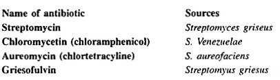

With this the bacteria are also used in the production of serums and vaccines. Vaccines are used against infection of many diseases such as cholera, typhoid, etc. Antibiotics obtained from bacteria are very effective to cure many diseases.
Streptomycin is generally used to cure TB, maningites and pneumonia; Aureomycin against osteomyelitis, whooping cough; Chloromycetin and terramycin against typhoid, pneumonia urinary infections and neomycin is used to cure T.B. Majority of the antibiotics are effective against Gram +ve bacteria.
While kanamycin and Polymixin B are effective against Gram -ve bacteria, aureomycin and Chloromycetin are effective against both Gram +ve and Gram -ve bacteria.
(b) Vitamins:
Clostridium butylicum is used in synthesis of riboflavin one of the B-vitamins.
(iv) Sewage Disposal:
Bacteria convert the solid or semisolid forms of waste products into simpler forms. The sewage is then filtered and the residue is used as a good quality manure.
(v) Pest Control:
Bacteria can also be used in place of pesticides in the biological pest control. The commonly used bacterium is Bacillus thuriengiensis (also called BT), a gram positive soil dwelling bacterium. This bacteria is used as a lapidopteran-specific insecticide-under trade names such as Dipel and Thuricide.
Some other bacteria are also helpful in biological control of insects pests. Bacillus papillae kills Japanese beetle, B. sphericus kills mosquitoes. Because of their specificity, these pesticides are regarded as environmentally friendly with little or no effects.
(vi) Genetic Engineering:
With the help of recombinant DNA technology, bacterial cells are transformed and used in the production of commercially important products, e.g., production of human insulin (used against diabetes) and human growth hormone (Somatotropin-used to treat pituitary dimorphism). Transfer of interferon gene into bacteria produces useful antiviral protein interferon’s. E. coli is the best material for work on genetics and molecular biology.
The bacteria are also helpful in so many ways. Some bacteria (Escherichia coli) live symbiotically in the intestine of man. They synthesize some of B vitamins and release into intestine. Some bacteria e.g., E. coli, Agrobacterium tumefaciens are used as test organisms for basic genetic researches (recombinant DNA technology). Methanobacillus produce biogas (methane) by fermenting the organic matter under anaerobic conditions.
Some bacteria e.g., Pseudomonas are used to check pollution after oil spillage. DDT is slowly decomposed by Acetobacter aero-genes. In ruminate animals cellulose is digested by ruminate bacteria Ruminococcus albus. In these animals rumen is inhabited by this bacteria. It secrete cellulase, an enzyme that help in the digestion of cellulose, of plant cell walls. Cellulose is the major source of energy of these animals.