In this essay we will discuss about grasshopper. After reading this essay you will learn about:- 1. Habit and Habitat of Grasshopper 2. External Structures of Grasshopper 3. Digestive System 4. Respiratory System 5. Excretory System 6. Circulatory System 7. Nervous System 8. Reproductive System 9. Development 10. Economic Importance 11. Control.
Essay Contents:
- Essay on the Habit and Habitat of Grasshopper
- Essay on the External Structures of Grasshopper
- Essay on the Digestive System of Grasshopper
- Essay on the Respiratory System of Grasshopper
- Essay on the Excretory System of Grasshopper
- Essay on the Circulatory System of Grasshopper
- Essay on the Nervous System of Grasshopper
- Essay on the Reproductive System of Grasshopper
- Essay on the Development of Grasshopper
- Essay on the Economic Importance of Grasshopper
- Essay on the Control of Grasshopper
1. Essay on the Habit and Habitat of Grasshopper:
The grasshoppers have chewing mouth parts. They occur throughout the world, mainly in grasslands. The locusts are related forms of grasshoppers and are well known for their migratory habits. The metamorphosis is incomplete, i.e. young forms gradually develop into adults and these young stages resemble the adult. It is also called direct metamorphosis.
2. Essay on the External Structures of Grasshopper:
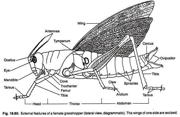
The body is slender, elongated and exhibits perfect bilateral symmetry. It may reach a length up to 8 cm. The colour may be yellowish-green, leaf-green or brown. Some forms have varied sopts and markings. The segments are distinct. Like that of cockroach the segments are arranged to form three tagmata—head, thorax and abdomen (Fig. 18.65).
All the three tagmata are enclosed by chitinous exoskeleton. The exoskeleton of each segment is called a sclerite and two adjacent sclerites are separated by a soft suture. Some segments of the body may be fused and the sclerites are indistinct.
A. Head:
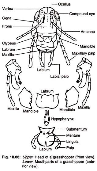
The head of a grasshopper is covered dorsally by vertex, laterally by genae or cheeks and anteriorly by frons. The ventral side of frons is united with the clypeus.
Following structures are present in the head region:
(a) One pair of large sessile compound eyes, which occupy the major part of the dorsolateral region of head.
(b) Three ocelli or simple eyes are present, one on the middle of the frons and one on the side of each compound eye.
(c) One pair of narrow filiform and jointed antennae is present, one on the anterior border of each compound eye.
(d) Mouth is present at the anterior terminal end of the body and directed ventrally. The mouth is provided with mouth- parts which are specialised for biting and griding leaves.
Mouth parts or trophi:
The mouth is bounded (Fig. 18.66) by an upper roughly rectangular labrum and a lower labium. The labrum remains attached to the anterior border of the clypeus. The labium which appears single is formed by the fusion of two. An elongated tongue or hypopharynx remains within the mouth cavity and is attached with the inner side of labrum.
The inner broad part of the labium is called submentum, middle portion as mentum and the two movable anterior parts are called lingulae. A pair of jointed labial palps arise one from each side of the mentum. The two lateral sides of the mouth are provided with mandibles.
Each mandible is strongly built. It has serrated inner border. On the outer side of each mandible lies a many-jointed maxilla. The proximal part of each maxilla is called the cardo, the middle stipes and the distal serrated part is lacinia. The outer border of stipes bears a palparifer and a five-jointed maxillary palp. The palparifer carries at one end a round galae.
B. Thorax:
The thorax has three segments—prothorax, mesothorax and metathorax. The outer covering of the thorax invaginates inside the body to form rigid endoskeleton for the insertion of muscles.
The dorsal covering of prothorax is called tergum or pronotum. It is large and subdivided into presculum, scutum, scutellum and postscutellum. Lateral coverings are known as pleuron. The pleuron is subdivided by a pleural suture into an anterior eipsternum and a posterior epimeron. The ventral side is covered by a single prosternum.
The mesothorax is covered dorsally by a single mesonotum and ventrally by a mesosternum. Each lateral side is covered by pleuron having divisions, called episternum and epimeron.
The metathorax is compact and larger than the other two divisions of the thorax. The exoskeletal coverings are same as in mesothorax, the dorsal one is called metanotum and the ventral one is called metasternum.
Structures present in the thorax:
Spiracle:
A pair of spiracles is present on the dorso-lateral sides of each meso and metathorax.
Leg:
One pair of legs is present in each thoracic segment. The prothoracic legs arise from the ventrolateral angle of the body and act to pull the body forward. The mesothoracic legs support the body during walking. The metathoracic legs are the largest and push the body forward during walking and are used for jumping.
Each leg consists of similar segments as that of cockroach. In the males, the third segment of the leg bears rows of chitinous hooks. These hooks are moved with pressure along the hard outer surface of the wing on prothorax to produce a sound. This sound producing organ is known as stridulatory organ.
Wing:
Two pairs of wings are present— one pair on the mesothorax and the other on the metathorax. The wings which remain attached on the dorso-lateral angle of mesothorax are called fore-wing or tegmina. The fore-wing is narrow and leathery. The wings at the dorso-lateral angle of the metathorax are called hind-wings.
These wings are broad and membranous. The hind- wings are used during flight. At the time of rest, these wings remain covered beneath the fore-wings. Each wing is composed of two cuticularised layers with branched tracheal tubes in between. These tracheal tubes are known as nervures or veins.
C. Abdomen:
The abdomen is made up of eleven embryonic segments. Of these segments, the anterior seven are easily recognised in adult grasshopper. The posterior segments undergo modifications. These modifications are different in male and female grasshoppers. Each ring-like segment is covered by a dorsal tergum and ventral sternum.
The inconspicuous inter-segmental membranes connect the exoskeletal coverings of the abdominal segments. The dorsal exoskeletal covering of the first abdominal segment is continuous with the same of metathorax. On the lateral surface of the first abdominal segment is present a tympanic membrane for receiving sound stimulus. Each abdominal segment bears a pair of spiracles.
Thus there are altogether eight pairs of abdominal spiracles. In males, the tergites of ninth and tenth abdominal segments are united and the sternite of ninth segment is considerably altered. The ninth segment bears the genitalia which include two pairs of gonapophyses. The outer pair of gonapophyses form the claspers and the inner small pair are known as parameres.
The gonapophyses cover the penis. The sternite of the tenth abdominal segment forms the subgenital plate and the tergite of eleventh segment works as supra-anal plate. The supra-anal plate bears one pair of cerci and two podical plates. In females, the sternites of ninth, tenth and eleventh segments are modified to form the genitalia which work as the ovipositor. The ovipositor has three pairs of valves.
The dorsal pair of valves arise from the ninth and the ventral pair from the eighth segments. The third pair of valves is placed inside to act as a channel for the outlet of eggs. The tergites of these three segments are incompletely united. The last segment bears—one pair of cerci (not so large as in males) and a pair of podical plates.
3. Essay on the Digestive System of Grasshopper:
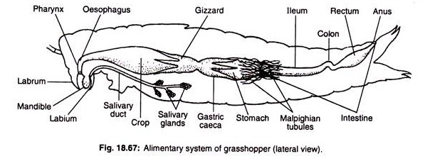
The digestive system (Fig. 18.67) consists of the alimentary canal and digestive glands. The alimentary canal is divisible into a foregut, midgut and hindgut. The foregut begins from mouth which leads into a small pharynx.
The mouth is provided with mouth parts for biting and chewing food. In the pharynx the salivary duct opens. The pharynx leads into a narrow oesophagus which runs upward to enter into a thin, dilated chamber called the crop.
The crop is followed by a small tube called proventriculus or gizzard which possesses in its inner wall chitinous plates raised to form teeth. The gizzard completely liquifies the food which then enters into the stomach or ventriculus. The opening is guarded by a sphincter. The stomach represents the midgut and its beginning is marked externally by the presence of six gastric or hepatic caeca.
Each is attached near the middle of caecum, i.e., each caecum has one anterior lobe and a posterior lobe. The opening between the midgut and the first part of the hindgut, called ileum is guarded by a sphincter.
This region of attachment between mid- and hindgut contains a circlet of numerous slender Malpighian tubules. The ileum leads to a slender colon which in turn enters to the enlarged rectum. The rectum ultimately opens to the exterior through an opening, called anus.
The Digestive glands include salivary glands, the lining of the midgut and hepatic caeca, all of which produce digestive juices. The salivary glands are small paired structures present on the outer surface of the crop. The salivary duct opens within the pharynx.
The grasshoppers are leaf-eaters. The mouth parts are used for cutting and crushing the leaf. The saliva moistens the food and at the same time partially digests it. The food remains temporarily stored within the crop and slowly flows into the gizzard. In the lumen of the gizzard the food is fully churned.
Then it goes to the midgut. Lining of the midgut and hepatic caeca secrete digestive juices. Various enzymes, present in the juices complete the digestion. Absorption takes place in these two areas. The residual matter passes into the rectum. The mineral salts and water are absorbed by the lining of the rectum and the faeces are ejected through the anus.
4. Essay on the Respiratory System of Grasshopper:
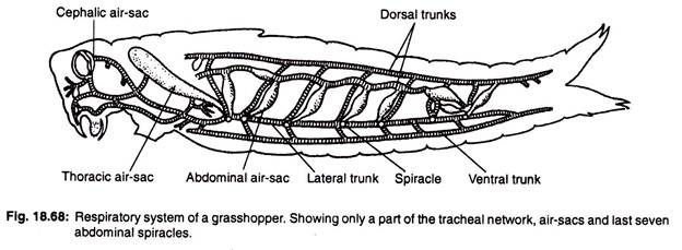
Respiration is aerial and the respiratory structures are tracheae (Fig. 18.68). There are two dorsal, two ventral and two lateral longitudinal tracheal tubes. These communicate with one another by transversely placed segmental tracheae. Some of these connecting tracheae are inflated as air-sacs.
From the trachea arises finer vessels, called the tracheoles, which form extensive network within the tissues. The inner wall of the trachea is provided with spiral cuticular thickening. Such thickenings are absent in tracheoles. The ultimate opening of the tracheole is immersed within body fluid, which conveys respiratory gases to and from the cells.
The lateral longitudinal vessels communicate with the exterior through ten pairs of spiracles. Of these ten pairs of spiracles, two pairs are on the thorax and the remaining eight pairs are on the abdominal segments. These are placed in a row of ten in each lateral side. Each spiracle is enclosed by a round sclerite, called peritreme and a valve to guard the opening. The valve is provided with muscles and nerves.
During inspiration or taking in of air, the first four spiracles open and the air rushes inside. At the time of expiration, last six pairs open and the air passes out of the body.
5. Essay on the Excretory System of Grasshopper:
The excretory organs are in the form of Malpighian tubules. These are present near the junction of stomach and ileum. Each tubule is thread-like and internally lined by a layer of striated epithelial cells.
The free blind end of these tubules extends within the haemocoel and removes metabolic wastes from the blood. These waste products are finally drained within the ileum, from where these are removed with the faeces.
6. Essay on the Circulatory System of Grasshopper:
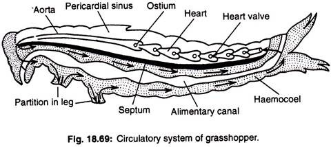
The heart is tubular and is placed on the dorsal side of the thoracic and abdominal cavities. It is drawn anteriorly into an aorta (Fig. 18.69). The heart contracts to drive blood anteriorly which flows through the aorta. The aorta breaks up into finer branches in the head and thoracic regions and ultimately opens to the haemocoelomic spaces (Haemocoel).
Through these spaces the blood returns to the posterior part of the body. The deoxygenated blood is finally accumulated within a large haemocoelomic space around the heart, called pericardial sinus. From the sinus the blood enters the heart through paired segmental openings of the heart, called ostia (singular—ostium).
The outer wall of the pericardial sinus is provided with segmentally arranged alary muscles. These muscles cause the pericardial wall to contract and force the blood to enter into the heart. The contraction of heart to drive the blood is possible due to the contractile nature of its wall.
7. Essay on the Nervous System of Grasshopper:
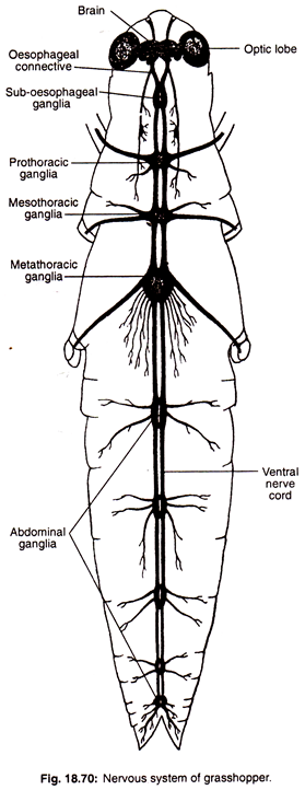
The nervous system has three usual divisions—central nervous system, peripheral nervous system and sympathetic nervous system (Fig. 18.70).
The central nervous system consists of brain or supra-oesophageal ganglion, oesophageal connectives, sub- oesophageal ganglion and ventral nerve cord. From brain paired peripheral nerves are supplied to the eyes and antenna. The sub-oesophageal ganglion supplies nerves to the mouth parts.
The ventral nerve cord originates from sub-oesophageal ganglion and runs up to the posterior end of the body along the mid-ventral line. The ventral nerve cord consists of the chains and throughout the length its double structure is visible.
The ventral nerve cord along its path bears three thoracic and five abdominal ganglia. These ganglia send paired peripheral nerves to the muscles and spiracles of the corresponding segment.
The sympathetic nervous system is represented by a thin stomatogastric nerve having oesophageal ganglion and visceral ganglion. It is connected with the brain and supplies branches to the heart and digestive tube.
Sense Organs of Grasshopper:
Following sense organs are present in the body of grasshopper:
(1) Organs for touch:
The structures like antennae, palps, distal segments of legs and cerci bear sensitive hairs which can feel the surrounding.
(2) Organs of smell:
The many-jointed antennae bear specialised sensory hairs for detecting the smell and thus help in finding the food.
(3) Organs of taste:
The surface of the palps and mouth parts are provided with special kinds of sensory bodies to determine the taste of food.
(4) Organs for detecting light:
Ocelli and compound eyes are the two organs for receiving stimuli in the form of light. Each ocellus consists of a lens formed by thick and transparent cuticle and a group of retinular cells. These ocelli are capable of differentiating light from darkness. The structure of compound eye is built up in same plan as that of cockroach. The image formation takes place in the identical way.
(5) Organs for receiving sound:
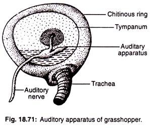
The tergum of the first abdominal segment bears on its each lateral side a circular area for brane is tightly stretched. In the inner wall of the tympanum lies a slender auditory apparatus (Fig. 18.71).
This auditory apparatus remains attached with its one end to the centre of the tympanum and by the other end to the auditory nerve. The sound waves hitting the tympanum are conveyed to the auditory apparatus to be communicated through auditory nerves.
8. Essay on the Reproductive System of Grasshopper:
Sexes are separate and sexual dimorphism is noted from the disposition of structures present in the posterior abdominal segments.
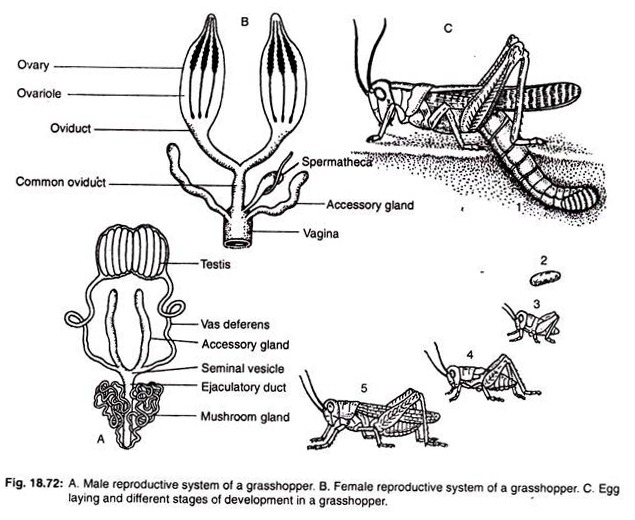
Male reproductive system (Fig. 18.72A):
The male reproductive organs or testes are paired structures placed on the ventral side of the alimentary canal. Each testis is divided into four regions—Germarium, Spermatocyte zone, Spermatid zone and Spermatozoa zone. The sperm cells from each testis are carried through a duct, called the vas deferens.
The two vasa deferentia open within an unpaired duct, called the ejaculatory duct, having cuticular lining. The last part of the vas deferens is swollen and is known as seminal vesicle. The ejaculatory duct opens to the exterior through a copulatory organ, called aedeagus or penis. The accessory glands, called mushroom glands, open within the ejaculatory duct.
Female reproductive system (Fig. 18.72B):
The female reproductive organs or ovaries are paired structures and present one on each side of the alimentary canal. Each ovary is made up of ovarioles. Each ovariole has following three zones—Terminal filament, Germarium and Vitellarium.
From each ovary reproductive cells or ova pass through a duct, called oviduct. The two oviducts unite to form a common oviduct which opens within a cuticularised chamber, called vagina. It opens to the exterior through ovipositor.
The vagina receives slender ducts from the spermathecae, where the sperm cells remain temporarily stored after copulation. The accessory glands, known as colleterial glands liberate a foamy product within the vagina. This substance forms a hard structure around the egg after deposition. The eggs are laid and burried within small holes which are dug up by the female with the help of ovipositor (Fig. 18.72C-1).
9. Essay on the Development of Grasshopper:
Early development continues for three weeks and then remains suspended for a certain period of time. This phase of arrested growth is called diapause; at this phase the individual tides over the adverse environmental condition.
After the return of favourable period, growth resumes and young grasshopper emerges out of the egg shell. It is then known as a nymph. Its body resembles the adult in all respects excepting the proportion of different parts (Fig. 18.72C, 2-5).
Further growth involves moulting or shedding off of the old exoskeleton. The grasshopper undergoes five moults before becoming an adult. Each phase between two moulting is called an instar. As in all other winged insects (excepting Mayfly) no moulting takes place in the grasshopper after the attainment of the adult form.
10. Essay on the Economic Importance of Grasshopper:
(i) As crop pests:
They eat many kinds of vegetation and cause serious damage to major crops like paddy, wheat. During favourable conditions and lack of enemies they can fly in huge number (June-July) causing swarms, called Plague of grasshoppers.
(ii) As food:
The eggs, nymphs and adults provide good food for several predatory insects, spiders, frogs-, reptiles, birds, etc. sometimes they are used by human as food and fish bait. Grasshopper is used in Japan, Phillipines, Mexico as dish of delicacy. They are commonly eaten by North American Indian and certain primitive tribes. The Greeks grind them by mortars, and the powder is then stored to be used as food.
11. Control of Grasshopper:
(i) Natural control:
Eggs are eaten by some beetles, bee, flies, moles, mice; nymphs by robber flies, digger wasps; both nymphs and adults by predatory large insects, frogs, reptiles, birds and mammals. Flesh flies (Sarcophaga) lay maggots on adults and tachinid flies lay eggs on adult and nymphs of grasshopper, thus maggots bore their way into hosts bringing bacteria and fungal diseases which may cause death.
(ii) Physical control:
During winter, the soil is exposed to sun by ploughing. Developing ones thus wither away in sunlight or eaten up by birds.
(iii) Chemical control:
Various insecticides are used in the form of dust, spray or poisoned bait. Insecticides like aldrin, dieldrin, chloradane, toxaphane are used very often. Methoxychlor is also now used for protecting vegetables and crop plants as it is a non- residual type of insecticide and is less harmful to man and domesticated animals.
