In this article we will discuss about:- 1. Giochidium Larva of Unio 2. T.S. Shell & Mantle of Unio 3. T.S. Anterior Region of Gills 4. T.S. Middle Region 5. T.S. through Posterior Region of Gills.
Giochidium Larva of Unio:
Fig. 136 GLOCHIDIUM LARVA
1. This is the slide of Giochidium larva of Unio.
2. It comprises a bivalve shell and a false mantle lining the shell.
3. Each shell valve is triangular, convex externally and concave internally and is attached to the other valve on the dorsal side only.
4. The free ventral ends of each valve are slightly curved inwards and bear few hooks or spines.
5. The mantle is projected into numerous protuberances bearing sensory bristles.
6. Between the two valves at the dorsal side is present a median adductor muscle which helps in closure of the valves.
7. Below the adductor is present a small median byssus gland horn which arises a long provisional byssus thread or adhesive filament.
8. The larva, prior to metamorphosis, leads a parasitic life where it is attached to a fish through hooked ventral ends.
T.S. Shell & Mantle of Unio:
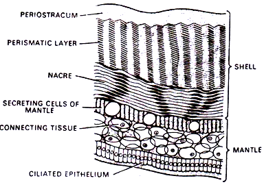
Fig. 137 T.S SHELL AND MANTLEOF UNIO
1. It is the slide of T. S. of shell and mantle of Unio.
2. It exhibits following features:
(a) The shell comprises of following three layers:
(i) Outer most and structure-less periostracum.
(ii) Middle and largest prismatic layer.
(iii) Innermost layer – the nacre.
(b) The periostracum is translucent, dark blue or brown in colour and is comprised of chitinous substance the choncholin.
(c) The prismatic layer is made up of numerous prism-like structures of crystalline Calcium carbonate which remains embedded in choncholin.
(d) The nacre or nacrous layer is comprised of alternate layers of choncholin and crystalline calcium carbonate.
(e) Below the shell lies a thin layer of mantle which is also comprised of three layers i.e.:
(i) Outer secreting epithelial layer,
(ii) Middle connective tissue layer and
(iii) inner most ciliated epithelial layer.
T.S. Unio Anterior Region of Gills:
Fig. 138 T.S UNIO ANT. REGION OF GILLS
1. It is the slide of T.S. unio passing through anterior region of gills.
2. The outer most layer is shell the inner surface of which is lined with mantle.
3. The two shell valves are open below and are connected above.
4. Above, the mantle cavity has a pericaridal cavity surrounding a pair of auricles and a ventricle. The rectum passes through the ventricle.
5. Below the pericardium inside the inner supra branchial chambers lies a pair of Kidney.
6. The foot is club-shaped and occupies major portion of mantle cavity in the centre and is filled with gonads and intestine cut in various shapes.
7. On either side of foot lies a pair of gill laminae the inner & outer.
8. Each gill lamina is made up of two gill lamellae.
9. The gill filaments and water tubes are absent.
10. The inner lamella of Inner lamina is not attached to foot but hangs freely in mantle cavity.
11. The lamella of outer lamina and outer lamella of inner lamina are attached to mantle.
12. The outer supra branchial chambers continue into outer gill laminae.
T.S. Unio through Middle Region:
Fig. 139 T.S UNIO MIDDLE REGION OF GILLS
1. It is the slide of T.S. of Unio passing through middle region of gills.
2. The outer most layer is shell, the inner surface of which is lined with mantle flap.
3. The two shell valves are open below and are connected above.
4. Above, the mantle cavity has a pericardial cavity in the middle which surrounds a ventricle through which runs the rectum.
5. Below the pericardium lies a pair of Kidney inside the inner supra branchial chambers.
6. The foot is a fusiform structure occupying the 3/4 of mantle cavity in the centre and is filled with gonads and Intestine cut in various forms and shapes.
7. On either side of foot are present one pair of gill laminae – the inner & outer.
8. Each gill lamina is connected with foot, with each other and with mantle flap at the base and are made up of gill filaments and water tubes.
T.S. Unio through Posterior Region of Gills:
Fig. 140 T.S UNIO POST REGION OF GILLS
1. It is the slide of T.S. of Unio passing through posterior region of gills.
2. The outer most layer is shell the inner surface of which is lined with mantle flap.
3. The two shell valves are open below and are connected above.
4. Above, the mantle cavity has rectum. Below the rectum the whole space is occupied by post adductor muscle. Below this muscle lies the visceral ganglion.
5. The foot heart, kidney, bladder, keber’s organ etc. are all absent.
6. The two gill lamellae of inner and outer gill laminae are without gill filaments and water tubes etc.
7. The inner gill lamellae of inner laminae are connected with each other in the middle.


