Epidermis:
i. The epidermis is outermost layer of the stem.
ii. It is protective in function.
iii. It is single cell thick.
iv. Multicellular hairs and stomata are present.
v. The outer wall of the cells is cuticularised.
Cortex:
i. It is present below the epidermis and several layers thick.
ii. It is divided into three regions hypodermis, middle cortex and endodermis.
Hypodermis:
i. It is made up of collenchymatous cells and is3-4 layered thick.
ii. These cells contain chlorcplast.
iii. The intercellular spaces are absent.
Middle cortex:
i. It is present between hypodermis and endodermis.
ii. It is made up of parenchymatous cells and is several layers thick.
iii. The cells are oval or spherical in shape with intercellular spaces.
iv. It mainly serves for food storage, besides providing mechanical strength.
Endodermis:
i. It is the innermost layer and present up to the stele.
ii. The cells are barrel-shaped and due to the accumulation of starch it is also called starch sheath (in uoung dicot stem).
iii. Casparian strips are present in these cells.
Pericycle:
i. It is formed of alternate bands of parenchymatous and sclerenchymatous cells.
Vascular bundles:
i. The vascular bundles are conjoint, collateral and open.
ii. These are arranged in a ring.
iii. Each vascular bundle contains xylem, phloem and cambium.
Cambium:
i. It is a thin strip of actively dividing meristemetic cells.
ii. It is made up of single layer of meristematic cells.
iii. The division of cambium cells adds to the phloem towards periphery and xylem towards centre.
Epidermis:
i. Epidermis is the outermost layer of the stem and is protective in function.
ii. It is single cell thick.
iii. Stomata may be present but hairs are absent.
iv. The outer wall of the cells is cuticularised.
Hypodermis:
i. It is made up of sclerenchymatous cells and is 2-4 layers thick.
ii. The intercellular spaces are absent.
Ground tissues:
i. These are present below the hypodermis.
ii. There is no differentiation between cortex, endodermis, pericycle and pith.
iii. The cells contain reserve food material.
iv. Vascular bundles are embedded in the ground tissues.
Vascular bundles:
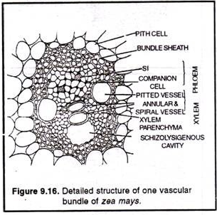
i. These are scattered throughout the ground tissue.
ii. Each bundle is surrounded by a sclerenchymatous sheath.
iii. The vascular bundles are conjoint, collateral, endarch and closed.
iv. Cambium is absent.
Anatomy of Root:
A root is characterized by following essential features:
i. The outermost layer is termed as epiblema or piliferous layer or rhizoaermis.
ii. The epiblema bears a large number of unicellular root hairs.
iii. Cuticle and stomata are absent.
iv. Cortex is formed of parenchymatous cells.
v. Endodermis is well developed.
vi. Pericycle is distinct.
vii. Vascular bundles are radialy arranged.
viii. Xylem is exarch (protoxylem towards periphery and metaxylem towards centre).
ix. Phloem consists of sieve tubes, companion cells and phloem parenchyma. (In monocots however, the phloem parenchyma is absent).
Anatomy of Dicot Root:
Epiblema:
i. It is the outermost layer of cells.
ii. The cells usually have fine tubular elongations, called root hairs.
iii. These help in the absorption of water.
Cortex:
i. It lies below the epiblema, up to endodermis.
ii. It is composed of circular or polygonal; parenchymatous cells with intercellular spaces.
iii. The innermost layer of cortex is called endodermis.
iv. Its cells have Casparian bands (special thickenings).
v. However some cells do not have these thickenings and are called passage cells.
Pericycle:
i. It is one layer thick structure that is present below the endodermis.
Vascular Bundles:
i. These are arranged in a ring (i.e., radial).
ii. Xylem and phloem are situated at separate radii.
iii. Xylem is exarch (i.e., the protoxylem towards outside and metaxylem towards centre).
Pith:
i. It lies in the centre but is poorly developed due to more development of metaxylem towards the centre.
Anatomy of Monocot Root
Epiblema:
i. It is the outermost layer of cells with large number of unicellular root hairs (Root hairs are the elongation of the cells of epiblema).
Cortex:
i. It lies below the epiblema, upto endodermis.
ii. It is multilayered.
iii. It is composed of circular or polygonal, parenchymatous cells with intercellular space.
iv. The innermost layer of cortex is called endodermis.
v. Its cells have Casparian bands (special thickenings).
vi. However some cells do not have these thickenings and are called passage cells.
Pericycle:
i. It is one layer thick partly sclerenchymatous structure that is present below the endodermis.
Vascular bundles:
i. Vascular bundles are polyarch, radial and exarch.
ii. Phloem parenchyma is absent.
Pith:
i. It lies in the centre and consists of loosely arranged parenchymatous cells.
Anatomy of Leaf:
Structure of a dicot leaf (dorsiventral leaf):
The anatomy of a dicot leaf exhibits following structures-
Upper epidermis:
i. It is the upper outer most layers and serves to protect the underlying tissues.
ii. It is single layer thick and made up of parenchymatous cells.
iii. Its outer walls are cuticularised.
iv. Stomata and chloroplasts are absent.
Lower epidermis:
i. This is like the upper epidermis but stomata are present on this surface. Chloroplasts are absent in the cells but the guard cells of the stomata contain chloroplast.
Mesophyll:
i. This tissue lies between the upper and lower epidermis and it is divided into two regions- Palisade parenchyma and spongy parenchyma.
(i) Palisade parenchyma:
i. The cells are elongated and perpendicular to the upper epidermis.
ii. Chloroplasts are present.
iii. These cells are mostly two layered and actively participate in photosynthesis.
iv. No intercellular spaces.
(ii) Spongy parenchyma:
i. It is present below the palisade parenchyma.
ii. The cells are spherical or oval and are irregularly arranged. They possess chloroplasts.
iii. Intercellular spaces are present which are interconnected and open to outside through stomata.
Vascular bundles:
i. These are scattered in spongy parenchyma.
ii. The vascular bundle of mid rib is the largest.
iii. The vascular bundles are collateral and closed.
iv. Around each bundle is present a parenchymatous sheath, called bundle sheath.
Structure of a monocot leaf (Isobilateral leaf):
The anatomy of a monocot leaf exhibits following structures:
Epidermis:
i. The upper and lower epidermises of leaf are similar and they consist of a single layer of cells.
ii. Some cells in the upper epidermis get enlarged in sized and are called bulliform cells or motor cells. It helps in leaf curling in grasses to check transpiration.
iii. Bulliform or Motor cells are found in groups.
iv. The cells of the epidermis are cuticularised.
Mesophyll:
i. It is not differentiated into palisade and spongy tissues.
ii. The cells are spherical and enclose intercellular spaces.
iii. The cells are irregularly arranged and contain chloroplasts.
Vascular bundles:
i. Many large and small vascular bundles are present in the leaf.
ii. Each bundle is surrounded by a layer of thin walled cells, called bundle sheath.
iii. The vascular bundles are conjoint, collateral and closed.
Normal Secondary Growth in Dicot Stem:
The permanent change in volume and weight of the plant or its organ is known as growth. It is of two types:
1. Primary growth:
Increase in length of root, shoot and leaves due to activity of primary meristem are called primary growth.
2. Secondary growth:
It refers to addition of extra amount of tissue in the stelar and extrastelar region due to activity of lateral meristem i.e., vascular cambium (fascicular cambium) and cork cambium respectively.
In a dicot stem, the process of secondary growth can be studied under the following headings:
Secondary growth in stelar zone
Secondary growth in extra stelar zone
Secondary growth in stelar zone
In a dicot stem, stelar zone extends from pericycle to pith and comprises following tissues:
(a) Parenchymatous pith
(b) Vascular bundles with primary vascular tissue (primary xylem and primary phloem)
(c) Vascular bundles are arranged in a ring
(d) Each vascular bundle is conjoint, collateral, open and endarch [i.e., centrifugal xylem)
(e) Primary medulary rays present in between two vascular bundles.
In stelar zone secondary growth takes place in the following steps:
I. Formation of inter fascicular cambium:
A parenchymatous cell of medulary rays in a line with vascular cambium becomes meristematic. These strips of meristem are known as inter fascicular cambium.
II. Formation of cambium ring:
Combination of vascular cambium (or intra fascicular cambium or fascicular cambium or cambium present in between primary xylem and phloem) with inter fascicular cambium results in the formation of cambium ring.
III. Activity of cambium ring:
A. Vascular cambium consists of two kinds of cells viz., fusiform initial (cells are elongated, spindle shaped and produces secondary xylem and phloem) and ray initial (with small, isodiametric cells, produces ray parenchyma in secondary xylem and phloem).
B. Due to activity of vascular cambium new secondary tissue is produced towards inner side as well as outer side. New cells cut off on the outer side of the cambium are gradually converted into phloem elements and inner tissue forms xylem elements. It is noted that cambium is more active on the inner side than the outer side. As a result, larger amount of secondary xylem is formed than the secondary phloem.
C. If secondary xylem is always produced towards centre and phloem towards periphery, it is known as normal secondary growth. Due to continuous formation of secondary xylem, pith is crushed and the space is occupied by primary xylem. This leads to the formation of annual ring.
D. Formation of annual rings or growth rings or growth layer takes place by the periodical activity of the cambium due to variation in environmental condition. Cambium is more active in spring and produces greater amount of xylem vessels with wider lumen. Cambium is less active in winter and produces small amount of xylem vessels with narrow lumen. The wood thus formed in spring is known as spring wood or early wood and the wood in winter is called autumn wood or late wood.
In transverse section of dicot stem, these two types of wood appear together as i concentric ring known as annual ring or growth ring. (Fig. 9.23 & 9.24) It is noted that each annual ring corresponds to one year growth (approximately). Hence, the age of plant can be determined roughly by counting the total number of annual rings and the branch is known as Dendrochronology.
E. Due to continuous normal secondary growth for several years formation of heart wood (duramen) and sap wood (alburnum) takes place. (Fig. 9.25)
Secondary growth in extra stelar zone:
This zone extends from hypodermis to endodermis and made up of primary ‘issues during primary growth.
Secondary growth takes place in following steps:
(a) Origin of cork cambium (Phellogen):
First or second layer of hypodermis or outer cortex becomes meristematic and known as cork cambium.
(b) Activity of cork cambium:
It divides in tangential plane cutting cells towards inner as well as outer side. The outer cells are compactly arranged and have thin wall in the beginning but as they mature there is gradual loss of living matter and the cell wall becomes thick due to deposition of fatty substance suberin. It is known as cork (phellem). Thin wall cell cuts of towards the inner side of the cork cambium are known as phelloderm (secondary cortex). Its cells are living with cellulose cell wall.
(c) Formation of periderm:
Cork (phellem), cork cambium (phellogen) and secondary cortex (phelloderm) together form periderm.
Bark:
All the tissues outside the vascular cambium are collectively known as Bark. It includes secondary phloem, primary phloem, pericycle, endodermis, primary cortex and periderm (Fig. 9.26)
(d) Lenticel:
It is a small portion of periderm where the activity of phellogen (cork cambium) is more. Cells are loosely arranged, thin walled with numerous inter cellular spaces known as complimentary cells. Lenticel (breathingpores) helps in gas exchange in woody part of the plant and helps in lenticular transpiration. (Fig. 9.27)

