List of four important early vascular plants:- 1. Cooksonia 2. Rhynia 3. Horneophyton 4. Trimerophyton.
Early Vascular Plant # 1. Cooksonia:
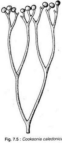
Cooksonia (Fig. 7.5) is the earliest known vascular plant, ranging in age from Middle- Upper Silurian to Lower Devonian. Several species of Cooksonia have been described from various places like Ireland, South Wales, Scotland, Germany, Czechoslovakia, Russia, North Africa and North America.
The plant has dichotomously branched aerial axis which are terminated in sporangia. There is no report of the basal parts of these plants. The sporangia exhibit various shapes from round to reni- form which contain isospores with trilete aperture.
Early Vascular Plant # 2. Rhynia:
Geological Occurrence:
Lower Devonian.
Geographical Distribution:
Mostly reported from Rhynie chert bed of Scotland.
Kidston and Lang described two species of Rhynia, namely Rhynia gwynne-vaughanii and R. major, from Rhynie chert bed (Lower Devonian). R. gwynne-vaughanii had prostrate rhizomatous stem and aerial axes (Fig. 7.6A). The plants attained a height of 7 inches (18 cm).
The prostate axes, bore numerous delicate rhizoids which performed the function of anchorage and absorption of nutrient and water from soil. The erect axes had both dichotomous and lateral (adventitious) branching. The dichotomizing axes terminated by ellipsoidal sporangia filled with isospores (homo- spores) (Fig. 7.6C).
The spores were trilete, thick- walled and about 40 µm in diameter. The entire surface of the plant was covered by thick cuticle. The aerial axes were photosynthetic having simple stomata in their epidermal layer. The lateral adventitious branches were shed and left scars on the aerial axes.
Both the rhizomatous and aerial stem showed protostelic (haplostele) configuration with a slender central xylem strand surrounded by a narrow phloem tissue (Fig. 7.6B). The cortex differentiated into two zones: the outer cortex with tightly packed cells and the inner cortex with loosely packed cells. The outer cortex provided mechanical strength to the stem (Fig. 7.6B).
The other species of Rhynia (Rhynia major) has been transferred to a new genus, Aglaophyton major, by D. S. Edwards (1986). He suggested that this was a non-vascular plant having a rather different branching pattern and no central xylem strand.
The erect dichotomising branches parted at a rather wide angle of over 60° and arose from the prostrate rhizoid-bearing axes that showed an unusual arched configuration (Fig. 7.7). The central conducting strand comprised of elongate thin-walled cells without having the characteristic secondary wall thickening of vascular plants.
These cells are very similar to the hydroid cells of moss plants. The sporangia were terminal and fusiform in shape and consisted of isospores (65 µm in diameter) with trilele aperture. Edwards concluded that Aglaophyton major was a non-vascular plant with a pteridophytic life cycle.
Gametophytes of Rhyniopsida:
Two gametophytic plants viz., Lyonophyton rhyniensis and Sciadophyton have been described from the Rhynie chert bed (Remy and Remy 1980). Lyonophyton rhyniensis (fig. 7.8C, D) had a rhizome with aerial axes.
Each terminal axis has a terminal bowl-shaped lobed gametangiophore bearing antheridia on the inner face of the gametangiophore lobe, and the groups of archegonia at the central position within the bowl.
It was suggested that Horneophyton lignieri might have been the corresponding sporophyte plant of Lyonophyton based on their lobed morphology and having common association in Rhynie chert bed. However, the gametophore of Lyonophyton are also reminiscent.of’ Aglaophyton major on the basis of their central conducting strand as well as having stomata and cuticle.
The other gametophyte called Sciadophyton is very much similar to Lyonophyton which has a variable number of vertical terete axes that occasionally dichotomise. The central conducting tissue of these vertical axes contained some amount of tracheids. These axes terminate in cup-shaped gametangiophore containing antheridia and archegonia. All the vertical axes radiate from a central corm (Fig. 7.8A, B).
The existence of such gametophytes in early vascular plants supports the theory of isomorphic alternation of generation where both the sporophyte and gametophyte were isomorphic, photo-synthetic and containing tracheids.
Early Vascular Plant # 3. Horneophyton:
Geological Occurrence:
Lower Devonian
Geographical Distribution:
Mostly reported from Rhynie chert bed of Scotland.
Characteristic Features:
Horneophyton lignieri was described for the first time by Kidston and Lang (1920) from Rhynie chert bed as Hornea lignieri. According to ICBN, the original generic name Hornea was changed later to Horneophyton as the original generic name was previously occupied by a flowering plant.
The plant was approximately 6-8 inches (15- 20 cm) high. The underground portion of the plant consists of a series of bulbous, corm-like structures bearing numerous unicellular rhizoids (Fig. 7.9A). The basal corm lacks any evidence of vascularization and is made up of dark, thick-walled parenchymatous cells. The aerial axes are naked and branched dichotomously several times.
Internally, the aerial axes have protostele with centrally placed protoxylem surrounded by metaxylem. The xylem is surrounded externally by phloem.
The sporangia are borne terminally at the apex of some of the aerial branches. Each sporangium is dichotomously branched and may have two to four lobes of varying length. The sporangial lobes are ellipsoidal-cylindrical in shape with truncated distal end (Fig. 7.9B).
Dehiscence of sporangia took place through an apical pore. Extending into the sporangial cavity is a column of sterile tissue around which a continuous layer of sporogenous tissue developed. Horneophyton is homosporous. The sporogenous tissue, composed of trilete isospores, overarched a central columella (Fig. 7.9B).
The sporangium of Horneophyton is unique among all vascular plants, because it consists of a branched fertile unit resulted from dichotomies of the stem apex. Moreover, each fertile lobe was produced by its own apex and remained meristematic for an extended period of time.
Evolutionary Significance:
The discovery of Horneophyton provides the evidence supporting the idea that primitive vascular plants had evolved from bryophytean (Anthoceros type) ancestor. The underground bulbous corm-like base bearing rhizoids and columellate sporangia of Horneophyton are interpreted as being transitional between Anthoceros type ancestor and vascular plants. (Rhynia type).
Early Vascular Plant # 4. Trimerophyton:
Geological Occurrence:
Lower Devonian.
Geographical Distribution:
The shore of Gaspe’ Bay, Canada.
Characteristic Features:
Trimerophyton robusticus, the type species of the class Trimerophytopsida, was established by Hopping (1956) and was initially described as a part by Dawson (1859) under the bionomian Psilophyton robusticus from the shore of Gaspe’ Bay, Canada. The plant was collected as compression-impression fossil — thus nothing is known regarding its internal structure.
The largest fragment of the main stem of Trimerophyton is about 4 inches (9.5 cm) long and approximately 0.4 inches (1.0 cm) wide and consists of numerous spirally arranged, trifurcate, lateral branches (Fig. 7.10). The primary and secondary branching patterns of the laterals are trichotomous.
The trichotomous secondary branches are unequal in size, of which the two upper ascending branches are smaller. The smaller branches dichotomize twice and the third and largest branch dichotomizes three times. All the ultimate branches are terminated by clusters of fusiform sporangia (Fig. 7.10). All the axes are smooth. Trimerophyton is homosporous containing trilete, smooth-wall isospores.
Evolutionary Significanze:
Trimerophyton, along with other members of Trimerophytopsida, exhibit almost every branching pattern to be found in megaphyllous pteridophytes and progymnosperms. Moreover, the sterile laterals can be regarded as dichotomising telome trusses which might have planated and webbed to form megaphylls.
Thus, trimerophytes are considered as the ancestor of megaphyllous leaves. Trimerophytes, in the course of evolution, diversified further for the evolution of all those megaphyllous plant groups such as Sphenopsida; Cladoxylales and Coenopteridales of Filicopsida and Progymnosperms which make their appearance in the Devonian.


