The following points highlight the five methods by which reproduction in bacteria takes place. The methods are: 1. Binary Fission 2. Conidia 3. Budding 4. Cysts 5. Endospore.
Method # 1. Binary Fission:
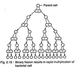
In binary fission, single cell divides into two equal cells (Fig. 2.19). Initially the bacterial cell reaches a critical mass in its structure and cellular constituents.
The circular double stranded DNA of bacteria undergoes replication, where both the strands separate and new complementary strands are formed on the original strands — results in the formation of two identical double stranded DNA (Fig. 2.20).
The new double stranded DNA molecule i.e., incipient nuclei, are then distributed into two poles of the dividing cell (no spindle formation takes place like mitotic division). A transverse septum develops in the middle region of the cell, which separates the two daughter cells.
The binary fission is a rapid process and cell undergoes division at an interval of 20-30 minutes. The division becomes gradually slow after certain time due to accumulation of toxic substance and exhaustion of nutrients.
Method # 2. Conidia:
Conidia formation takes place in filamentous bacteria like Streptomyces etc., by the formation of a transverse septum at the apex of the filament (Fig. 2.21 A). The part of this filament which bears conidia is called conidiophore. After detachment from the mother and getting contact with suitable substratum, the conidium germinates and gives rise to new mycelium.
Method # 3. Budding:
The bacterial cell develops small swelling at one side which gradually increases in size (Fig. 2.21 B). Simultaneously the nucleus undergoes division, where one remains with the mother and other one with some cytoplasm goes to the swelling. This outgrowth is the bud, which gets separated from the mother by partition wall, e.g., Hyphomicrobium vulgare, Rhodomicrobium vannielia, etc.
Method # 4. Cysts:
Cysts are formed by the deposition of additional layer around the mother wall. These are the resting structure and during favourable condition they again behave as the mother, e.g., many members of Azotobacter.
Method # 5. Endospore:
Spores are formed during unfavourable environmental condition like desiccation and starvation. As the spores are formed within the cell, they are called endospores. Only one spore is formed in a bacterial cell. On germination, it gives rise to a bacterial cell.
A. Some endospore forming bacteria:
1. Gram-positive
(a) Bacilli
(i) Obligate aerobes, e.g., Bacillus subtilis, B. anthracis.
(ii) Obligate anaerobes, e.g., Clostridium tetani, C. botulinum.
(b) Cocci, e.g., Sporosarcina.
2. Gram-negative
(i) Bacillus, e.g., Coxiella burnetii
(ii) Cocci, e.g., Escherichia coli
B. Some non-sporing anaerobic bacteria:
1. Gram-positive
(a) Bacilli
(i) Lactobacillus
(ii) Propionibacterium
(iii) Bifidobacterium
(b) Cocci
(i) Peptococcus
(ii) Sarcina
(iii) Peptostreptococcus
2. Gram-negative
(a) Bacilli
(i) Fusobacterium
(ii) Leptotrichia
(iii) Bacteroides
(b) Cocci
(i) Acidoaminococcus
(ii) Veillonella
C. Shape and position of endospore:
Spores may be oval or spherical in shape. The position, relative size and shape remain constant in a particular species. The position of spore may be central, subterminal or terminal (Fig. 2.22). In diameter, it may be the same or wider (Clostridium) or less (Bacillus) than the width of the specific bacterial cell.
D. Structure:
Endospore consists of a central protoplast, the core (Fig. 2.23). The core is mainly composed of DNA, ribosome, t-RNA, enzymes etc. The core is covered by a thin membrane, called core membrane or inner membrane or germ cell membrane, from which the cell wall of future vegetative bacterium develops.
It is covered by a thick layer, the cortex and then a multilayered thin and tough outer spore coat, which may be differentiated into outer and inner coat layer. In some species (Bacillus thuringiensis), it is covered by an additional covering, called exosporium or exosporium basal layer, which is apparently loose.
E. Formation of endospore:
The endospore formation does not take place during active phase of growth. The sporulation starts in conditions unfavourable for the growth due to starvation, desiccation, high temperature etc. The sporulation can also be induced by depleting S, C, N, Fe and PO4 from culture medium.
During sporulation, the first detectable change is the conversion of compact nucleoid into an axial chromatin filament (Fig. 2.24). Then a transverse septum is laid down towards one pole, which separates into small and large portion. The small portion with its cytoplasm and DNA forms the fore spore, which later on develops into a spore.
The membrane of large portion gradually grows around the fore spore. The fore spore increases in size, which becomes opaque and highly refractive, called the endospore. The entire process of sporulation takes place within 16 to 20 hours. The cell in which spore is formed, called sporangium, which remains viable for short period of time after maturation of spore. The spore is liberated by autolysis of sporangium.
The spores can survive in different adverse conditions like heat, drying, freezing, toxic chemicals and radiations. Some bacilli can resist a temperature higher than 150°C.
The endospores germinate under favourable condition which consists of three stages:
(i) Activation:
It takes place by the induction of one or more factors such as acidic pH, heat (60°C for 1 hour), compounds containing free SH groups or through abrasion.
(ii) Initiation:
Binding of any effector substance like L-alanine, adenosine etc. of the medium with the spore coat activates to form an autolysin. The autolysin destroys the peptidoglycan of the cortex. Thereafter, water is taken up and calcium dipicolonate is released. Dipicolonic acid helps to stabilise the spore protein, and both dipicolonic acid and Ca ions provide resistance to heat.
(iii) Outgrowth:
The swelling of spore wall and disintegration of the cortex help to emerge a germ cell after breaking the spore coat, which behaves like a vegetative cell.
Each cell forms only one endospore and persists during unfavourable condition. During favourable condition, it germinates and gives rise to a single bacterial cell. So the spore is a perennating organ and the process is called perennation rather than multiplication.


