1. Rheumatoid Arthritis (RA) Test:
Rheumatoid arthritis (RA) factor is a chronic symptomatic disease of unknown etiology. It is frequently characterized by swelling, pain in joints, by inflammatory and degenerative process involving cartilage and muscle tissues.
Rheumatoid factors (RF) are a group of proteins present in the blood of the patient, suffering from rheumatoid arthritis. It is believed that rheumatoid factors are autoantibodies, present against human globulin. The presence of these autoantibodies is the indication of disease.
The clinical significance of RA determination consists of differentiation between rheumatoid arthritis in which RF has been demonstrated in the serum of approximately 80 percent of the cases and rheumatoid fever in which RF are almost always present.
Principle:
The RA test is a serological test, based on the principle of agglutination. Latex reagent, containing latex particle coated with human IgG, is mixed with test serum. If agglutination results, it indicates the presence of RF more than 8 IU/ml.
Requirement:
Test slide, disposable dropper, disposable mixing stick, latex reagent and blood sample.
Procedure:
1. Pipette out one drop of serum test sample to the ring of test slide with the disposable dropper.
2. Mix the latex reagent by gently shaking of the vial. Add one drop of RA latex reagent to the drop of serum test sample added on the circle of the test slide.
3. Thoroughly mix the sample with the latex reagent using a separate mixing stick uniformly within the area of circle of the test slide.
4. Slowly tilt the slide exactly for two minutes and observe for agglutination.
Observation:
If the test is positive, clumps are seen within two minute.
2. Venereal Disease Research Laboratory (VDRL):
The Venereal Disease Research Laboratory (VDRL) is a test to diagnose syphilis.
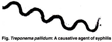
Syphilis is a venereal disease caused by spirochete Treponema pallidum.
The VDRL is an agglutination reaction, which detects anti- syphilis antibody in the patient’s serum. The VDRL antigen used in the test is cardiolepin antigen.
Requirement:
Cavity slide, disposable dropper, disposable mixing stick, antigen reagent and blood sample.
Procedure:
1. Pipette out one drop of serum test sample to the ring of cavity slide with disposable dropper.
2. Add one drop of antigen to it.
3. Rotate the slide for four minutes.
4. Observe for needle-like clumps.
Observation:
In case of VDRL, the results are reported as reactive or non- reactive, depending upon the size and amount of clumps observed.
If clumps are not observed, the reaction is nonreactive. Reactive serum has average clumps. Weakly reactive clumps are those which are seen under the microscope only. Highly reactive has larger clumps, which can be seen by the naked eye.
3. Reactive Protein (CRP) Test:
The C- reactive protein (CRP) is a serum protein synthesized in the liver. The name of protein is derived from the fact that it has capacity to precipitate C carbohydrate of Pneumococcus.
The rate of synthesis and secretion of CRP increases within an hour of acute injury or the onset of inflammation. The finding can be useful as an index of disease activity and treatment status. Apart from indicating inflammatory disorders, CRP measurement helps in differential diagnosis in the management of neonatal septicemia and meningitis.
Principle:
The test is based on the principle of agglutination. Test serum is mixed with CRP latex reagent, if CRP concentration is greater than 0.6 mg/dl, a visible agglutination is observed.
Requirement:
Test slide, disposable dropper, disposable mixing stick, latex reagent and blood sample.
Procedure:
1. Dilute the patient’s serum 1:5 with normal saline.
2. Place one drop of diluted serum to the ring of test slide with the disposable dropper.
3. Mix the latex reagent by gently shaking of the vial. Add one drop of CRP latex reagent to the drop of serum test sample added on the circle of the test slide.
4. Thoroughly mix the sample with the latex reagent using a separate reagent stick uniformly within the area of circle of test slide.
5. Slowly title the slide exactly for two minutes and observe for agglutination.
Observation:
If the test is positive, clumps are seen within two minutes.
4. Antisreptolysin O (ASO) Test:
Group-A Streptococcus produces soluble oxygen liable hemolysin known as streptolysin 0. This has a lethal effect on human beings and especially toxic action to the heart muscle. The pathological changes to the heart and joints produce non-purulent arthritis.
Principle:
The ASO test reagent contains polysterene particles, coated with purified, stabilized streptolysin O. This will react with antistreptolysin 0 (antibody) in the test sample, resulting in agglutination of particles.
Requirement:
Test slide, disposable dropper, disposable mixing stick, ASO test reagent and blood sample.
Procedure:
1. Pipette out one drop of serum test sample to the ring of test slide with the disposable dropper.
2. Mix the ASO test reagent by gently shaking of the vial. Add one drop of reagent to the drop of serum test sample added on the circle of the test slide.
3. Thoroughly mix the sample with the ASO test reagent using a separate mixing stick uniformly within the area of circle of the test slide.
4. Slowly tilt the slide exactly for two minutes and observe for agglutination.
Observation:
Agglutination within two minutes confirms that the ASO title is above 200 IV.
5. Widal Test:
Widal test is a diagnostic test for typhoid. The causative agent of typhoid is Salmonella typhi. The test is based on antigenic reaction of the bacteria.
Principle:
The killed bacterial suspension of Salmonella carries specific o and H antigens. This will react with immune-specific antibodies, which are present in the patients serum if he/she is suffering from the disease. The reaction results in agglutination.
Requirement:
Widal rack with four rows of six tubes (total 24 tubes), Antigen S. typhi O, S. typhi H, S. paratyphi A, S. paratyphi B, waterbath, pipette, beaker, etc.
Procedure:
1. For each test sample, arrange four rows of six tubes in a Widal rack.
2. Take five tubes in another rack for preparation of master dilution.
Prepare master dilution as:
(i) Keep seven rnl of normal saline in tube one and 3.5 rnl in other four tubes.
(ii) Add 0.5 rnl of serum in tube one and mix well.
(iii) Take 3.5 rnl from tube one and add to tube two.
(iv) Mix well and continue the transfer till the last tube.
3. Transfer 0.5 rnl from master dilution tube to each of the corresponding vertical row in the test rack.
4. Place 0.5 rnl of normal saline in each tube in the sixth row to serve as control.
5. Add 0.5 rnl of S. typhi 0 antigen to each tube in the first horizontal row.
6. Add 0.5 ml of S. paratyphi A antigen to each tube in the third horizontal row.
7. Add 0.5 ml of S. paratyphi B antigen to each tube in the fourth horizontal row.
8. Add 0.5 ml of S. paratyphi B antigen o each tube in the fourth horizontal row.
9. It gives dilutions as- 1: 30, 1: 60, 1: 120, 1: 240, and 1: 480.
10. Shake the rack well to mix and incubate the rack with tubes at 37°C for 24 hours.
Observation:
The highest dilution in which there is evidence of agglutination is noted. The agglutination is observed with the help of hand lens or mirror. Agglutination from tube two (1: 60) indicates positive test. Widal’s test can also be done by routine slide agglutination method. But the tube method gives exact titer of the antibodies, hence the severity of the disease.
6. Human Immunodeficiency Virus (HIV):
AIDS is characterized by changes in the population of T -lymphocytes. In an infected individual, virus causes depletion of helper T-cell which leaves the person susceptible to opportunistic infections and some malignancies. The virus that causes AIDS exists in two related types-HIV-1 and HIV-2.
Principle:
Sample is added to sample pad as the sample migrates through conjugate, it reconstitutes and mixes with the selenium colloid antigen conjugate. The mixture continues to migrate through the solid phase to the immobilized recombined antigens and synthetic peptides at the patient’s window site. If antibodies to HIV-1 and HIV-2 are present in the sample, the antibodies bind to the antigen selenium colloid and the antigen at the patient’s window shows a red line.
If antibodies to HTV-1 and HTV-2 are absent, the antigen selenium colloid flows fast the patient’s window and no red line is formed.
Requirement:
Blood sample, HIV test strip.
Procedure:
1. Remove the test strip from the protective cover.
2. Apply 0.05 ml of sample to sample pad and wait for 15 minutes.
3. If the reaction is negative after 15 minutes, observe till one hour.
Observation:
Observe for red lines on the strip. The test is negative if only one line appears on control window. The test is positive if lines appear on test window as well as on control window.
7. Hepatitis B Surface Antigen (HbsAg) Test:
He HBsAg test is attest t&-diagnose viral hepatitis/Australia antigen/hepattis B. Viral hepatitis is a symptomatic disease primarily involving the liver. Hepatitis A virus, hepatitis
B virus and hepatitis C virus cause most cases of acute viral hepatitis. The complex antigen found on the surface of hepatitis B virus is also called HBsAg or Australia antigen.
The presence of HBsAg in serum is an indication of active hepatitis B antigen, may be acute or chronic. In a typical hepatitis B infection, HBsAg will be detected after two to four weeks.
Principle:
The test is an immunoassay for the detection of hepatitis B surface antigen in serum. The membrane is pre-coated with anti- HBs antibody on the test line and antimouse antibody on the control line. During testing, the serum or plasma reacts with the dye, which has been pre-coated in the test strip.
The mixture migrates upwards on the membrane chromatographically by capillary action to react with anti-HBs antibodies on the membrane and generate red lines. Presence of this red line indicates positive test, while its absence indicates negative test. Regardless of the HBsAg as the mixture continues to migrate across the membrane to the immobilized antimouse region, red line at control appears.
Requirement:
Blood sample, HBsAg test strip.
Procedure:
1. Remove the strip from the pouch and use within one hour.
2. Immerse the test strip in the serum sample within arrows pointing towards sample.
3. Be sure that the sample level is below stop line on the test strip.
4. Wait for red lines to appear. Observe for one hour in case of negative test.
Observation:
Observe for red lines on the strip. The test is negative if only one line appears on control window. The test is positive if lines appear on test window as well as on control window.


