In this article we will discuss about the dissection of toad. Also learn about:- 1. Dissection of Alimentary System 2. The Circulatory System 3. Dissection of Venous System 4. Dissection of Arterial System 5. Dissection of Cranial Nerves 6. Dissection of Spinal and Sympathetic Nerves 7. Dissection of Brain 8. Dissection of Urinogenital System 9. The Urinary (Excretory) System 10. The Genital System.
In structural organisation the toad (Fig. 16.1) or frog is a typical vertebrate. To gain a first-hand knowledge in vertebrate anatomy students are asked to dissect toad or frog at the very beginning. Both are most common throughout India.
Killing:
Toads are placed in a glass jar. A certain amount of cotton wool, depending on the size of the container is soaked with chloroform and placed in the jar. The jar is covered with a glass lid. Within a short period the toads become unconscious. Such specimens are used for dissection.
Dissection:
Place the toad on a dissecting tray keeping the ventral surface upward. Stretch the limbs and fix them on the tray by pushing down pins through the fore and hind limbs. Hold the skin of the abdomen with a pair of forceps. Lift it a little and make a small cut.
Pushing the blunt arm of a pair of scissors through the cut, give an incision in the skin along the mid-ventral line extending anteriorly up to the lower jaw, and posteriorly to the end of the trunk. With lateral incisions cut the skin of the limbs along the length. Separate the skin from underlying muscles of the ventral surface and the limbs. Pin down the flaps of the skin.
Give an incision on the abdominal wall at about its middle at a distance of about 6 mm either on the right or the left side of the mid-ventral line. Proceed for a short distance both anteriorly and posteriorly. Lift the cut end of the abdominal wall with a pair of forceps and find out the anterior abdominal vein attached to the dorsal surface of the wall.
Separate it carefully from the abdominal wall. Continue the longitudinal incision up to the anterior limit of the coelome cutting the pectoral girdle. Proceed backwards up to the posterior limit of the coelom.
Turn the flaps of the abdominal wall and pin them down. Push the cut ends of the pectoral girdle outwards taking care that blood vessels are not damaged, and fix them with pins. The organs of the viscera are fully exposed.
Dissection of Alimentary System:
Remove the pericardium with heart and adjacent tissues to expose the oesophagus. Cut the mesentery, as required, to uncoil the intestine. Push the liver to the right. The whole of the alimentary canal is exposed (Fig.16.2). Display the organs properly by fixing them with pins.
Mouth:
A large, semicircular opening at the anterior end of the head. It is bounded by two toothless jaws.
Buccal cavity:
A dorsoventrally compressed spacious chamber with a fleshy tongue on the floor. The tongue is attached with the front end.
Pharynx:
The narrowed posterior region of the buccal cavity.
Oesophagus:
A short, narrow, muscular tube joining pharynx with the stomach.
Stomach:
A large, elongated, sac-like muscular structure lying on the left side in the abdominal cavity and dorsal to the liver. The anterior wide portion is the cardiac, and the posterior narrow part is the pyloric stomach.
Small intestine:
A constriction, the pyloric constriction, is present at the junction of the stomach and the intestine. The intestine has two zones. The first portion parallel to the stomach is the duodenum and the rest long, slender, much coiled part, the ileum.
Rectum:
A short but wide tube, opening posteriorly into the cloaca.
Liver:
A large, reddish-brown, bilobed gland connected by a short, narrow, median portion. The left lobe is larger than the right one and sub-lobed. The small, spherical gall bladder is median in position. The common bile duct opens into the duodenum.
Pancreas:
A large, cream white, irregular gland surrounding the common bile duct. It lies between the stomach and the duodenum.
The Circulatory System:
The heart, veins, arteries and capillaries constitute the circulatory system. The heart is partly venous and partly arterial. It is the major organ, both in the venous and arterial systems.
The Heart:
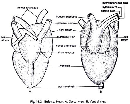
Dissection. Open the viscera following the procedure described previously. Remove the semitransparent pericardium and the heart is exposed. It is a reddish, muscular structure and consists of five parts (Fig. 16.3).
Sinus venosus:
A large, dorsally placed, thin walled, triangular sac.
Atria (auricles):
Two thin walled chambers in front of the ventricle. The right one is larger in which the sinus venosus opens. The left atrium receives pulmonary veins.
Ventricle:
A highly muscular thick walled conical structure posterior to the atria.
Conus arteriosus:
A very short but stout, whitish vessel, arises from the right side of the base of the ventricle. It continues forward and divides into two truncus arteriosus.
Dissection of Venous System:
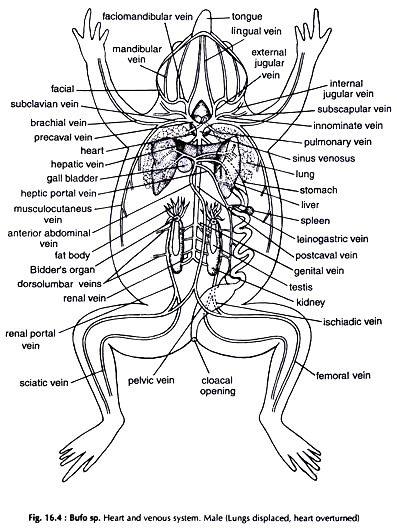
Vessels bringing back blood to heart are veins. All the veins together and the heart constitute venous system (Fig. 16.4).
Remove the pericardium and carefully turn the heart upwards. The narrow posterior end of the heart is now directed anteriorly. Find out the sinus venosus. Locate the two precavals anteriorly and the postcaval vein which is posterior.
The branches joining to form the precavals are identical. Trace a precaval and the veins it receives. Expose the veins received by the median postcaval, and the veins of the hepatic portal and renal portal systems. To expose the femoral and sciatic veins coming from the hind limbs, split the disc formed by the fusion of two halves of the pelvic girdle into two, with a sharp scalpel. This makes removal of femur easier.
A. Precaval vein:
It receives the following veins:
External jugular:
The anteriormost vein formed by the union of lingual from the tongue and faciomandibular coming from the facial region and lower jaw.
Innominate:
It is the middle one and receives internal jugular from the head region and subscapular from the shoulder.
Subclavian:
Largest and posteriormost of the series. Formed by the union of brachial from the fore limb and musculocutaneous from the skin and muscles of the back.
B. Postcaval vein or vena cava:
It is a large, median vein, proceeds anteriorly from near the posterior end of the kidneys, lying between them.
It receives the following veins:
i. Renal:
A few pairs from the kidneys.
ii. Genital:
A pair from the gonads—testes or ovaries.
iii. Hepatic:
A pair from two lobes of the liver.
Renal portal system:
It consists of two veins and their tributaries opening in the two kidneys. The veins of the two sides are similar.
Femoral:
Runs from the outer side of the hind limb. In the coelom it gives out a branch, the pelvic and continues forward. The pelvic joins its fellow of the other side to form the anterior abdominal vein.
Sciatic:
Runs from the inner side of the femur.
Renal portal:
Formed by the union of femoral and sciatic. Runs forward and opens laterally in the kidney.
Dorsolumbar:
Two to four veins from the dorsal body wall join the renal portal vein along the side of the kidney.
Hepatic portal system:
It consists of veins opening in the liver.
Hepatic portal vein:
Formed by the union of veins running from the stomach, intestine, rectum and spleen.
Anterior abdominal vein:
Formed by the union of two pelvic veins. Pelvic veins are branches of femoral veins. The hepatic portal and the anterior abdominal join together. The common vein divides into branches to enter the two lobes of the liver.
C. Pulmonary vein:
Small but wide vessels not associated with the major veins. Two veins from two lungs unite to a common vein just before opening in the left atrium. They bring oxygenated blood from lungs.
Dissection of Arterial System:
Vessels carrying away blood from heart are arteries. All the arteries together and the heart constitute arterial system (Fig. 16.5).
To dissect arterial system some of the veins are to be removed. Expose the heart by removing the pericardium. Trace the arteries along their courses.
Conus arteriosus:
A stout vessel arising from the base of the ventricle at its right side, runs obliquely forward and divides into two truncus arteriosus.
Truncus arteriosus:
Each truncus divides into three vessels or arches.
A. Carotid arch:
The anterior most one. It runs anterolaterally, forms a swelling, the carotid labyrinth and divides into two.
External carotid ends in throat and tongue.
Internal carotid goes to head.
B. Systemic arch:
The middle arch. It turns round the alimentary canal, courses dorsally and unites with its fellow of the other side to form dorsal aorta. The branches of the systemic arches are similar except oesophageal.
Laryngeal:
A small vessel at a short distance from the origin of the arch. It supplies larynx.
Occipitovertebral:
Springs at about the middle of the arch and Courses dorsomedially. Supplies head and vertebral column.
Subclavian:
Origin close to the occipitovertebral. Runs outward to supply the fore limb and shoulder.
Oesophageal:
Origin slightly anterior to the beginning of the dorsal aorta. Supplies blood to the oesophagus. It is present only in the left systemic arch.
Dorsal aorta:
It runs posteriorly and sends following branches.
Coeliacomesenteric:
Origin close to the beginning of the dorsal aorta. It divides into two. The anterior branch coeliac supplying stomach, pancreas, gall bladder and liver, and the posterior branch mesenteric supplies spleen, intestine and liver.
Urinogenital:
A number of small, paired arteries ending in the kidneys and the gonads.
Iliac:
Posteriorly the dorsal aorta bifurcates to two iliacs. The iliac sends a branch, the epigastric to the ventral body wall and divides into two, the femoral and sciatic supplying the hind limb.
C. Pulmocutaneous arch:
It sends deoxygenated blood through two branches. The vessel loops over the heart anterodorsally and divides into two branches.
Cutaneous to the skin of the back.
Pulmonary to the lung of the side.
Dissection of Cranial Nerves:
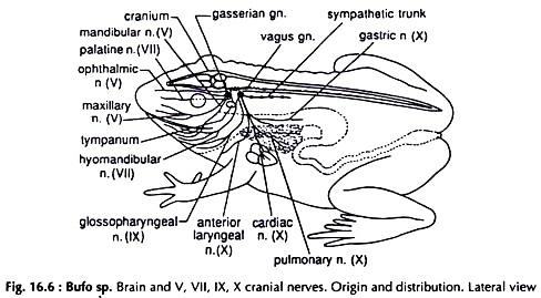
The cranium or brain box is bony. To expose the brain and the roots of cranial nerves follow the technique used in case of Lata fish (Channa punctatus). Trace the nerves from origin to distribution along their courses (Fig. 6.6).
Carefully remove the eye-ball from the orbit to trace the branches of fifth and seventh cranial nerves.
Fifth (V) cranial nerve:
The fifth or trigeminal is the first nerve of the medulla oblongata and arises from the ventrolateral side at its anterior end. A swelling, the gasserian ganglion is present at its base.
It has three branches:
a. Ophthalmic runs across the dorsal border of the orbit and supplies the skin above the eye-ball.
b. Maxillary courses along the postero- ventral border of the orbit and innervates the upper jaw.
c. Mandibular is posterior to the maxillary and supplies the lower jaw.
Seventh (VII) cranial nerve:
The seventh or facial is the third nerve of the medulla oblongata. It arises from the side of the medulla behind the fifth nerve. The facial nerve fuses with the gasserian ganglion of the fifth nerve. After emergence from the ganglion it divides into two branches.
a. Palatine runs through the floor of the orbit and innervates the roof of the mouth cavity.
b. Hyomandibular proceeds outward and backward around the auditory capsule, bifurcates at the angle of the jaws and supply head and lower jaw.
Ninth (IX) cranial nerve:
The ninth or glossopharyngeal nerve arises from the lateral side of the medulla oblongata just behind the auditory nerve. It is the fifth nerve of the medulla. Immediately after its origin the nerve fuses with the vagus ganglion.
After emergence from the ganglion it divides into two branches. The small anterior branch joins the hyomandibular branch of the VII or facial nerve, and the large posterior branch innervates the floor of the buccal cavity, tongue and pharynx.
Tenth (X) cranial nerve:
The tenth or vagus springs from the side of the hindermost part of the medulla oblongata. A swelling, the vagus ganglion is present at the base. It has four branches, which course posteroventrally.
a. Laryngeal innervates the larynx.
b. Pulmonary innervates the lung.
c. Cardiac innervates the heart.
d. Gastric innervates the oesophagus and stomach.
Dissection of Spinal and Sympathetic Nerves:
Displace the viscera to one side, the spinal and sympathetic nerves of the side are exposed. The nerves of the other side can be exposed in similar way. The spinal nerves run outward from the vertebral column. The sympathetic trunks are closely set to the vertebral column (Figs. 16.7 and 16.8).
The spinal nerves:
The spinal nerves are ten pairs. Each pair come out of the vertebral column through intervertebral foramina (sing, foramen) between the adjacent vertebrae, surrounded by calcareous body. From anterior to posterior, they are named first to tenth spinal nerves.
First spinal nerve:
The hypoglossal nerve, comes out of the vertebral column behind atlas and courses along the glossopharyngeal (ninth cranial) nerve.
Second and third spinal nerves:
The thick and stout second spinal nerve joins with a thin, third spinal nerve to form brachial plexus, supplying fore limb as brachial nerve.
Fourth, fifth, sixth spinal nerves:
Innervate skin and muscles of the trunk region.
Seventh, eighth, ninth, tenth spinal nerves:
They join with one another and form sciatic plexus, from which arises the sciatic nerve supplying hind limb.
The Sympathetic Nerves:
Sympathetic trunks:
The sympathetic trunks are two thin nerves, run from the posterior end of the vertebral column lying close to it, one on each side. The posterior ends of trunks are near the end of the spinal cord.
Anteriorly each sympathetic trunk accompanies the systemic arch of the side. Further forward it splits to encircle the subclavian artery, enters the cranium, forms the communicating twig to the vagus ganglion and terminates by joining the gasserian ganglion of the fifth cranial nerve.
Sympathetic ganglia:
A sympathetic trunk bears ten pairs of ganglia, the sympathetic ganglia. Each ganglion is connected to the corresponding spinal nerve by a short ramus communicans.
Solar plexus:
Three slender nerves arising from fifth, sixth and seventh sympathetic ganglia of each side join to form solar plexus around coeliacomesenteric artery and innervates the stomach, intestine and related organs as splanchnic nerve.
Dissection of Brain:
Take out the brain (Fig. 16.9) following the technique used for Channa punctatus. Put it under water in a watch glass.
Dorsal View of the Brain:
Olfactory lobes:
Paired, anteriormost outgrowths of the brain. They unite mesially and each gives out an olfactory nerve.
Cerebral hemispheres:
Two large, ovoid structures, unite mesially and form the cerebrum.
Diencephalon:
A rhomboidal area between the cerebrum and optic lobes.
Pineal body:
A small mass springing from the dorsal surface of the diencephalon.
Optic lobes:
Two large, almost round bodies behind the diencephalon.
Cerebellum:
A transverse band-like structure posterior to the optic lobes.
Medulla oblongata:
It is swollen in front and narrows posteriorly to continue as the spinal cord.
Ventral View of the Brain:
Optic chiasma:
It is really not a part of the brain. This is formed by the crossing of the two optic nerves in the posterior region of the cerebrum.
Infundibulum:
It is a mesial downward process from the ventral surface of the diencephalon, behind optic chiasma.
Pituitary body:
An ovoid body attached to the infundibulum.
The olfactory lobes, cerebral hemispheres, optic lobes and medulla oblongata are visible from ventral side also.
Dissection of Urinogenital System:
The urinary and genital systems are not completely separate in toad and the two are collectively described as urinogenital system. The interdependence of the two systems are, however, restricted only to males.
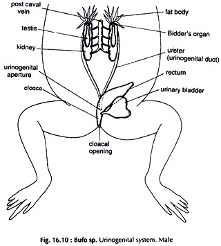
The organs are located in the posterior half of the coelom. Remove the alimentary system, the heart and major portion of the circulatory system, leaving only the posterior part of the dorsal aorta and the postcaval vein. The urinogenital system is exposed (Figs. 16.10 & 16.11). Remove finger-like fat bodies attached to the anterior end of the kidneys.
The Urinary (Excretory) System:
(Figs. 16.10 & 16.11)
Kidneys:
Two flat, somewhat elongate oval, slightly lobulated reddish bodies, one on each side of the vertebral column.
Ureters:
Thin walled ducts, arise from the outer border of the kidneys and run backward. Posteriorly the two ureters join, form a common duct and opens into the cloaca on its dorsal surface.
Urinary bladder:
A thin walled, bilobed median sac anteroventral to the cloaca. It opens into the cloaca on the ventral surface.
The Genital System:
Male Genital System:
(Fig. 16.10)
Testes:
Whitish, slightly elongated, paired bodies, attached to the ventral face of the kidneys near the anterior end.
Bidder’s organ:
Rounded bodies, one attached to the anterior end of each testis.
Urinogenital ducts:
In male, the ureter functions as both vas deferens and ureter.
Female Genital System:
(Fig. 16.11)
Ovaries:
Two large, much folded, membranous, dark bodies with numerous white dot like structures, one on each side of the coelom. The ovaries are slung to the dorsal wall of the coelom by mesentery.
Oviducts:
Two large, convoluted tubes. The oviduct ends in a funnel at the anterior end opening in the coelom.
Uterus:
The posterior dilated portion of the oviduct. The two uteri join posteriorly and the common genital duct opens into the cloaca on its dorsal surface.


