In this article we will discuss about the dissection of Prawn. Also learn about:- 1. Dissection of Alimentary System 2. Dissection of Nervous System.
Dissection:
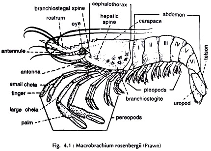
Put the specimen (Fig. 4.1) on the dissecting tray. Lift the carapace from the lateral sides and cut it loose at the anterior end. Cut the inner lining membrane and the carapace is separated. Remove the abdominal terga with the pleura by making two parallel cuts along the ventrolateral lines of the abdomen starting from the anterior end and running up to telson. The dorsal surface of the animal is completely exposed.
Put clear water in the dissecting tray and pin down the specimen properly. The organs lodged beneath the dorsal wall of the cephalothorax from dorsal to ventral surface in succession are, the pericardial sinus containing the heart, the gonads-testes or ovaries, the renal sac, the cardiac and pyloric stomach and the ventral thoracic ganglionic mass. The hepatopancreas is a large orange-yellow mass in which some of the above organs are embedded.
Dissection of Alimentary System:
Remove the dorsal wall of the cephalothorax, pericardial sinus, gonads and the renal sac. The major part of the cardiac stomach, the pyloric stomach and the anterior part of the mid gut (intestine) are embedded in the hepatopancreas (Figs. 4.2, 4.3).
Give a superficial incision with a pair of fine scissors along the mid-dorsal line of the abdomen up to its posterior end. A narrow groove is exposed in which lies the intestine. The intestine blends with the surrounding muscles in colour but very often it stands out prominently due to its contents (Fig. 4.2). The muscles of one side at the base of the telson should be cut to expose the rectum (Fig. 4.3).
The half of the hepatopancreas should be removed from one side (Fig. 4.3) proceeding from the dorsal to ventral surface to expose whole of the cardiac stomach, the pyloric stomach and the anterior part of the intestine.
Mouth:
A large elliptical aperture, situated ventrally in the head region. Laterally, it is guarded by two mandibles.
Buccal cavity:
A small anteroposteriorly compressed chamber next to the mouth.
Oesophagus:
A broad, vertically oriented tube joining buccal cavity with the cardiac stomach.
Stomach:
A spacious sac divided into two:
a. cardiac stomach:
large, bag-like, constituting the dorsal part, and
b. pyloric stomach:
a small chamber located ventrally. The opening between the two chambers is guarded by a valve.
Intestine:
A long slender tube. Arising from the posterior end of the pyloric stomach it runs backward ascending between the two lobes of the hepatopancreas to reach the dorsal groove in the abdomen beyond cephalothorax and runs posteriorly to end. in rectum in the last abdominal segment.
Rectum:
The last part of the gut. It is a small chamber, wider anteriorly and narrows down posteriorly to open on the ventral surface at the base of the telson.
Hepatopancreas:
A large, yellow-orange mass, consists of two lobes and occupies major portion of the cephalothorax. Two ducts from each lobe of the hepatopancreas open into the pyloric stomach.
Dissection of Nervous System:
Remove the carapace and the terga with pleura. The brain is located below the base of the rostrum. Remove the chitinous endo-pharyngeal skeletal plates overlying the brain. The brain is covered by a thick layer of fat. Remove the fat to expose the brain. Remove the hepatopancreas. Cut the oesophagus and pull it out with the stomach.
Expose the nerves arising from the brain and follow their courses. Trace the two circumoesophageal connectives running downwards from the lateral sides of the brain to meet the ventral sub-oesophageal ganglionic mass.
To expose the ventral nerve cord cut the large flexor muscles of the abdomen along the middle line with a scalpel. Press the muscles laterally and pin them down to the wax of the dissecting tray. The nerve cord with its ganglia is clearly visible (Figs. 4.4, 4.5).
Brain or supraoesophageal ganglia:
A bilobed ganglionic paired mass at the base of the rostrum.
Following paired nerves arise from the brain:
a. Optic supplying the eyes.
b. Ophthalmic supplying eye muscles.
c. Antennulary supplying antennules.
d. Antennary supplying antennae.
e. Tegumental supplying labrum.
Circumoesophageal connectives:
Arising from the lateral sides of the brain the two nerves run backward and downward round the oesophagus and join the large thoracic ganglionic mass.
Post-oesophageal loop:
The circumoesophageal connectives are joined by a fine, transverse commissure before they join the thoracic ganglionic mass.
Thoracic or sub-oesophageal ganglionic mass:
Formed by the fusion of 11 segmental ganglia of the cephalothorax and sends 11 pairs of nerves, supplying:
(1) Mandibles,
(2) Maxillulae,
(3) Maxillae, (4-6) 3 pairs of maxillipeds, (7-11) 5 pairs of walking legs (pereopods).
Ventral nerve cord:
Runs along the mid-ventral line of the abdomen and bears 6 ganglia. The last or the stellate ganglion is the largest. Each ganglion sends nerves to musculature, pleopods and the last one to the uropod.
Visceral Nervous System:
Two small visceral ganglia, one behind the other; a small nerve connecting the ganglia with the brain and two connectives joining the two visceral ganglia with the circumoesphageal connectives (Fig. 4.5).


