In this article we will discuss about the dissection of crab. Also learn about:- 1. Dissection of Alimentary System 2. Dissection of Nervous System 3. Dissection of Reproductive System.
Dissection:
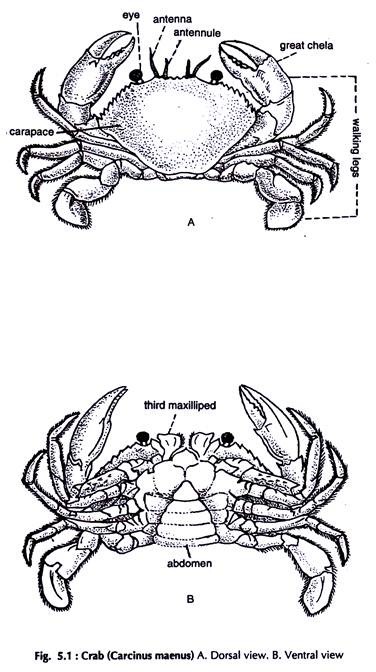
Crabs (Fig. 5.1) are usually dissected live in most cases, after clipping the last one or two segments of the walking legs. Hold the crab with left hand in its walking position. Turn it upside down and clip off portion of legs with a pair of stout scissors or a bone cutter.
Cut along the margin of the dorsal carapace joining the ventral sternites. Turn the dorsal surface upwards, carefully separates the carapace from the lining membrane below it and the carapace is free.
Release the flexed abdomen off the groove formed by sternites on the ventral surface and straightens it. Place the specimen on the dissecting tray keeping the dorsal surface upwards.
Fix it on the tray with the help of pins passing through the walking legs and lateral sides of the abdominal sternites. Cut the tergites of the abdomen along the lateral borders proceeding from the junction of the cephalothorax and the abdomen. Remove the tergites. The organs in the thoracic and abdominal cavities are exposed.
Dissection of Alimentary System:
The structures present in the cephalothorax from the dorsal to the ventral surface in succession are the dorsal wall, the pericardial sinus with the heart, the gonads, the renal sac, major part of the alimentary canal and the ventral thoracic ganglionic mass.
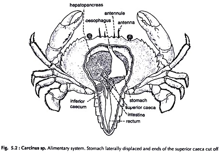
The hepatopancreas is a large orange-yellow organ in which some of the above organs are embedded. Partly remove the hepatopancreas from the dorsal surface to expose the fore gut and anterior part of the mid gut (Fig. 5.2).
Mouth:
A square opening, median in position, on the ventral side of the cephalothorax at its anterior border, guarded by two jaws.
Oesophagus:
A short tube, moving upwards from the mouth and widens posteriorly to open in the stomach.
Stomach:
A large, oval, muscular sac just beneath the gastric region of the carapace.
It is divided into:
(a) A large anterior portion-the cardiac chamber, and
(b) A small posterior portion-the pyloric chamber.
A valve is present between the two. The pyloric chamber is drawn out ventrally and joins the intestine.
Intestine:
A narrow tube, runs postero- medially in the abdomen and ends in rectum.
Superior caeca:
A pair of narrow, blind diverticula at the junction of the pyloric chamber of the stomach and the intestine.
Inferior caecum:
A narrow, blind, tubular projection at the junction of the intestine and rectum.
Rectum:
Wider than intestine, runs backward in the abdomen, narrows down posteriorly to open in the anus in the last abdominal segment.
Hepatopancreas:
A large, yellow-orange mass occupying major portion of the cephalothorax. The ducts from it open in the pyloric chamber of the stomach.
Dissection of Nervous System:
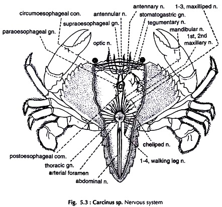
Remove the carapace and the tergites of the abdomen. Remove also the digestive system including the hepatopancreas, other structures dorsal to the nervous system and fat, to expose major portion of the nervous system (Fig. 5.3).
Brain or supraoesophageal ganglia:
Mid- dorsal in position, at the anterior end of the cephalothorax, formed by the fusion of two ganglia.
Following major paired nerves arise from the brain:
a. Optic supply eyes.
b. Ophthalmic supply eye muscles.
c. Antennulary supply antennules.
d. Antennary supply antennae.
Circumoesophageal connectives:
Arising from the lateral sides of the brain the two nerves run backward and downward round the oesophagus and join, the large ventral thoracic ganglonic mass.
Post-oesophageal loop (post-oesophageal connective):
The circumoesophageal connectives are joined by a fine, transverse connective before they join the ventral thoracic ganglionic mass.
Ventral thoracic ganglionic mass:
A large mass formed by the fusion of sub-oesphageal, thoracic and abdominal ganglia and major part of the ventral nerve cord. Nerves arising from the ganglionic mass supply maxillulae, maxillipeds, walking legs and muscles of the abdomen.
Ventral nerve cord:
Runs posteriorly from the ventral thoracic ganglionic mass to the posterior end of the abdomen and sends a few branches at the posterior extremity. It is devoid of ganglia.
Dissection of Reproductive System:
The sexes are separate. Sexual dimorphism is present. The abdomen in male crab is narrow, while in female it is broad.
Remove the carapace and hepatopancreas carefully. Remove also the posterior portion of gills.
Male Reproductive System:
(Fig. 5.4)
Testes:
Paired, tubular, cream coloured, located on either side of the stomach, extending from anterior part of the stomach to the middle of the cephalothorax, where they are joined by a transverse testicular isthmus.
Vas deferens:
Paired, long, narrow, partly convoluted, cream coloured tubes, continuation of testes behind testicular isthmus. The vas deferens is divided into three regions.
Anterior vas deferens:
The anterior convoluted part.
Median vas deferens:
The less convoluted middle portion.
Posterior vas deferens:
The straight posterior part.
Seminal vesicle:
The saccular last part of posterior vas deferens opening in the genital pore.
Genital pores:
Small, round openings located at the base of the fifth walking legs.
Female Reproductive System:
(Fig. 5.5)
Ovaries:
Paired, cream coloured, located on both sides of the stomach, extending from anterolateral to the posterior part of the cephalothorax, joined by a transverse ovarian isthmus behind the stomach.
Oviducts:
Paired, one arises from the ventral side of each ovary, posterior to ovarian isthmus and runs backward.
Seminal receptacles:
The dilated middle portion of the oviducts. Posteriorly the oviducts open in the genital pores.
Genital pores:
Small, round openings, located at the base of the third walking legs.


