In this article we will discuss about the killing and dissection of silk worm larva.
The silkworm larvae are easily collected from the farmers rearing silk worms or silk worm rearing centre. The 5th instar larva is suitable for dissection.
Killing:
The larva is immobilised with a few drops of 50% alcohol squeezed out on it from a piece of cotton wool soaked in it.
Dissection:
Fix the freshly killed specimen with fine pins (Fig. 8.1) in a dorsoventral position on a paraffined petri dish. Give incision carefully with a pair of fine scissors through the dorsal midline from posterior to anterior end. Carefully pin down the skin on the lateral sides. The viscera of the specimen is exposed.
The Silk Gland:
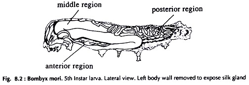
The silk or modified labial glands are tubular glands with characteristically branched nuclei, situated on the ventrolateral sides of the mid intestine (Fig. 8.2). Partially uncoil the tubular glands with a needle at the anterior end up to the spinneret. Anteriorly, the paired ducts unite and open into the spinneret.
The silk gland (Fig. 8.3) is a paired structure, divided into three distinct regions: anterior, middle and posterior.
Anterior region:
Two straight tubes joining at the fore end open in the spinneret. Posteriorly the tubes open into the middle region.
Middle region:
The largest and thickest of the three regions and with three definite flexons. This region again is divided into three sub regions. The beginning of the middle region is narrow but widens suddenly; the mid-part is the widest and the end of the rear part is narrow. The posterior part is crooked and curved between the tracheae and dorso-visceral muscles.
Posterior region:
A narrow coiled tube ending blindly. This part remaining coiled in mid portion to posterior portion of the body, above and sides of the gut.
Fillipi’s or Lyonnet’s gland:
A pair of glands, situated at the junction of the two tubes at the anterior region, secreting viscous fluid of unknown function.


