In this article we will discuss about the dissection of apple snail. Also learn about:- 1. Dissection of Alimentary System 2. Dissection of Nervous System 3. Dissection of Reproductive System.
Killing:
Pila may be killed in a stretched condition. They can also be killed with hot water, taking care that the flesh is not cooked. This practice may be resorted to only in emergency.
Dissection:
Remove the shell piece by piece with a bone cutter cutting along the suture. Breaking the shell by striking against hard object causes damage to soft internal organs. Detach the collumellar muscle from the collumella with a sharp scalpel and free the snail from the columella by slowly twisting the body in a direction opposite to the turn of the columella.
Wash out mucus from the specimen. The body of Pila is enclosed in a mantle or pallum. In the body whorl it forms a cloak over the body enclosing a mantle cavity (Fig. 10.1). In the rest it is thin and adherent to the visceral mass.
Make a longitudinal cut starting from the edge of the mantle on the right side and left to the ctenidium. Proceed up to the posterior limit of the mantle cavity. Turn the flap to the left and the pallial complex is exposed. Pin down the mantle and fix the specimen on the dissecting tray with pins passing obliquely through the foot.
Dissection of Alimentary System:
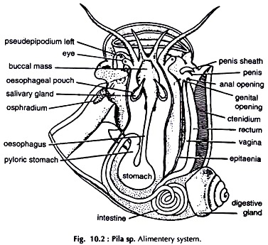
Give a longitudinal incision along the middle of the diaphragm forming the floor of the mantle cavity. Proceed anteriorly up to the head. Turn the flaps and the fore gut is exposed. Note the buccal mass. Follow the oesophagus.
It ends in the stomach partially embedded in the digestive gland. Trace the intestine by removing tissues of the digestive gland. The rectum is exposed to the mantle cavity in its right side. It is adherent to the mantle and runs by the side of the ctenidium (Fig. 10.2).
Mouth:
A vertical slit at the tip of the snout.
Buccal mass:
A fairly large, pyriform structure in the head region. A pair of jaws and an odontophore are present at the anterior border and in the posterior part of the buccal cavity respectively.
Salivary glands:
A pair of white glands, one on either side of the buccal mass. The glands open into the dorsal aspect of the buccal cavity.
Oesophagus:
Tubular, runs postero- medially from the buccal mass, turns left and enters the visceral mass to join the stomach.
Oesophageal pouches:
A pair of sac-like glands at the origin of the oesophagus.
Stomach:
A somewhat globular, dark red, muscular sac embedded partially in the digestive gland. It has two chambers.
a. Cardiac chamber. The oesophagus opens into it.
b. Pyloric chamber. The intestine arises from it.
The stomach with the oesophagus and the intestine is a U-shaped body. The base of the U is formed by the stomach, and the arms by the oesophagus and the intestine.
Caecum:
A small, blind diverticulum at the origin of the intestine, not clearly discernible from outside.
Intestine:
A narrow tube. It forms usually 2½ coils in the mass of the digestive gland. Emerging from the gland it enters the mantle cavity and joins the rectum.
Rectum:
A wide tube, adherent to the inner surface of the mantle on the right side, parallel to the ctenidium. It runs anteriorly to open through the anus.
Digestive gland:
A large, bilobed, brownish or dull green mass. It is roughly a spirally coiled triangular plate. Two ducts from two lobes unite to form a common duct before opening in the stomach.
Dissection of Nervous System:
Expose the mantle cavity. Expose the organs of the perivisceral region by cutting the diaphragm longitudinally. Be careful not to damage the visceral ganglion close to the posterior end of the mantle cavity. Remove the muscular covering of the buccal mass and a thick, band-like, light yellowish cerebral commissure is exposed. Trace it laterally and the cerebral ganglia are seen on the sides of the buccal mass.
One fine cerebrobuccal connective, and two stout connectives, cerebropleural and cerebropedal, arise from each cerebral ganglion. Trace them backward, to buccal, pleural and pedal ganglion of the side respectively. The buccal ganglia are embedded in the muscles of the buccal mass, close to the origin of the oesophagus.
The two pleuropedal ganglionic masses are ventrolateral in position. Cut the oesophagus at its base and remove the major part of it. The pedal commissure is exposed. The infraintestinal nerve lies close to it.
Trace backwards the infraintestinal and supraintestinal visceral connectives from the right and left pleuropedal ganglia to the visceral ganglion, placed close to the heart. Expose the nerve running obliquely backwards and connecting the right pleuropedal ganglia with the supraintestinal ganglion (Fig. 10.3).
Cerebral ganglia:
Paired, roughly triangular, located on the dorsolateral sides of the buccal mass at its anterior end. Each ganglion sends nerves to the snout, eye, tentacles and statocyst of the side.
Cerebral commissure:
Band-like, dorsal to the buccal mass and connects the two cerebral ganglia medially.
Buccal ganglia:
Two small, oval ganglia, dorsolateral in position at the junction of the buccal mass and the oesophagus. They send nerves to the buccal mass.
Buccal Commissure:
A slender nerve connecting the two buccal ganglia, located ventral to the oesophagus.
Cerebrobuccal connectives:
Slender nerves, connecting the cerebrals with the buccal ganglia.
Pleuropedal ganglia:
A pair of ganglionic masses, one on either lateral side, posteroventral to the buccal mass. Each ganglionic mass is formed by the fusion of pleural and pedal ganglion of the side. Pleural ganglia send nerves to the mantle complex and the pedal to the foot.
Cerebropleural connectives:
Two. Each runs posteroventrally from the cerebral ganglion around the oesophagus and joins the pleural region of the pleuropedal ganglion of the side. Its origin is medial to that of the cerebropedal connective.
Cerebropedal connectives:
A pair. Each runs posteroventrally from the cerebral ganglion around the oesophagus and joins the pedal region of the pleuropedal ganglion of the side.
Pedal commissure:
One, thick and connects the pedal regions of the two pleuropedal ganglia.
Infraintestinal nerve:
A slender nerve, close to the pedal commissure, joining the pleural regions of the two pleuropedal ganglia.
Supraintestinal visceral connective:
Running backwards from the pleural part of the left pleuropedal ganglia joins the visceral ganglion. It bears a supraintestinal ganglion at about its middle. The ganglion sends nerves to the gills and pulmonary sac.
Infraintestinal visceral connective:
Runs backward from the pleural part of the right pleuropedal ganglion and joins the visceral ganglion.
The supra and infraintestinal visceral connectives are also designated as left and right pleurovisceral connectives respectively.
Supraintestinal nerve:
Runs obliquely backwards and connects the right pleuropedal ganglion with the supraintestinal ganglion.
Visceral ganglion:
A large, unpaired ganglion close to the posterior border of the mantle cavity, near the heart. It sends nerves to kidney, digestive gland, reproductive organs, heart and intestine.
Dissection of Reproductive System:
The sexes are separate.
The reproductive system should always be dissected in freshly killed specimens, as the digestive gland in which the gonad is partially embedded becomes brittle in preserved snails. Carefully uncoil the spirally coiled digestive gland, which constitutes the major part of the visceral mass.
The gonad, testis or ovary, occupies the same position. It is restricted to about half the height of the inner concave border of the first 2 to 3 whorls of the digestive gland. The gonad is covered by the mantle, which is very thin and transparent.
Male Reproductive System:
(Fig. 10.4)
Testis:
Much branched and cream coloured; clearly visible through the transparent membraneous mantle. It enlarges posteriorly. Remove the membrane and expose it.
Vas deferens:
Many fine ducts, the vasa efferentia emerging from the testis join to form the vas deferens, which leaves the testis at the posterior end and runs along the inner border of the posterior renal chamber.
The vas deferens has three distinct zones:
a. Tubular part:
The slender first part runs along the inner edge of the posterior renal chamber.
b. Glandular part:
Terminal part of the vas deferens. Fairly large, swollen at the beginning and narrows distally. Lodged in the mantle cavity, parallel to the rectum and opens at the tip of the genital papilla.
c. Vesicula seminalis:
A curved blind tube at the junction of the above two.
Penis:
A long, whip-like flagellum, partially en-sheathed; an outgrowth of the mantle, on the right side of the mantle cavity.
Female Reproductive System:
(Fig. 10.5)
Ovary:
Orange red in colour, smaller than the testis and occupies the same position as the testis in male.
Oviduct:
Formed by the joining of a number of narrow ducts from the lobes of the ovary and leaves it at the posterior end.
The oviduct is divisible into two distinct zones:
a. Tubular part:
The slender first part running along the border of the digestive gland, turns downward above the level of the kidney and joins the bean-shaped receptaculum seminis at the junction of the glandular and tubular part.
b. Glandular part:
It lies in the mantle cavity parallel to the rectum and divisible into two zones:
i. Uterus:
A bluntly rounded, yellowish sac, lying at the junction of the two renal chambers and narrows down to form the vagina.
ii. Vagina:
Tubular, light cream coloured, narrows down anteriorly and opens through a slit-like aperture on the genital papilla.


