In this article we will discuss about the dissection of fresh water mussel. Also learn about:- 1. Dissection of Alimentary System 2. Dissection of Nervous System 3. Dissection of Reproductive System.
Fresh water mussel (Fig. 9.1) can be kept alive for quite a long period in aquaria. Dissections are best done in specimens preserved for a few days in a mixture of 50% glycerol and 50% alcohol at 1: 1 ratio.
Killing:
Bivalves are best killed by leaving the specimens overnight in 1% chloral hydrate solution. They can also be killed with hot water, taking care that the flesh is not cooked. This practice may be resorted to only in emergency.
Pallial complex:
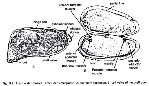
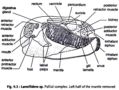
Insert the handle of a scalpel between the two valves of the shell at about the middle. Slowly turn the handle to widen the gap between the two valves to about 1 cm. Take another scalpel. Carefully separate the mantle from one of the valves of the shell at the posterior end. Keeping the blade of the scalpel against the inner surface of the shell cut the posterior adductor muscle.
The valves are partially released and the gap between the two increases. Working in the same way cut the anterior adductor muscle. Separate the mantle from the inner surface of the shell. Separate the other valve of the shell in the same way and the mussel (Figs. 9.2, 9.3) 9.3 is free from it.
It is not necessary to separate the animal from the shell for dissection. Many workers prefer to dissect organ-systems by removing only one valve leaving the other undisturbed.
Dissection of Alimentary System:
Alimentary system is best dissected in a preserved specimen. In a fresh specimen colour of the stomach and the intestine blends with the colour of the digestive gland and the gonad respectively, making dissection difficult.
Fix the specimen on a dissecting tray with pins passing through the edge of the foot and the upturned mantle. Remove the gills of the side. The labial palps should not be removed. Locate the mouth at the meeting point of the labial palps of both the sides, anterodorsal to the foot.
Give an incision on the skin, a little posterodorsal to the mouth. Hold the skin with a pair of forceps in your left hand and carefully separate it from the underlying digestive gland in the visceral mass, with a sharp scalpel.
Insert a blunt seeker through the mouth and ascertain the exact location and boundaries of the oesophagus and the stomach. Remove a part of the light grey digestive gland, bit by bit with a pair of fine forceps.
The oesophagus and the stomach are exposed. Arising from the posterior end of the stomach the intestine enters the visceral mass dorsal to the foot. Carefully remove the gonad and other tissues and the tubular intestine in the form of loops is exposed.
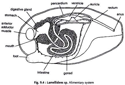
Start working from the posterior end. Cut open the supra-branchial chamber, the rectum is exposed. Remove the pericardial sinus with the heart and trace the rectum anteriorly up to the point where it joins the intestine (Fig. 9.4).
Mouth:
A transverse slit; the labial palps forming the upper and lower lips.
Oesophagus:
A short tube ending in stomach.
Stomach:
A fairly large oval sac, embedded in the digestive gland.
Intestine:
A narrow tube. Leaving the stomach it descends in the visceral mass above the foot, form two incomplete loops, ascends to the level of the stomach, anterior to the pericardial sinus.
Rectum:
Terminal part of the gut. Runs parallel to the dorsal surface of the mussel piercing the pericardial sinus and the ventricle to open through the anus.
Digestive gland:
A pair of light grey, irregular mass in which the stomach is lodged. They open in the stomach by short, narrow ducts.
Dissection of Nervous System:
The two cerebral ganglia are separate and located at the base of the labial palps, just under the skin. The two pedal ganglia are partially fused and lodged in the muscles of the foot, close to its junction with the visceral mass at about 1 cm above the ventral border of the foot, posterior to its middle. The visceral ganglia are large, partially fused and located ventral to the posterior adductor muscles. They are covered by a thin peritoneum.
Remove the labial palps and the gill. Carefully separate the skin from the underlying tissue at the base of the labial palp with the help of a fine needle and the cerebral ganglion of the side is exposed. Cut and remove a strip of about 0.5 cm of the anterior and ventral border of the foot. Give a vertical incision along the middle line of the cut border of the foot.
Hold each half with a pair of stout forceps and separate them slowly. The pedal ganglia are visible at about 0.5 cm from the cut margin. The visceral ganglia can be seen without removing any tissue. By carefully removing the covering peritoneum they are fully exposed.
Locate the origin of the cerebropedal connective at the posterior border of the cerebral ganglion. Trace the nerve to the pedal ganglion of the side running through the visceral mass and muscles of the foot. Follow the cerebrovisceral connective from its origin at the posterolateral border of the cerebral ganglion. Running through the visceral mass it joins the visceral ganglion of the side (Fig. 9.5).
The nerves become more prominent on exposure to aceto-alcohol. A few drops of the mixture may be put on the nerves with a pipette.
Cerebral ganglia:
Two, triangular in shape, pale yellow in colour, located at a distance from each other. A few nerves arise from each ganglion and innervate the adjacent region.
Cerebral commissure:
A slender nerve, runs around the oesophagus at the anterior part of it and connects the two cerebral ganglia.
Pedal ganglia:
Oval in shape; pale yellow in colour. The two ganglia fuse medially to form a bilobed mass. A few nerves arise from each pedal ganglion and innervate the foot.
Cerebropedal connectives:
Each connects cerebral ganglion with the pedal ganglion of the side at its anterior end.
Visceral ganglia:
Flattened, pale yellow in colour, triangular in shape. The two in vivo assume x-shape. Quite a few nerves arise from each visceral ganglion.
Cerebrovisceral connectives:
Each joins the cerebral ganglion with the anterior end of the visceral ganglion of the side.
Dissection of Reproductive System:
The sexes are separate. Gonads are paired. The male and female gonad and their ducts are almost similar. They occupy the same relative positions.
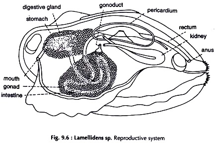
Give an incision on the skin of the visceral mass dorsal to the middle of the foot. Hold the skin with a pair of forceps and carefully separate it from the underlying mass with a sharp scalpel, working dorsally, anteriorly and posteriorly. The gonad of the side is exposed (Fig. 9.6). It occupies the major portion of the visceral mass in which the loops of the intestine are lodged.
Male Reproductive System:
Testes:
A pair of large racemose glands, white in colour.
Vasa deferentia:
Two narrow ducts. A vas deferens arises from the posterodorsal border of each testis. It is short and opens just in front of the renal aperture of the side through a round male genital pore.
Female Reproductive System:
Ovaries:
A pair of large racemose glands, reddish in colour.
Oviducts:
Two. An oviduct arises from the posterodorsal border of each ovary. It is short and opens just in front of the renal aperture of the side through a round female genital pore.


