In this article we will discuss about Vorticella Campanula:- 1. Habit and Habitat of Vorticella Campanula 2. Culture of Vorticella Campanula 3. Structure 4. Locomotion 5. Nutrition 6. Respiration, Excretion and Osmoregulation 7. Behaviour 8. Reproduction.
Contents:
- Habit and Habitat of Vorticella Campanula
- Culture of Vorticella Campanula
- Structure of Vorticella Campanula
- Locomotion of Vorticella Campanula
- Nutrition of Vorticella Campanula
- Respiration, Excretion and Osmoregulation in Vorticella Campanula
- Behaviour of Vorticella Campanula
- Reproduction in Vorticella Campanula
1. Habit and Habitat of Vorticella Campanula:
Vorticella campanula is found in freshwater ponds, lakes, rivers and streams with aquatic vegetation. It is worldwide in distribution. Vorticella is solitary and not colonial but usually social, several of them being found together.
Vorticella Campanula is sedentary (fixed) form. It is commonly attached by a long highly contractile stalk to some submerged objects like weeds, animals and stones, etc. Vorticella is often found in large groups.
All the individuals in the group, however, remain free and independent of each other. They are found in abundance in stagnant water rich in decaying organic matter and feeds largely on bacteria. But V. campanula and V. nebulifera live only in uncontaminated water where bacterial growth is not good.
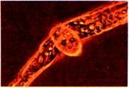
Vorticella microstoma.
2. Culture of Vorticella Campanula:
Culture of Vorticella Campanula is prepared in the same way as that of Amoeba. Make an infusion of hay and dead leaves in rain or distilled water, let this stand for some days, a brownish scum will be formed on the surface, below which many Vorticella will be found; this shows the presence of cysts in Vorticella.
3. Structure of Vorticella Campanula:
Shape, size and colour:
Vorticella Campanula is a microscopic stalked form with an inverted bell-shaped asymmetrical body. Due to the bell-shaped body it is often called bell-animalcule. The largest species is Vorticella campanula, the bell of which measures up to 157 microns in length and 99 microns in width and stalk varies from 53 to 4150 microns in length which is highly contractile.
The colour of the animalcule is yellowish, greenish or bluish. The smallest species is V. micro-stoma, which measures about 55 microns in length and 35 microns in width.
Body or Bell:
The body of Vorticella campanula is like an inverted bell.
The detailed structure of the bell is described below:
1. Peristome:
The margin or rim of the broad free end of the bell is thickened and is termed as peristomial collar or lip. Inside the peristomial collar is a narrow, shallow, circular and marginal depression called the peristome or oral groove. The peristome surrounds a broad, slightly convex circular central disc, the peristomial disc or oral disc that seems to close the opening of the bell.
The peristomial disc is fused with the collar on one side with the result the peristome does not form a complete ring. The peristomial disc can be withdrawn when the peristome contracts and covers it.
2. Yestibule:
Between the peristome and the peristomial disc is a permanently open sunken space on the left side, it is called a vestibule or infundibulum. From the vestibule a narrow cytopharynx leads inwards. The cytopharynx has no cilia and it opens into the endoplasm. Between the vestibule and cytopharynx is a cytostome which can open or close.
3. Cilia:
There is an adoral wreath of cilia on the peristomial region and the rest of the body is devoid of cilia. The peristomial groove bears three concentric rows of adoral cilia arranged into circlets. The inner circlet has two row circles of cilia which are closely associated forming a double row, its cilia standing straight up and keep up constant undulations.
The outer circlet is single and has short cilia which are inclined outwards over the collar like a shelf and guide the food into the vestibule. Each circle of cilia forms more than a complete ring which slightly overlaps. All cilia lie anti-clockwise, they are fused at their bases but are free distally.
The circles of cilia turn at the margin of the disc and are continued in a counter clockwise direction into the vestibule where the cilia of the outer circle become long and fuse together to form a triangular undulating membrane along the outer wall of the vestibule, while the cilia of the two inner circles lie along the inner wall of the vestibule.
In feeding, the food particles pass along the outer cilia and are driven down by the undulations of the two inner rows of cilia. The body and the stalk are devoid of cilia, but their kinetosomes are present in circles showing that their cilia are lost, besides which there are circular striations on the body where the cilia might have been.
4. Pellicle:
The entire animal is covered by a pellicle which is ringed transversely by parallel striae, it is very thick at the base of the bell and continued with stalk. In V. monliata the pellicle has nodular warts of paraglycogen. In the stalk the pellicle is covered by an external cuticle.
5. Cytoplasm:
The body cytoplasm is differentiated into an outer layer of clear and firm ectoplasm or cortex and an inner fluid and granular endoplasm or medulla.
(i) Ectoplasm:
The ectoplasm is modified to form a myoneme system having longitudinal, oblique and circular myonemes, they are more visible in the base of the bell. Longitudinal myonemes shorten the body, oblique myonemes pull the disc inwards, and the circular myonemes contract the peristome and close it over the disc.
(ii) Endoplasm:
The endoplasm is granular and fluid-like. It contains the nuclei, contractile vacuole and food vacuoles. Near the cytopharynx is a clear space called the reservoir which joins the cytopharynx by a narrow tube.
Close to the reservoir is a contractile vacuole having a lining membrane, thus, it is a permanent vacuole which is osmoregulatory and it pours its contents into the reservoir at each systole, from where they pass out through the vestibule (In V. picta and V. monliata there are two contractile vacuoles.) Near the reservoir is a cytoproct opening into the vestibule, it is a temporary or permanent aperture in different species.
There is a large, long, horse-shoe-shaped macronucleus and a micronucleus, both lying in the endoplasm. The macronucleus is highly polyploid with a large amount of chromatin material scattered in the nucleoplasm. The micronucleus, which can be seen only in a stained condition, lies in close association to macronucleus.
Stalk:
The bell-shaped body of Vorticella is attached to weeds or stones by a long, thin, un-branched and highly contractile stalk. It is a prolongation of the pellicle and ectoplasm of the bell. The body myonemes combine and run through the centre of the stalk as a single loose spiral known as an axial filament or spasmoneme. In V. campanula there are the coplastic granules on the spasmonema.
When the stalk is contracted the spiral spasmonema coils up tightly and looks like a spring. Around the spasmoneme the stalk has a protoplasmic sheath covered by the pellicle and an external cuticle. Vorticella is extremely sensitive to any mechanical stimulus, slightest contact causes instantaneous coiling of the stalk into a tight spiral, the body becomes rounded, the disc is pulled in and the peristome closes over it.
4. Locomotion of Vorticella Campanula:
Vorticella Campanula does not move freely because it is usually found fixed aborally by its long highly contractile stalk. However, with the help of stalk and myonemes, the bell sways to and fro in the surrounding water like a flower in a breeze. The individuals of a group move in their own way. The detached individuals swim freely by means of cilia and are referred to as telotrochs.
5. Nutrition of Vorticella:
Nutrition is holozoic as in Paramecium. It is omnivorous but its food generally consists of small Protozoa, bacteria and organic bits. The cilia of the peristome and the disc produce a current of water by which small organic particles fall on the disc, from where they are carried into the vestibule; then the undulating membrane carries them to the cytopharynx, movement of food is aided by undulations of the two inner rows of cilia.
At the base of the cytopharynx the food particles along with some water form food vacuoles one after the other. Movements of food vacuoles in the endoplasm are in an irregular cyclosis (unlike Paramecium). Digestion occurs as in Paramecium’, the food vacuoles at first acidic and then they become alkaline.
The digested material is absorbed in the endoplasm and assimilated. The excess digested food forms refractile glycogen granules in the endoplasm. The undigested remains are expelled in the vestibule through the cytoproct. Thus, in Vorticella the food and faecal matter pass through the same passage.
6. Respiration, Excretion and Osmoregulation in Vorticella Campanula:
Respiration and excretion in Vorticella Campanula is exactly performed in the same way by the process of diffusion through general body surface as in Amoeba and Paramecium.
Osmoregulation in Vorticella is performed by the contractile vacuole, as in other freshwater Protozoa. In Vorticella, contractile vacuole is single, large and pulsating usually situated between the disc and vestibule in the endoplasm. The contractile vacuole opens in the vestibule by a permanent opening; it pulsates rhythmically showing diastolic and systolic phases.
In fact, during diastolic phase, the excess of water drawn in the endoplasm by the process of osmosis is secreted into the contractile vacuole and during the systolic phase, the contractile vacuole expells out the excess of water into the vestibule. Thus, contractile vacuole helps in maintaining the water content in the body.
7. Behaviour of Vorticella Campanula:
Vorticella Campanula exhibits high degree of contractility and irritability; it is extremely sensitive to any mechanical stimulus and also responds to external stimuli. When irritated, its all activities cease at once; the stalk is retracted and becomes coiled into closed spiral to reduce its size, then the disc is withdrawn and covered over by peristomial lip.
Due to all these activities, its body becomes somewhat globular and remains motionless till normalcy is restored.
8. Reproduction in Vorticella Campanula:
Vorticella Campanula normally reproduces asexually by longitudinal binary fission, but at intervals conjugation also occurs which is sexual mode of reproduction. Encystment has also been reported to occur during un-favourable conditions.
Longitudinal Binary Fission:
Binary fission of Peritrichia differs from that of other ciliates in being generally unequal and in a plane that is longitudinal running along the oral-aboral axis or nearly so. Vorticella closes its peristome over the disc, the body becomes depressed and transversely elongated.
Endoplasmic circulation continues and the contractile vacuole pulsates throughout division. The long macronucleus becomes condensed and short, it then becomes straight and lies transversely in the middle, then it divides into two amitotically.
The micronucleus divides by an elongated mitosis.
A constriction begins in the centre of the anterior end, and dividing the peristome, it passes down the length of the cell, just to one side of the stalk; this constriction divides the animal into two unequal parts, the slightly smaller part has no stalk, it has a ring of oral cilia and it develops a contractile vacuole and forms a ring of aboral cilia at its posterior end, it becomes cylindrical and gets detached, it is now called a telotroch.
The telotroch swims away with its aboral pole foremost, it settles down by its aboral end which has a short scopula. The scopula is a circlet of stiff protoplasmic processes derived from cilia, it secretes a stalk by which the telotroch gets fixed, then it loses it scopula, its bell expands, a new disc is formed and it metamorphoses into an adult. Binary fission takes 20 to 30 minutes.
The larger product of division retains the old disc and stalk and it may be called the parent, while the smaller telotroch is the offspring.
No such distinction is seen in other Protozoa. In un-favourable conditions the normal Vorticella also grows a posterior ring of cilia to become a telotroch which breaks from its stalk and swims away to some favourable spot and then grows a stalk. At times Vorticella Campanula encysts in a two-layered cyst while still fixed to its stalk, then the cyst falls from the stalk, on excystment it swims away as a telotroch.
Conjugation:
Conjugation is a mode of sexual reproduction, which is very characteristic in Vorticella. The process of conjugation and syngamy was studied by Maupas (1888) in Vorticella nebulifera and by Finlay (1943) in Vorticella micro-stoma.
However, the process of sexual reproduction in Vorticella Campanula described hereunder is a generalised description; it can be studied in the following two phases:
(a) Formation of micro and macrogametes
(b) Conjugation of micro- and macro-conjugants and their fusion.
(a) Formation of Micro- and Macro-Conjugants:
In sexual reproduction, Vorticella Campanula divides by binary fission into two very unequal parts, the larger cell is the ordinary individual, while the very small cell is called a micro-conjugant. In some species more than one micro-conjugant is produced by repeated fission. The micro-eonjugants acquire a-girdle of cilia at the posterior end of each.
The microeonjugants become detached and swim about; their swimming is an adaptation for conjugation in sessile species; microeonjugants differ from telotrochs in being smaller and in the fact that they never metamorphose into adults nor do they form stalks. Micro-conjugants never feed or encyst, they live for about 24 hours, after which they die.
A stalked Vorticella Campanula undergoes nuclear modifications, though it appears normal, it is then known as a macroconjugant. The macroconjugant is morphologically the same as a normal trophic individual, stationary and passive but it is specialised physiologically and it can attract microconjugants for about 2 hours.
(b) Fusion of Conjugants:
While swimming, when a micro-conjugant approaches a macro-conjugant then it gets attached to the lower end of macro-conjugant near its stalk (Fig. 21.6). Thus, both the conjugants come together. Soon, cilia and pellicle of the micro-conjugant is thrown off and nuclear changes start taking place simultaneously in both the conjugants; the nuclear changes occur in the following way (Fig. 21.6).
Fig. 21.6 Vorticella, Stages in Conjugation
(i) The macronuclei of both the conjugants degenerate and finally absorbed in the cytoplasm.
(ii) The micronucleus of macroconjugant divides two times to form four daughter micronuclei. But the micronucleus of microconjugant divides three times to form eight daughter micronuclei. In these divisions, the last being reduction division.
(iii) Three, out of four daughter micronuclei of macroconjugant and seven out of eight daughter micronuclei of microconjoint degenerate. Thus, the left out one micronucleus in macroconjugant becomes female pronucleus, while the one micronucleus in microconjugant becomes the male pronucleus which are haploid.
(iv) The partition wall in between two conjugants disappears, thus, complete union occurs between the two conjugants.
(v) The male and female pronuclei fuse together to form zygote nucleus or synkaryon.
(vi) The synkaryon divides three times to form eight daughter nuclei, seven of these become macronuclei and one micronucleus.
(vii) This micronucleus divides into two and the zygote also divides, so that two daughter individuals are formed; one with four macronuclei and one micronucleus, and the other with three macronuclei and one micronucleus.
(viii) Then the micronucleus of each individual goes on dividing with the body of individual until the daughter individuals with one micro- and one macronucleus is formed.
(ix) Thus, the daughter individual with four macronuclei divides twice forming 4 offspring’s (each with one macro- and one micronucleus) and the daughter individual with three macronuclei produces 3 offspring’s (each with one macro- and one micronucleus). Therefore, total 7 individuals are formed which later develop stalk and metamorphose into adults.
Encystment:
Von Brand (1923) has reported the formation of cyst around the body of Vorticella during un-favourable conditions. The encysted body breaks off from the stalk. In an encysted condition, the myonemes become indistinct, the pulsating rate of contractile vacuole is slowed down and finally it disappears, the peristome is also absorbed. In this condition, the Vorticella tides over the un-favourable conditions.
After the return of favourable conditions, the cyst breaks and the individual emerges out (Fig. 21.7, E to G) develops contractile vacuole and becomes enlarged. It grows an aboral circlet of cilia to become a teiotroch. It swims freely for some time and then settles at some substratum, develops a stalk and grows into an adult Vorticella.
Sometimes, during un-favourable conditions, Vorticella Campanula develops a posterior ring of cilia to become a teiotroch. It separates away from the stalk and swims away to some favourable place where it develops stalk and starts leading normal life (Fig. 21.7, A to D). The teiotroch helps in its dispersal.
Conjugation in Vorticella Campanula shows an advance over that in Paramecium. In Paramecium the conjugants are similar, conjugation is a temporary union of two individuals with an exchange of nuclear material, but there is no fusion of cytoplasm, both ex-conjugants reproduce by fission.
In Vorticella Campanula conjugating gametes are dissimilar anisogametes, conjugation is permanent with a fusion of both cytoplasm and nuclei of gametes to form a zygote which reproduces by fission. Vorticella Campanula also shows a differentiation of sex in its dimorphic gametes, hence, the sexual process in Vorticella is somewhat intermediate between conjugation (in Paramecium) and syngamy (in Plasmodium).


