In this article we will discuss about Monocystis:- 1. Habit and Habitat of Monocystis 2. Structure of Monocystis 3. Locomotion 4. Nutrition 5. Respiration 6. Excretion 7. Reproduction 8. Life Cycle 9. Mode of Transmission 10. Host Parasite Relationship 11. Laboratory Study.
Contents:
- Habit and Habitat of Monocystis
- Structure of Monocystis
- Locomotion of Monocystis
- Nutrition of Monocystis
- Respiration in Monocystis
- Excretion in Monocystis
- Reproduction in Monocystis
- Life Cycle of Monocystis
- Mode of Transmission of Monocystis
- Host Parasite Relationship in Monocystis
- Laboratory Study of Monocystis
1. Habit and Habitat of Monocystis:
Monocystis lives as an intracellular parasite in its young stage when it lives in the bundle of developing sperms and becomes extracellular in its mature stage when it lives in the contents of seminal vesicles of earthworms. Its infection is so wide that practically all mature earthworms are found parasitized by this parasite.
2. Structure of Monocystis:
The adult mature Monocystis is called trophozoite which is a feeding stage. The young trophozoite lives in the sperm morula (sperm morula is a group of developing sperms) of the host; it feeds and grows at the expense of the protoplasm of the developing sperms until all the protoplasm is exhausted.
So, it is now seen to be surrounded by the tails of the dead sperms. In this stage, it is sometimes mistaken to be a ciliated organism. But, soon the sperm tails are detached from its body and the trophozoite becomes free.
Shape and size:
The fully grown mature trophozoite is elongated, flattened, spindle- shaped, worm-like and pointed at both the ends. It measures up to 500 microns in length and nearly 65 microns in width at its broadest portion. It is large enough to be seen with naked eyes, though its structural details can only be observed with the help of microscope.
Pellicle:
It marks the external boundary of the body of trophozoite. The pellicle is thick, smooth, firm, permeable and modified in different ways. It contains longitudinally arranged contractile microtubules.
Cytoplasm:
The cytoplasm is clearly differentiated into an outer clear, dense ectoplasm or cortex and an inner, fluid-like endoplasm or medulla. The cortex is further divided into an outer epicuticle, middle sarcocyte and inner myocyte. The myocyte consists of longitudinal and transverse contractile fibrils called myonemes.
The action of both sets of myonemes is coordinated and they act like muscles and bring about sluggish gliding movements and metaboly due to contractions. The endoplasm contains numerous granules of a starch paraglycogen as reserve food, fat globules and sometimes volutin granules and nucleus.
Nucleus:
The nucleus is large, vesicular, spherical or ellipsoidal in shape. It is placed more anterior in the upper half the body. The nucleus is surrounded by a nuclear membrane and the nucleoplasm contains a distinct endosome; sometimes more nucleoli or karyosomes are seen. The nucleoplasm contains four chromosomes, reticulum which represents the haploid number.
Contractile vacuoles, mouth and organelles of locomotion are absent due to a parasitic mode of life. A related genus of Monocystis, Nematocystis magna (frequently parasitising Pheretima) is larger in size but narrower, it may have fine root-like cytoplasmic processes at one or both ends.
Electron structure of sporozoite:
The electron structure of sporozoite (Fig. 17.3) reveals that it possesses all typical structures seen in protozoans, i.e., the Golgi apparatus, mitochondria, nuclear components, etc., are as usual. In addition, pellicle shows longitudinally arranged contractile microtubules.
In the anterior end, a pair of elongated reservoir-like roptries are seen which secrete some secretion that helps the trophozoite in penetrating through the host tissues. The anterior end also shows the presence of conoids and micronemes, whose functional nature is not definitely known.
3. Locomotion of Monocystis:
Monocystis is sluggish in locomotion. The wriggling movement is brought about by contractions and expansions of myonemes which are arranged in lines below the pellicle. These slow movements are called gregarine movements which are like euglenoid movements and are accompanied by marked circulation of the endoplasmic contents.
4. Nutrition of Monocystis:
Nutrition is saprozoic. Monocystis secretes digestive enzymes from its body which digest cytoplasm and developing sperms of the seminal vesicles. The digested products are absorbed by osmosis through the pellicle (osmotrophy). The excess food is stored in endoplasm as reserve paraglycogen.
5. Respiration in Monocystis:
In Monocystis, respiration is performed by diffusion.
Since it lives as parasite within the body of the earthworm, it has no direct contact with the exterior and the chance of getting free oxygen is very remote. It is presumed that Monocystis gets its oxygen supply either from the seminal vesicle which gets oxygen from the blood of earthworm or by the process of fermentation of carbohydrates occurring in the body of earthworm.
The carbon dioxide produced diffuses out from the body and are finally eliminated through the blood of the host.
6. Excretion in Monocystis:
Nitrogenous wastes diffuse out of the body of Monocystis into the body of the host and are eliminated by the excretory organs of the host.
7. Reproduction in Monocystis:
Monocystis reproduces sexually and the process is always followed by asexual reproduction. As a matter of fact, both the processes are interdependent.
8. Life Cycle of Monocystis:
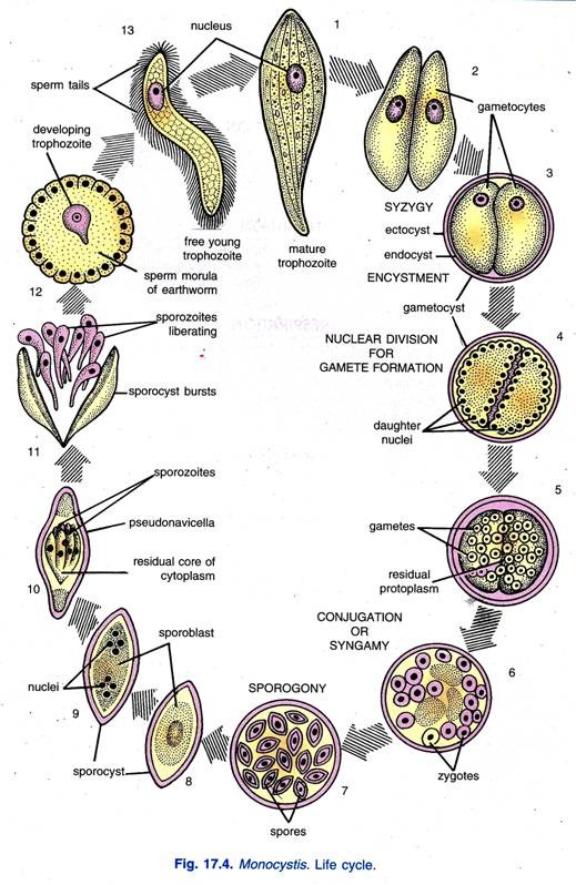
The life cycle of Monocystis is completed within a single host, i.e., monogenetic and characterised by the absence of asexual reproduction by schizogony. After a short free extracellular trophic phase, the mature trophozoite enters the reproductive phase of its life cycle. Life cycle of Monocystis includes gametogony, syngamy and sporogony.
Gametogony:
Two adults or trophozoites, after leading a considerable period of feeding and wandering about, come together in the cavity of the seminal vesicles by their anterior ends (Hawes, 1961) or in some species side by side. The long bodies of trophozoites become shortened and rounded referred to as gamonts or gametocytes. Now they secrete a common protective two- layered cyst called gametocyst.
The outer layer of the cyst is a thicker hard ectocyst and the inner thin soft layer is the endocyst. The two trophozoites in the gametocyst never fuse or conjugate but remain together in pair. This stage is known as syzygy.
The nucleus of each gametocyte divides mitotically several times giving rise to numerous daughter nuclei. The daughter nuclei are haploid because the gametocytes themselves are haploid having 4 chromosomes.
The nuclei of a gametocyte migrate to the periphery and project outwards so that the gametocytes look like a berry in which the endoplasm is dense and opaque and the surface projections are transparent. The daughter nuclei get surrounded by some cytoplasm to form gametes, but residual cytoplasm remains un-segmented in the middle, and it contains vacuoles and Para glycogen.
The walls between two gametocytes break and they coalesce.
Marshall and Williams (1972) have suggested the terms gamontogony for gametogony and gamontocyst for gametocyst.
Syngamy:
All the gametes produced by both gametocytes are identical morphologically and are called isogametes (M.A. Sleigh, 1973). They move about within the gametocyst and come together in pairs. Two isogametes of a pair fuse together to form a zygote. It is presumed that the two gametes uniting in pairs are from different gametocytes.
This union and fusion of two gametes of the same species is called syngamy. If the two gametes are isogametes, then, their syngamy is spoken of as isogamy. Thus, numerous larger, nucleated, diploid zygotes are formed within the gametocyst by syngamy.
Sporogony:
The developing zygotes are also called sporoblasts. A covering called sporocyst is secreted around each zygote, it is then known as spore. The spore with its sporocyst becomes spindle-shaped or boat-shaped with a mucoid plug at each end.
The spore resembles a diatom Navicella (Gr., novum = boat) in shape, consequently it is often called a pseudonavicella (Gr., pseudo = false; navum = boat). The nucleus and cytoplasm of the spore divide three times, first being reduction division to form eight spindle-shaped or fusiform sporozoites which lie like flakes of an orange around a central core of residual cytoplasm.
The sporozoites have been formed by asexual fission and constitute an asexual generation. The sporozoites can develop further only if the spores containing them are transferred from the host to another earthworm by oral infection. When the spores enter the intestine of a new earthworm, the sporocyst is digested and the sporozoites are set free.
Most probably the sporozoites bore through the intestinal wall and come to the coelom from where they penetrate the sperm mother cells of sperm morula of the seminal vesicles. But the method by which the sporozoites find their way from the gut to the seminal vesicles is quite unknown.
In the seminal vesicles, the sporozoite of Monocystis agilis begins its intracellular phase by penetrating a cytophore (a cytophore is a cytoplasmic mass about which developing sperms are arranged).
In the cytophore, the parasite inhibits the development of spermatogonia which do not mature, but the testes are unaffected. In case of Nematocystis magna, the sporozoites penetrate an epithelial cell of the ciliated funnel of vasa efferentia. In some species development is entirely extracellular in the cavity of the seminal vesicle.
The sporozoite grows into a young trophozoite which is falciform or like a bent spindle. It feeds on sperms and cytoplasm, and degenerating sperms are seen attached to it. Then it grows and becomes an adult trophozoite which comes to lie freely in the cavity of the seminal vesicles.
However, the life cycle of Monocystis exhibits an alternation of a sexual generation of gametocytes with an asexual generation of sporozoites.
9. Mode of Transmission of Monocystis:
The transmission of sporocysts from one earthworm to another is not known with certainty. It may be brought about in one of the following ways:
By Contaminated Soil:
When an earthworm, infested with the parasites, dies, the sporocysts are released in the soil which are eaten by other earthworms.
By Birds:
When an infested earthworm is eaten up by a bird, the resistant sporocysts are not digested and come out to the soil along with the faeces of the bird. When the soil containing the sporocysts is eaten by other earthworms, the cyst wall is dissolved in the intestine of earthworm and the sporozoites make their way into the seminal vesicles.
During Copulation:
The sporocysts are transferred from one host to another during copulation or exchange of sperms from the seminal vesicles of one earthworm to another.
10. Host Parasite Relationship in Monocystis:
Though nearly all earthworms are found to be infected with Monocystis, even then they cause no appreciable damage to the earthworms. The parasite destroys the sperms of the earthworms, no doubt, but this does not affect the fertility of worms because they produce sperms in abundance.
The earthworms appear to be able to combat the parasites by forming resistant protective envelope around the trophozoites and also by killing the spores by phagocytosis. On the other hand, the parasite shows various structural, physiological and reproductive adaptations so that it leads its life successfully and maintains the continuity of its race.
11. Laboratory Study of Monocystis:
Monocystis can be easily obtained in laboratory and studied. For this, take a sexually mature earthworm and pin it down in a dissecting tray keeping its dorsal surface in your front. Cut the integument from segment 10 to 15 to make a slit. Whitish body will extrude out through the slit, which is seminal vesicle.
Pinch it off with the help of forceps and keep it in a watch glass containing 0.7% sodium chloride solution. Stir it to make a thin paste of the contents of seminal vesicle. Make a thin smear of these pasty contents on a clean glass slide and cover it with a cover slip.
Examine under microscope to observe the trophozoites, spores, gametocysts and other stages of the life cycle of Monocystis. For preparing a permanent slide, the smear is allowed to dry and usual method of staining, dehydration, etc., are processed.


