In this article we will discuss about Euglena Viridis:- 1. Habit and Habitat of Euglena Viridis 2. Culture of Euglena Viridis 3. Structure 4. Locomotion 5. Nutrition 6. Respiration 7. Excretion 8. Behaviour 9. Reproduction 10. Position 11. Some Other Euglenoid Flagellates.
Contents:
- Habit and Habitat of Euglena Viridis
- Culture of Euglena Viridis
- Structure of Euglena Viridis
- Locomotion of Euglena Viridis
- Nutrition of Euglena Viridis
- Respiration in Euglena Viridis
- Excretion of Euglena Viridis
- Behaviour of Euglena Viridis
- Reproduction in Euglena Viridis
- Position of Euglena Viridis
- Some Other Euglenoid Flagellates
1. Habit and Habitat of Euglena Viridis:
Euglena viridis (Gr., eu = true; glene = eye-ball or eye-pupil; L., viridis = green) is a common, solitary and free living freshwater flagellate. It is found in freshwater pools, ponds, ditches and slowly running streams. It is found in abundance where there is considerable amount of vegetation.
Ponds in the well maintained gardens containing decaying nitrogenous organic matter, such as twigs, leaves and faces of animals, etc., are good source of this organism. It generally lives with the other species of the genus. They are sometimes so numerous as to give a distinct greenish colour to the water or at times forming a green film of scum on the surface of the pond water.
2. Culture of Euglena Viridis:
The culture of Euglena Viridis can be easily prepared in the laboratory by the following method. Boil some cow or horse dung in distilled water in ajar and allow it to cool for two days. Then put some weeds from a pond containing Euglenae into the jar and place the jar near the well-lighted window. In a few days, Euglenae will appear in this nitrogenous infusion.
3. Structure of Euglena Viridis:
Shape:
Euglena viridis is elongated and spindle-shaped in appearance. The anterior end is blunt, the middle part is wider, while the posterior end is pointed.
Size:
Euglena viridis is about 40-60 microns in length and 14-20 microns in breadth at the thickest part of the body.
Pellicle:
The body is covered by a thin, flexible, tough and strong cuticular periplast or pellicle which lies beneath the plasma membrane. It has oblique but parallel striations called myonemes all round. But according to Chadefaud (1937), the pellicle is made of an outer thin layer epicuticle and inner thick layer cuticle.
Both the layers of pellicle are present all over the body but only the epicuticle ends into an anteriorly placed cytopharynx and reservoir.
The pellicle is composed of fibrous elastic protein but not of cellulose. The pellicle maintains a definite shape of the body, yet it is flexible enough to permit temporary changes in the body shape, these changes of shape are spoken of as metabody or euglenoid movements.
Electron structure of pellicle:
Electron microscopic study of pellicle reveals that it is made of helically disposed strips. These strips are fused at both the ends of the cell body and each has a groove along one edge and a groove along the other. The edges of neighbouring strips overlap and articulate in a way that the ridge of one strip fits into the groove of the other.
In fact, the articulating ridges give the pellicle striated appearance. Just beneath and parallel to the strips, a row of mucus-secreting muciferous bodies and bundles of microtubles are found arranged (Fig. 12.3).
Cytostome and cytopharynx:
At the anterior end is a funnel-shaped cytostome or cell mouth slightly to one side of the centre. Cytostome leads into a short tubular cytopharynx or gullet which, in turn, joins a large spherical vesicle, the reservoir or flagellar sac. The cytostome and cytopharynx are not used for ingestion of food but as a canal for escape of fluid from the reservoir.
Contractile vacuole:
A large osmoregulatory body, the contractile vacuole lies near the reservoir on one side. It is surrounded by several minute accessory contractile vacuoles, which probably fuse together to form the larger vacuole. The contractile vacuole discharges the excess of water and some waste products of metabolism into the reservoir from where it goes out through the cytostome.
Flagellum:
A single, long, whip-like flagellum emerges out of the cytostome through cytopharynx. The length of flagellum differs in different species of Euglena but in Euglena viridis it is as long as the body of the animalcule. It arises by two roots from the base of the reservoir from the side opposite to the contractile vacuole.
Each root springs from a blepharoplast (Gr., blepharon = eyelid; plastos = formed) or basal granule which lies embedded in the anterior part of the cytoplasm.
According to some workers, there are two flagella, one long and other short, each arising from a basal granule located in the cytoplasm at the base of the reservoir. The short flagellum does not extend beyond the neck of the reservoir and it often adheres to the long flagellum producing the appearance of bifurcation.
The flagellum consists of an outer contractile protoplasmic sheath and an inner elastic axial filament, the axoneme. The distal portion of the flagellum contains numerous minute fibres known as mastigonemes which project along one side of the sheath and, therefore, the flagellum is stichonematic type.
Electron structure of flagellum:
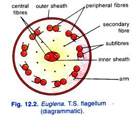
Electron microscopic study of the flagellum reveals that it consists of two central and nine peripheral fibrils. Each central fibril is single, while the peripheral fibrils are paired having two sub-fibrils in each. One of the two sub-fibrils of each peripheral fibril bears a double row of short projections called arms; all the arms being directed in the same direction.
The two central fibrils are found enclosed in an inner membranous sheath. All the fibrils are enclosed within an outer protoplasmic sheath continuous with the cell membrane. There are nine secondary fibrils between central and peripheral fibrils.
All these fibrils fuse to join the blepharoplast or basal granule. Manton (1959) has suggested that mastigonemes, hair-like contractile fibres, arise from two of the nine peripheral fibrils.
Stigma:
Near the inner end of the cytopharynx close to the reservoir is a red eye spot or stigma. It consists of a plate of lipid droplets, a carotenoid pigment as red granules of haematochrome which stains blue with iodine. Stigma is cup-shaped with a colourless mass of oily droplets in its concavity which function as a lens. The stigma is sensitive to light.
Paraflagellar body or photoreceptor:
A small swelling known as paraflagellar body lies either on one root or at the junction of two roots of the flagellum. The paraflagellar body is sensitive to light and it is regarded to be photoreceptor. Recent studies of Chadefaud and Provasoli have shown that the stigma and paraflagellar body together form the photoreceptor apparatus.
Cytoplasm:
The cytoplasm of Euglena Viridis is differentiated into an outer layer of ectoplasm and inner layer of endoplasm. The ectoplasm is thin, clear or non-granular, while the endoplasm is more fluid-like and granular. The endoplasm contains nucleus, chromatophores and paramylum bodies.
Nucleus:
Euglena has a single, large, round or oval and vesicular nucleus lying in a definite position usually near the centre or towards the posterior end of the body. There is a distinct nuclear membrane. The nucleus contains a central body known as endosome (which is also known as nucleolus or karyosome).
Chromatin forms small granules in the space between nuclear membrane and the endosome. There is a large amount of nucleoplasm.
Chromatophores or chloroplasts:
Radiating from the centre of the body of Euglena, there are several, slender, band like elongated chromatophores. The chromatophores contain the green pigment, chlorophyll a and b, along with β-caroteneand are also known as chloroplasts.
Euglena Viridis derives its green colour from these chromatophores. Chloroplasts are arranged in a stellar fashion or like the rays of the stars. Each chromatophore or chloroplast consists of a very thin central part known as pyrenophore which is enclosed by a pyrenoid.
The pyrenoid is enclosed between a pair of hemispherical structures made of paramylum. Paramylum is a polysaccharide (β-1, 3 glucan) starch which gives no colour with iodine. A careful observation of chloroplasts suggests the presence of groups of chlorophyll bearing lamellae or thylakoids in them.
Each thylakoid bears three lamellae; the thylakoids are placed in the stroma or matrix of the chloroplasts and also contain ribosomes and fat globules. A chloroplast is bounded by a triple membrane envelope.
Paramylum bodies:
Paramylum bodies of various shapes and sizes are found scattered throughout the endoplasm. These are refractile bodies and contain stored food material in the form of paramylum which is a product of photosynthesis.
Other cytoplasmic contents:
The cytoplasm also contains other cellular components like Golgi apparatuses, endoplasmic reticulum, mitochondria whose number is more near the reservoir and the ribosomes which are found scattered in the endoplasm, on the endoplasmic reticulum and in the chloroplasts.
4. Locomotion in Euglena Viridis:
There are two methods of locomotion in Euglena Viridis, viz,:
(i) Flagellar movement
(ii) Euglenoid movement
(i) Flagellar Movement:
Vickerman and Cox (1967) have suggested that the flagellum makes direct contribution to locomotion. However, several theories have been put forth to explain the mechanism of flagellar movement. Butschli observed that the flagellum undergoes a series of lateral movements and in doing so, a pressure is exerted on the water at right angles to its surface.
This pressure creates two forces one directed parallel, and the other at right angles, to the main axis of the body. The parallel force will drive the animal forward and the force acting at right angles would rotate the animal on its own axis.
Gray (1928) suggested that a series of waves pass from one end of the flagellum to the other. These waves create two types of forces, one in the direction of the movement and the other in the circular direction with the main axis of the body. The former will drive the animal forward and the latter would rotate the animal.
For quite a long time it was generally presumed that the flagellum is directed forwards during flagellar movement but now it is generally agreed that the flagellum is straight and turgid in effective stroke and dropped backwards in the recovery stroke.
Recently Lowndes (1941-43) has pointed out that the flagellum is directed backwards during locomotion. According to Lowndes, a series of spiral waves pass successively from the base to the tip of the backwardly directed flagellum at about 12 per second with increasing velocity and amplitude.
The waves proceed along the flagellum in a spiral manner and cause the body of Euglena to rotate once in a second. Thus, in its locomotion, it traces a spiral path about a straight line and moves forward. The rate of movement is 3 mm per minute.
However, movement of flagellum is related to the contraction of its all fibrils. The energy for the contraction of these fibrils is derived from ATPs formed in the mitochondria of blepharoplasts.
(ii) Euglenoid Movement or Metaboly:
Euglena sometimes shows a very peculiar slow wriggling movements. A peristaltic wave of contraction and expansion passes over the entire body from the anterior to the posterior end and the animal moves forward. The body becomes shorter and wider first at the anterior end, then in the middle and later at the posterior end.
This type of movement is called euglenoid movement by which slow and limited movement occurs. Euglenoid movements are g brought about by the contractions of cytoplasm or by the contractions of myonemes present in the cytoplasm below the pellicle.
5. Nutrition of Euglena Viridis:
The mode of nutrition in Euglena, is mixotrophic, i.e., the nutrition is accomplished either by holophytic or saprophytic or by both the modes.
(i) Holophytic or Autotrophic Nutrition:
In Euglena, the chief mode of nutrition is holophytic or plant-like. The food is manufactured photosynthetically, as in plants, with the aid of carbon dioxide, light and chlorophyll present in the chromatophores. The chlorophyll decomposes the carbon dioxide into carbon and oxygen in the presence of sunlight.
The oxygen is set free and carbon is retained and combined with the elements of water to form carbohydrate (polysaccharide) like paramylum. The paramylum differs from starch because it does not become blue with iodine solution. In Euglena the reserve food is stored in the form of refractile paramylum bodies and their number is abundant in a well fed Euglena.
(ii) Saprophytic or Saprozoic Nutrition:
In the absence of sunlight, Euglena derives its food by another mode of nutrition known as saprophytic, osmotrophic or saprozoic. In this mode, the animal absorbs through its general body surface some organic substances in solution from decaying matter in the environment of animal. They require ammonium salts, instead of nitrates, for their sources of nitrogen.
Euglena can subsist on saprozoic nutrition when it loses its chlorophyll in complete darkness. Usually, the chlorophylls lost in darkness are regained in light. But in forms like E. gracilis, the change is permanent, i.e., the chlorophylls once lost are not regained. The saprophytic nutrition may also supplement the normal holophytic nutrition.
Pinocytosis has also been reported to occur at the base of the reservoir for the intake of proteins and other large molecules. When an organism exhibits by using more than one method, then it is said to exhibit mixotrophic mode of nutrition.
Euglena exhibits both holophytic and saprozoic nutrition, therefore, it exhibits mixotrophic mode of nutrition. Digestion is carried on by enzymes secreted into the food vacuoles by the surrounding cytoplasm.
6. Respiration in Euglena Viridis:
In Euglena Viridis, the exchange of gases (intake of O2 and giving out of CO2) takes place by diffusion through the body surface. It absorbs dissolved oxygen from the surrounding water and gives out carbon dioxide by diffusion.
There is every reason to believe that during the day time, the oxygen released during the photosynthesis is utilised for the purpose of respiration and carbon dioxide given out in respiration can be utilised for photosynthesis.
7. Excretion in Euglena Viridis:
The elimination of carbon dioxide and nitrogenous waste product (ammonia) takes place through the general body surface by diffusion. At least some excretion, however, is carried out by the contractile vacuole.
Osmoregulation:
Since Euglena Viridis has a semi-permeable pellicle and lives in water so that water continuously enters in its body by endosmosis. The removal of excess of water from the body is known as osmoregulation. The elimination of excess of water is done by the contractile vacuole.
The accessory contractile vacuoles collect excess of water from the surrounding cytoplasm and liberate their contents into the main contractile vacuole which gradually increases in size and finally bursts and forces the water into the reservoir. From the reservoir water, escapes out by cytosome through the cytopharynx. Along with this, water soluble wastes are also thrown out of the body.
Recently Chadefaud has pointed out that the contractile vacuole is surrounded by a specialised granular and excretory cytoplasm. The contractile vacuole periodically attains its maximum size and collapses to discharge its contents into the reservoir (i.e., systole).
Simultaneously, several small accessory vacuoles appear in the excretory cytoplasm. These vacuoles then fuse together to form a new large vacuole (i.e., diastole) which attains the maximum size and collapses to discharge the water like the former one.
8. Behaviour of Euglena Viridis:
Euglena Viridis responds to a variety of stimuli and is very .sensitive to light. It swims towards an ordinary light such as that from a window and avoids strong light. If a culture of Euglena is examined, most of the animals will be found on the side towards the light. This is of distinct advantage to the animal, because light is necessary for the assimilation of carbon dioxide by means of its chlorophyll.
Euglena will swim away from the direct rays of sun. Direct sunlight will kill the organism if allowed to act for a long time. If a dish containing Euglenae is placed in the direct sunlight and then one half of it is shaded, the animals will avoid the shady part and also the direct sunlight and will remain in a small band between the two in the light best suited for them (Fig. 12.9), that is, their optimum.
A swimming Euglena moves in a spiral manner rotating and gyrating around its own axis but it shows a shock reaction whenever the direction of light is changed.
It has been found that the region in front of the eye spot is more sensitive to light than any other part of the body. Euglena orientates itself parallel to rays of light whenever the paraflagellar body (photoreceptor) is shaded by the stigma or eyespot. The animal adjusts its position to the direction of light moving either towards or away from it.
When the animal rotates, the stigma acts as a screen, the paraflagellar body is alternately exposed or shielded when light falls on it from the side. The animal adjusts itself until the paraflagellar body is continuously exposed, this happens when the source of light is either straight in front or behind.
Euglena gives avoiding reaction to mechanical, thermal and chemical stimuli on a trial and error pattern (phabotaxis). When stimulated by a change, Euglena, in majority of cases, stops or moves backward, turns strongly towards the dorsal surface, but continues to revolve on its long axis.
The posterior end then acts as a pivot, while the anterior end traces a circle of wide diameter in the water. The animal may swim forward in a new direction from any point in this circle. This is avoiding reaction.
9. Reproduction in Euglena Viridis:
Euglena Viridis reproduces asexually by longitudinal binary fission and multiple fission. Encystment also takes place. Sexual reproduction does not occur, although a primitive form of it is reported in some species.
(i) Longitudinal Binary Fission:
During active periods, under favourable conditions of water, temperature and food availability, Euglena reproduces by longitudinal binary fission. The fission is always symmetrogenic, i.e., the parent Euglena divides into two daughter euglenae, which are exactly identical to one another.
The nucleus divides by mitosis. The endosome elongates transversely and becomes constricted into two approximately equal parts. Nuclear division takes place within nuclear membrane.
The organelles at the anterior end such as stigma, blepharoplasts, reservoir, cytopharynx and chromatophores and paramylum bodies are also duplicated. The body begins to divide lengthwise, from the anterior end downwards to the posterior end resulting in the formation of two daughter individuals.
The old flagellum is retained by one half, whereas a new flagellum is developed by the other, contractile vacuole and paraflagellar body do not divide but they disappear and are made again in the daughter individuals.
(ii) Multiple Fission:
Multiple fission usually takes place in encysted condition. Sometimes during resting or inactive periods, encystment occurs in Euglena. The mass of cytoplasm and the nucleus inside the cyst undergo repeated mitotic divisions giving rise to 16 or 32 small daughter individuals.
On the return of favourable conditions, the cyst breaks and the daughter individuals escape out from the cyst. Each daughter individual develops the various organelles and starts the normal life. Some workers considered the daughter individuals as the spores and this process as sporulation.
(iii) Palmella Stage:
Sometimes,usually under unfavourable conditions, large number of euglenae come close together, lose their flagella and become rounded. They secrete gelatinous covering or mucilaginous matrix within which they remain embedded. This condition is called palmella stage which is often seen as green scum on the water surface of ponds.
Individuals of palmella stage carry on metabolic activities and reproduce by binary fission. On the arrival of favourable conditions, the gelatinous covering swells by the absoprtion of water and the euglenae are released. They regenerate their flagella and start normal active life.
(iv) Encystment:
During unfavourable conditions such as drought, extreme cold or extreme hot, scarcity of food and oxygen Euglena undergoes encystment. First of all Euglena becomes inactive, loses its flagellum and secretes a cyst around it. The cyst is secreted by the muciferous bodies lying below the pellicle.
The cyst is thick-walled, rounded and red in colour due to the presence of a pigment called haematochrome. This cyst is of the protective type.
During the encysted condition the periods of unfavourable conditions are successfully passed. During encystment, binary fission may occur one or more times, resulting in 2 to 32 small daughter euglenae within the cyst. On the return of favourable conditions, cyst wall breaks, the animals become active and emerge from the cyst to lead a normal free swimming life.
In fact, encystment occurs only to tide over the unfavourable conditions and during this condition dispersal of Euglena occurs to a wide area.
10. Position of Euglena Viridis:
Euglena Viridis shows many characters of plants such as chloroplasts with chlorophyll and holophytic nutrition but it is regarded as an, animal due to the following facts:
(i) Its pellicle is made of proteins and not of cellulose as in plants.
(ii) Presence of blepharoplasts, comparable to centrioles.
(iii) Presence of stigma and paraflagellar body, the photosensitive structures.
(iv) Presence of contractile vacuoles, which are not found in plants.
(v) Saprozoic mode of nutrition and also holozoic as has been claimed by certain zoologists.
(vi) Presence of longitudinal binary fission, which is not found in plants.
11. Some Other Euglenoid Flagellates:
(i) Euglena Gracilis:
It is small, elongated, spindle-shaped measuring about 50 microns in length. The chloroplasts are large, flat, plate-like and about ten in number. Each chloroplast bears a proteinaceous pyrenoid. Unlike Euglena viridis, its chloroplasts once lost in darkness cannot be regained. Its cytoplasm contains many paramylum bodies in association with the chloroplasts (Fig. 12.14 A).
(ii) Euglena Spirogyra:
It is large-sized measuring about 95 microns in length and 18 microns in width. Its body is elongated, spindle-shaped and posteriorly its body is drawn out as a tail. There are numerous, small, disc-like chloroplasts without pyrenoids. It is characterised by the paramylum bodies in its cytoplasm (Fig. 12.14 B).
(iii) Astasia Longa:
It is a typical euglenoid form and generally supposed to be the bleached form of Euglena gracilis. Chloroplasts, stigma and paraflagellar body are not found. It exhibits nutrition by osmotrophy due to the absence of chloroplasts and its cytoplasm contains many paramylum bodies (Fig. 12.14 C).
(iv) Paranema Trichophorum:
It is an euglenoid flagellate having somewhat stumpy body. It is believed to feed holozoically by phagotrophy upon quite large microorganisms. Of its two flagella, one is locomotory and long, while the other is trailing and found attached to its body surface. The stigma and paraflagellar body are not found. Its cytoplasm contains food vacuoles and many small paramylum bodies.
It is characterised by the presence of an accessory rod-like apparatus called trichites in its cytopharynx (Fig. 12.14 D).

