
Here is a compilation of notes on Gene Mutation. After reading these notes you will learn about: 1. Introduction to Gene Mutation 2. Origin of Gene Mutation 3. Effects 4. Direction 5. Types 6. Induction 7. Molecular Basis 8. Detection 9. Importance.
Contents:
- Notes on Introduction to Gene Mutation
- Notes on the Origin of Gene Mutation
- Notes on the Effects on Gene Mutation
- Notes on the Direction of Gene Mutation
- Notes on the Types of Gene Mutation
- Notes on the Induction of Gene Mutation
- Notes on the Molecular Basis of Gene Mutation
- Notes on the Detection of Gene Mutation
- Notes on the Importance of Gene Mutation
Note # 1. Introduction to Gene Mutation:
Inheritance is based on genes that are faithfully transmitted from parents to offspring’s during reproduction. Different mechanisms have evolved to facilitate the faithful transmission of genetic materials (information) from generation to generation. Nevertheless, ‘mistakes’ or changes in the genetic material do occur. Such sudden, heritable changes in the genetic material are called mutations.
Hugo de Vries used the term ‘mutation’ to describe phenotypic changes which were inheritable. The term ‘mutation’ refers both to the, change in the genetic material and to the process by which the change occurs.
An organism exhibiting a novel phenotype as a result of the presence of mutation is referred to as mutant. However, the term mutation is often used in a rather strict sense to cover only those changes which alter the chemical structure of the gene at the molecular level.
These are commonly called gene mutations or point mutations. A gene which represents a particular segment of DNA with characteristic base sequence transcribes m-RNA with particular code sequence, codon is triplet to be translated into protein of definite amino acid sequence. Mutation involves the change in the base sequence of DNA which is reflected in amino acid sequence of protein through RNA.
Note # 2. Origin of Gene Mutation:
A. Spontaneous mutation — mutation occurs during normal celjular activities, primarily DNA replication and repair.
B. Induced mutation — mutation occurs as a result of treatment with a mutagenic agent or environment; mutation rate is usually higher than background levels.
i. Ionizing radiation — α-, β-, y- or X-rays; usually results in deletions or insertions of DNA.
ii. Non-ionizing radiation — UV light; causes adjacent thymines on one DNA strand to bond together (thymine dimer) resulting in a structure that must be repaired in order for DNA replication to proceed; inefficient repair can lead to point mutations.
iii. Chemicals — chemical substances that interact with DNA to create base changes.
(a) Base analogues — chemicals that are structurally similar to bases in DNA, but may have different base pairing properties; bromouracil (BU) is structurally similar to thymine so will be incorporated in a growing DNA strand in place of T, but due to its properties it base pairs more frequently with G than with A. The mutagenic effect is mostly due to incorrect base pairing with G, leading to GC-AT transitions.
(b) Base modifiers — chemicals that make changes to a specific base changing its ability to base pair properly; e.g., deamination of cytosine creates a uracil base that will pair with an A instead of the G previously designated by the original C, or alkylating agents that add a methyl group causing guanine to mispair with thymine.
(c) Intercalating agents — chemicals that insert themselves into the DNA helix causing DNA replication and transcription problems; usually results in deletions or insertions.
C. Mutator mutations — mutations that influence the mutability of other genes.
i. Specific mutators — limited to one locus.
ii. Nonspecific mutators — effect is not specific to one locus; these mutations are generally in genes that control DNA repair.
Note # 3. Effects on Gene Mutation:
i. Effect on Protein (codons):
A. Silent mutation — change in a codon (usually in the third position) that does not change the amino acid coded for.
B. Nonsense mutation — change in a codon from amino acid specificity to a stop codon; results in premature amino acid chain termination during translation.
C. Missense mutation — change in a codon that changes the specificity to a different amino acid; changes the primary sequence of the polypeptide chain and alters the function of the protein.
D. Neutral mutation — change in the codon such that a different amino acid is specified however, the new amino acid behaves similarly to the original one (e.g., has a similar functional group) and does not alter the function of the protein.
E. Frame shift mutation — a shift of the reading frame caused by a deletion or insertion of one or a few nucleotides; creates numerous missense and nonsense codons downstream of the mutational event.
ii. Effect on Gene Function:
A. Loss-of-function mutation — a mutation that results in a lack of gene function, this can result from a number of different types of mutations and is recessive in nature.
B. Gain-of-function mutation — a mutation that results in a new or different gene function; this can result from a number of different types of mutations and is dominant in nature.
iii. Effect on DNA:
A. Structural mutations — changes in the nucleotide content of the gene.
1. Base substitution mutations — substitution of one nucleotide for another.
(a) Transition mutations substitute one purine for another purine or one pyrimidine for another pyrimidine.
(b) Transversion mutations substitute one purine for a pyrimidine or vice versa.
2. Deletion mutations — loss of some portion of DNA.
3. Insertion mutations — addition of one or more extra nucleotides.
B. Chromosomal rearrangements — changing the location of a piece of DNA within the genome can result in large structural changes (translocations or inversions) in genes or may change the expression of a gene by placing it under the control of a different promoter (called a “position effect”).
1. Translocations — movement of DNA to a nonhomologous chromosome; usually an exchange occurs between two nonhomologous chromosomes.
2. Inversions — movement of DNA within the same chromosome; a 180° rotation or “flip”.
iv. Magnitude of phenotypic effect:
A. Change in mutation rate — alleles mutate at different rates; some can be distinguished based on their rate of mutation.
B. Isoalleles — produce identical phenotypes in homozygous or heterozygous combinations with each other, but prove to be distinguishable when in combination with other alleles.
C. Mutants affecting viability
1. Subvitals — relative viability is greater than 10% but less than 100% compared with wild type.
2. Semilethals — cause more than 90% but less than 100% mortality.
3. Lethals — kill all individuals before adult stage.
Note # 4. Direction of Gene Mutation:
A. Forward mutation — creates a change from wild type to abnormal phenotype.
B. Reverse or back mutation — changes an altered nucleotide sequence back to its original sequence.
C. Suppressor mutations — produces a change from abnormal (i.e., mutated) phenotypes back to wild type. There are two types of suppressor mutations.
1. Intragenic suppressor — a mutation in the same gene as was originally mutated, but at a different site, that results in restoration of wild-type function (e.g., if an arginine codon CGU was originally mutated to serine codon-, AGU, the suppression causes a change back to an arginine codon, AGA; also, restoration of a reading frame by additions or deletions)
2. Intergenic suppressor — a mutation in another gene that results in restoration of wild- type function (e.g., a nonsense mutation may be suppressed by a mutation in the tRNA for that codon so that it now inserts an amino acid). These are sometimes referred to as suppressor genes or extragenic suppressors.
Note # 5. Types of Gene Mutation:
i. Morphological Mutation:
This involves changes in morphology including colour, shape, size, etc., e.g., albino ascospores in Neurospora, kernel colour in corn, curly wings in Drosophila, and dwarfism in pea.
ii. Lethal Mutation:
This involves genotypic changes leading to death of an individual. Example includes albino mutation resulting from chlorophyll deficiency in plants.
iii. Biochemical Mutation:
Biochemical mutations are identified by a deficiency, so that the defect can be overcome by supplying the nutrient or any other chemical compound, for which the mutant is deficient. Such mutation has been studied in bacteria and fungi, as well as blood disorders in human.
iv. Resistant Mutation:
Resistant mutations are identified by their ability to grow in presence of an antibiotic (e.g., streptomycin, ampicillin, cycloheximide) or a pathogen, to which wild type is susceptible.
v. Conditional Mutation:
Conditional mutations are those which permit the mutant phenotype to be expressed only under certain restrictive conditions (e.g., high temp.). Under normal condition termed as permissive condition the mutants express normal phenotype.
vi. Somatic and Germinal Mutation:
During development of organisms, mutation may occur in any cell at any stage of the cell cycle. If a mutation occurs in the somatic cell of the organism, it immediately reproduces other cells like itself resulting chimera, but not the whole organism being mutated. When the new individual develops from such cells through vegetative means, it is said to be somatic mutation.
When, however, mutation occurs in the germ cells, it can produce entirely a new organism and the type of mutation is known as germinal mutation.
vii. Missense Mutation:
A missense mutation is one which results in replacement of one amino acid in a polypeptide chain by another. As a result of mutation, one base of a codon may be substituted by another base. The changed codon may then code for another amino acid.
viii. Nonsense Mutation:
Of the 64 codons 61 code for amino acids, while three are termination codons which do not specify any amino acid. The three termination codons are UAA, UGA and UAG. Any mutation resulting in the alteration of a codon specifying an amino acid to a termination codon is called nonsense mutation. Thus if the codon UAC (for tyrosine) undergoes a one base substitution (C G) it becomes UAG, a termination codon.
A nonsense mutation leads to termination of polypeptide synthesis. As a result the polypeptide is incomplete. Such chains are likely to be biologically inactive. A nonsense mutation causes a relatively drastic change in the enzyme synthesized and as such likely to have a deleterious effect on the phenotype.
ix. Silent Mutation:
A mutation which does not result in phenotypic change is called a silent mutation. Silent mutations are of different types.
(a) The genetic code is degenerate, i.e., more than one codon may specify an amino acid. Therefore, when a mutated codon codes for the same amino acid as the original, there is no change in amino acid.
(b) The codon change may result in one amino acid substitution but this is not sufficient to modify the function of protein appreciably.
(c) The mutation may occur in a nonfunctional gene.
x. Suppressor Mutation:
The effect of a mutation on the phenotype can be reversed so that original wild type phenotype is brought back. A second mutation at a different site neutralizes the effects of the first mutation.
xi. Spontaneous and Induced Mutation:
Mutations may arise spontaneously in nature. They may be artificially induced, or may be caused by environment agents. Induced mutations are thus resulting from exposure of organisms to mutagenic agents such as ionizing irradiation, ultraviolet light or various chemicals which react with genes. Mutations can be classified on the basis of several criteria (Table 13.1).
Note # 6. Induction of Gene Mutation:
Mutagens:
Mutations can be artificially induced with the help of mutagenic agents or mutagens which can be broadly grouped into physical mutagens and chemical mutagens (Table 13.2).
Physical Mutagens:
These include various kinds of radiations including X-rays whose mutagenic effect was first demonstrated by Muller and Stadler. Radiations may be ionizing or non-ionizing. Ionizing radiations will cause ionization and will force ejection of an electron from the atom it attacks. X-rays, gamma rays, beta rays and neutrons are common ionizing radiations used for inducing mutations.
Non-ionizing radiations like UV do not cause ionization, but cause excitation through energy transfer.

Chemical Mutagens:
Auerbach in Drosophila was the first to demonstrate that mutation can be induced by certain chemicals. Later Oehelkers demonstrated the same effect in plants. At present, there are a variety of chemical substances known which cause mutations in plants and animals.
Majority of chemicals, even water from certain sources may cause dis-balance in metabolism and mutation. As such, any commercial or medical products, before being released, are tested for the mutagenic effects. Mustard gas, EMS have been most extensively used for induction of mutation.
Other Mutagens:
In addition, low pH as well as high temperature may cause mutation. Mutation rate is also influenced by aging of the organism.
Clastogens, Carcinogens and Teratogens:
Clastogens are agents causing effects include chromosomal alterations — breaks, gaps, fragments, lagging, sticky bridge, pulverization, stickiness, waviness, wooliness, polyploidy, inversion, translocation, sister chromatid exchanges and associated aberrations. Clastogens include both chemical (gammexene) and physical (X-ray) agents.
The clastogenic effects are often used as parameters of genotoxicity. The end points for the test of genotoxicity at the microscopic level are chromosomal changes, fragments and micronuclei, mostly observed at the metaphase and later stages.
The standard test system used in higher plants are Allium cepa, Tradescantia virginiana, Vicia faba, Hordeum vulgare and Zea mays. All clastogens, in general, are mutagens as well, but all mutagens are not necessarily clastogens. Specially those which cause point mutation like X-rays in low dosage is a mutagen whereas in high dosage it may be clastogen.
Carcinogens are a group of chemicals which causeancer in animals and humans. The carcinogens affect DNA, preventing it from giving the necessary directions for the synthesis of substances which control cell growth. Most carcinogens act as mutagens and both kinds of effects are related to DNA damage.
Radiation and many chemical carcinogens act by damaging DNA and inducing mutations. Most common carcinogens are aflatoxin, dimethyl nitrosamine, nickel- carbonyl, benzo (α-) pyrene, α-naphthylamine, vinyl chloride, etc.
Teratoma are considered as abnormal body developments in early embryogenesis. The agents either physical (X-ray) or chemical (cocaine) which cause such abnormalities are termed as teratogens. The susceptibility to teratogens is also a factor controlled by individual genetic system.
Ames Test:
Ames test, developed by Bruce Ames, is a rapid inexpensive and easy test for mutagens. This is a screening method for measuring the ability of potential carcinogens to induce mutations (most carcinogens act as mutagens). He worked with a strain of Salmonella typhimurium that requires histidine to grow.
However, it will grow in presence of a carcinogenic mutagen that causes the defective gene in histidine pathway to revert to the wild type.

Histidine auxotrophic bacteria are plated onto agar containing very little histidine and treated with substance under test. Only those bacteria that revert to the wild type, be able to synthesize histidine, will form colonies (Fig. 13.1A). Colony counting indicates the mutagenicity of the substance to be tested.
The test can be modified to detect pro-carcinogens by adding rat liver homogenates to the medium which causes biochemical modifications of the chemical to become carcinogenic.
Note # 7. Molecular Basis of Gene Mutation:
The mutation may arise out of:
A. Base-pair Substitution:
Base-pair substitution results in the incorporation of wrong bases during replication or repair of DNA. In base-pair changes, one base of triplet codon is substituted by another, resulting in changed codon. If the original message or reading frame is CAC GAC CAC GAC CAC, after A being substituted by G in the third codon, it will be CAC GAC CGC GAC CAC.
Sickle cell anaemia in human is due to base- pair substitution, a kind of point mutation. The RBC of such individual contains an abnormal haemoglobin and are elongated filamentous sickle shaped. It is inheritable and recessive in nature.


B. Frame Shift Mutation:
A mutation in which there is deletion or insertion of one or a few nucleotides is called frame shift mutation. The name is derived from the fact that there is shift in the reading frame backward or forward by one or two nucleotides. Addition or deletion of one or two bases results in new sequence of codons which may code for entirely different amino acids and the proteins often become nonfunctional.
If shift involves three nucleotides, the resulting protein is with minor changes in amino acid sequence (only against the region from first base change to third base change of the reading frame). If the original message or reading frame is CAC GAC CAC GAC CAC GAC, then deletion of base C at seventh position will change the sequence to CAC GAC ACG ACC ACG AC.
Similarly, the insertion of base G at same position will make the message out of frame — CAC GAC GCA CCA CCA CGA C.
Note # 8. Detection of Gene Mutation:
Different methods have been devised for detection of mutations in different organisms.
A. Sex-linked Lethals Detection (Drosophila):
Lethal mutations induced on sex chromosome of Drosophila have been detected by the following methods:
(a) CIB Method:
This method involves use of a GIB stock of Drosophila which carries
(i) An inversion in heterozygous state to work as crossover suppressor (C)
(ii) A recessive lethal (I) on X-chromosome in heterozygous state, and
(iii) A dominant marker Bar (B) for barred eye. One of the two X-chromosomes in a female fly carried all these 3 features and the other X chromosome was normal.
Male flies irradiated for induction of mutations were crossed to GIB females (Fig. 13.20). Male progeny receiving GIB X-chromosome will die. The GIB female flies obtained in progeny can be detected by barred phenotype.

These are crossed to normal males. In the next generation 50% of males receiving GIB X-chromosome will die. The other 50% males will receive X-chromosome which may or may not carry induced mutation. In case lethal mutation was induced, no males will be observed.
On the other hand, if no lethal mutation was induced, 50% males will survive. Thus, GIB method of Muller was the most efficient method for detecting sex linked lethal mutations.
(b) Muller-5 Method:
Muller-5 Drosophila stock carries two marker genes-barred and apricot eye on X chromosome and a complex inversion with better crossover suppressor. Muller-5 stock when crossed with irradiated normal male, the F1 generation shows that 50% males are barred, apricot and remaining 50% are normal. But if lethal mutation is induced in X-chromosome of irradiated male, no wild male would appear.
Therefore, absence of wild type males in F2 is an indication of an induced lethal mutation (Fig. 13.21).

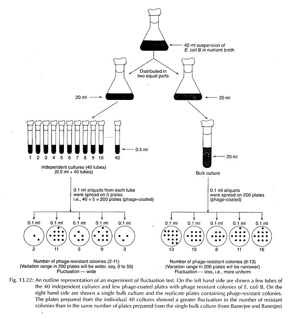
B. Fluctuation Test (Bacteria):
Variations occurring in bacteria, e.g., resistance to phage or antibiotics is due to genetic changes through mutation or due to adaptation to environmental condition, was confirmed by fluctuation test carried out by Luria and Delbruck. They allowed the growth of E. coli cells (103 cells per ml) in two sets: independent culture – 40 tubes each with aliquots of 0.5 ml; bulk culture – one tube with 20 ml.
After an incubation of 36 hours at 37°C; small aliquots (0.1 ml) from each tube of the independent cultures, as well as bulk culture were spread over a large number of replica plates coated with T, phage.
Number of phage-resistant colonies growing on each plate was counted which revealed a much greater fluctuation (i.e., a wider variation) exists among the plates prepared from independent cultures than the plates prepared from bulk culture.
The greater fluctuation in independent cultures is mainly due to origin of spontaneous mutation arising independently in different tubes at different times during growth.
The number of each resistant mutant arising at different times in independent culture multiplied during incubation and final number of resistant bacteria in different tubes was widely variable at the time of plating (before coming in contact with phage).
In contrast, the bulk culture contained a uniform population of both sensitive and resistant bacteria at the time of plating, i.e., before the bacteria come in contact with phage. Development of resistance due to adaptation will occur only after the bacteria come in contact with phage. This experiment thus proved that resistance appeared due to random mutation, not due to physiological adaptation (Fig. 13.22).
Note # 9. Importance of Gene Mutation:
(a) Role in Evolution:
Mutation is the major source of genetic variation; it provides raw material for evolution. Without mutation all genes will exist in only one form, alleles would not exist. Different organisms would not be able to evolve and adapt to environmental changes.
(b) Application in Plant Breeding:
Mutations are normally deleterious. Gustaffsson estimated that less than one in thousand mutants produced, may be useful in plant breeding. Several important mutants have, however, been obtained in different crops.
(i) In wheat, several useful mutations, viz, branched ears, lodging resistance, amber seed colour and awned spikelet were obtained and utilized in plant breeding. The most remarkable mutation obtained by Swaminathan is Sharbati Sonora. Other important varieties released in India are Pusa Lerma, NP 836.
(ii) In rice, several high yielding elite varieties – Reimei, Japonica, Indica have been obtained through mutations. Mutants were also obtained in rice for increased protein and lysine content. Jagannath, I/T48, l/TGO are the products of induced mutation in India.
(iii) In barley, mutant known as erectoides is of high yield. RBD-1, DL-253 are induced mutants in India.
(iv) In Legumes, Hans-pea, Ranjan-lentil, MUM 2-mung bean are mutants developed in India.
(v) Other important mutant varieties released in India are S 12-tomato, Rasmi-cotton, RLM 514-mustard, Co997-sugarcane, JRC 7447-Jute.
Regular survey by joint FAO and IAEA reported that there has been a highly significant increase in the number of mutant varieties developed in different crops.
(c) Another valuable application of induced mutation is the increased production of antibiotics, such as penicillin from species of Penicillium.
(d) Somatic mutations have also been found useful in many ornamentals. In tissue culture as well, several somaclonal mutants leading to somatic mutants have been obtained in horticultural species.