In this article we will discuss about Unio:- 1. Habit and Habitat of Unio 2. External Features of Unio 3. Internal Structures 4. Coelom 5. Locomotion 6. Respiratory System 7. Digestive System 8. Blood Vascular 9. Excretory System 10. Nervous System 11. Sense Organs 12. Respiratory System 13. Fertilisation 14. Development 15. Metamorphosis.
Contents:
- Habit and Habitat of Unio
- External Features of Unio
- Internal Structures of Unio
- Coelom of Unio
- Locomotion of Unio
- Respiratory System of Unio
- Digestive System of Unio
- Blood Vascular of Unio
- Excretory System of Unio
- Nervous System of Unio
- Sense Organs of Unio
- Respiratory System of Unio
- Fertilisation of Unio
- Development of Unio
- Metamorphosis of Unio
Contents
- 1. Habit and Habitat of Unio:
- 2. External Features of Unio:
- 3. Internal Structures of Unio:
- 4. Coelom of Unio:
- 5. Locomotion of Unio:
- 6. Respiratory System of Unio:
- 7. Digestive System of Unio:
- 8. Blood Vascular System of Unio:
- 9. Excretory System of Unio:
- 10. Nervous System of Unio:
- 11. Sense Organs of Unio:
- 12. Reproductive System of Unio:
- 13. Fertilisation in Unio:
- 14. Development of Unio:
- 15. Metamorphosis in Unio:
1. Habit and Habitat of Unio:
Unio is found in freshwater ponds, lakes, streams and rivers. The animal is sedentary but ploughs slowly through the mud or sand by its wedge-shaped muscular foot at the bottom of the pond or river. It does not go deep in the burrow because the posterior extremities of the valves remain exposed for the ingress and egress of respiratory water current.
Unio usually stays in shallow water during night but migrates to deeper water during day time. The food of Unio comprises microscopic organisms, both plants and animals, which are fed upon by filter-feeding mechanism.
2. External Features of Unio:
(i) Shape and Size:
The body of Lamellidens is laterally flattened. The anterior side of the body is roughly oval in outline and the posterior end is slightly narrower. Lamellidens has a bilaterally symmetrical body. The size varies from 5 to 10 cm in length.
(ii) Shell:
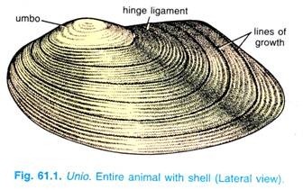
The soft body of Unio is completely enclosed by a hard calcareous shell. Shell is composed of two symmetrical and equal halves called valves and known as right and left valves.
The two valves are united by a dorsal elastic band called a hinge ligament which is continuous with the two shell valves but is made of un-calcified conchiolin, it is elastic and causes the valves of the shell to open. Near the hinge ligament are teeth and sockets which fit into each other to form an efficient inter-locking arrangement to prevent a fore and aft displacement of the two shell valves.
At the anterior end of the hinge ligament on each side, is a swelling called umbo which is the oldest part of the shell and is first formed in a young animal. Below the umbo are concentric lines of growth of the shell valves. The shell valves are rounded anteriorly but somewhat pointed posteriorly.
The umbo is directed anteriorly making it possible to determine the right and left shell valves of the animal. In most Pelecypoda the two shell valves are similar and equal in size, but in some sessile families (oysters) the upper or left valve is always larger than right valve by which the animal is attached.
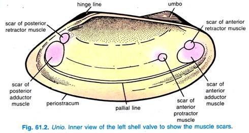
If a shell valve is removed from the mantle lobes, its inner surface is seen which shows marks of insertion of muscles running transversely between two valves.
The insertion of the edge of the mantle marks a pallial line. Anteriorly is an impression of an anterior adductor muscle, posteriorly is a larger impression of a posterior adductor muscle; close to these impressions are marks of an anterior retractor muscle and a posterior retractor muscle, Near the anterior adductor is also an impression of a protractor muscle.
The adductor muscles close the shell valves tightly by pulling them together, the retractors pull in the foot, and the protractor pushes out the foot. The hinge ligament acts antagonistically to the adductor muscles and causes the shell valves to open when the adductors relax.
Primitively the two adductor muscles are equal in size, but in many families the anterior adductor becomes reduced, and in oysters and scallops it disappears completely, then the posterior adductor moves to the centre of the shell valves. All muscles are un-striped, they gradually shift with growth of the animal, their faint lines may be traced to the umbo.
Microscopic Structure of Shell:
In section the shell has three layers, an outer brown, horny layer, the periostracum which is protective and is made of a horny organic material called conchiolin. Below it the middle layer is a thick prismatic layer made of vertical crystals or prisms of CaCO3 separated by conchiolin. The innermost nacreous layer or the “mother-of-pearl” layer is made of alternate layers of CaCO3 and conchiolin.
The hinge ligament is made of un-calcified conchiolin, it is continuous with the periostracum. Reserve calcium carbonate for the two inner layers of the shell is stored in certain cells of the digestive gland.
The nacreous layer is thickest at the umbo and thinnest at the shell margin, it is used for manufacturing buttons. In formation of the shell the periostracum is laid down by the outer lobe of the mantle, while the prismatic and nacreous layers are secreted by the entire outer surface of the mantle, though the nacreous layer is also secreted by the thickened lower edge of the mantle.
3. Internal Structures of Unio:
(i) Body:
The body is elongated but laterally compressed. The head is lost, in the upper half is a visceral mass which passes into a mid-ventral, wedge-shaped, laterally-compressed foot directed anteriorly, this is an adaptation for burrowing.
The foot has a large sinus which gets filled with blood when the foot is extended by blood pressure and muscular action of a pair of pedal protractor muscles, the protractors extend transversely from each side of the foot to the opposite shell valve.
The foot becomes swollen and turgid, it is then used for ploughing through mud in burrowing. Withdrawal of the foot is effected by pair of anterior and a pair of posterior retractor muscles attached to the foot on one side and to the shell valves on the other, and also by the muscle fibres within the foot itself. A pair of fleshy, flattened lobes, called labial palps are situated just below the anterior protractor muscles.
In the depression formed by labial palps, between anterior adductor muscle and the foot, is a transverse slit-like aperture, the mouth.
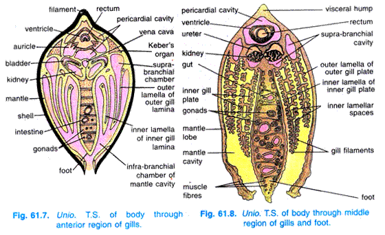
On each side the body is produced into a mantle lobe, the space between the mantle lobes is mantle cavity into which hangs a ctenidium on each side. The mantle cavity is large and extends on each side of the body, it protects the ctenidia and prevents their clogging with silt, and it allows a current of water to pass in and out in definite directions. 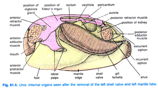
(ii) Mantle:
Lining the inner surface of the shell valves is a semi-transparent mantle or pallium which is made of two lobes continuous dorsally.
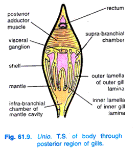
It is like skin, it encloses the soft parts and also hangs down like a skirt. Enclosed by the mantle is a mantle cavity which extends the entire length of the body on each side. The mantle cavity can be divided into two chambers, a large ventral infra-branchial chamber and a small dorsal supra-branchial chamber.
The bases of ctenidia mark the partition between these chambers. The mantle encloses the body in the upper half, and a muscular foot on the mid-ventral side. The lower border or edge of each mantle lobe is thickened and contains muscles, the muscles attach the mantle to the shell valve along a pallial line.
The thickened lower border of the mantle has three parallel lobes or folds, the innermost fold is the largest and it is this fold which contains muscles which are both radial and circular, it controls the flow of water. The middle fold is sensory in function.
The outer fold secretes the shell, the inner surface of the outer fold lays down the periostracum, while its outer surface secretes the prismatic and nacreous layers, but the nacreous layer is also secreted by the entire outer surface of the mantle.
The mantle secretes a pearl in many bivalves when any foreign particle lodges between the shell and the mantle, the pearl is formed in concentric layers around the foreign particle. The posterior edges of the mantle lobes are also thickened and they project outside the shell as two short tubes, a dorsal exhalant siphon and a ventral inhalant siphon.
The exhalant siphon is an actual tube formed by the fusion of the two lobes of the mantle, but the inhalant siphon is a temporary tube formed by approximation of mantle lobes, it has delicate fimbriae at its edges. Water enters through the inhalant siphon and after circulating it passes out through the exhalant siphon.
Histologically, the mantle has outer epithelium of one layer of cells in contact with the shell, it has branches of blood vessels, inside this is connective tissue with blood vessels, nerve cells and fibres, unicellular mucous glands, and nacreous glands which secrete the nacreous layer of the shell. On the innermost side is a single cell layer thick ciliated epithelium.
4. Coelom of Unio:
Coelom is schizocoelic having been formed in molluscs by splitting of the mesoderm, but the coelom is reduced to the paired cavities of kidneys and gonads and a pericardium which encloses a heart.
Originally all three cavities are intercommunicated, but there has been a progressive separation so that only the cavities of kidneys and pericardium communicate, the cavities of gonads have separated completely. However, the general body cavity is the haemocoel filled with haemolymph.
5. Locomotion of Unio:
The muscular foot is the chief locomotory organ. When the Unio walks, the foot is thrust forward between the two valves of the shell and burrows like plough-share through the mud. This permits blood to flow into the many sinuses of the foot, causing it to swell and, thus, form an anchor.
As the retractor muscles contract, the Unio is drawn forward an inch or so. The blood then is forced out of the foot so that it thins down again and can be withdrawn from the mud or sand. The process is repeated with each step and wedge-shaped path is left behind.
6. Respiratory System of Unio:
Lamellidens is an aquatic animal, hence, it respires by the oxygen dissolved in water, i.e., the mode of respiration is aquatic exclusively. So, for performing this life activity, Lamellidens possesses a pair of ctenidia or gills; mantle also helps in this activity. The gills of Lamellidens are, in fact, supposed to be highly modified and specialized derivatives of its mantle.
(i) Ctenidia:
There is a single pair of elongated ctenidia or gills, one on each side of the foot and are of eulamellibranch type. The large mantle cavity has made it possible for the great length of ctenidia which lie laterally. Each ctenidium appears double but it is made of two gill plates or demibranchs or laminae, an outer and an inner gill plate which have been derived by the folding of a single ctenidium.
The ctenidia divide the mantle cavity on each side into a large ventral inhalant chamber or infra-branchial chamber and a smaller dorsal exhalant chamber or supra-branchial chamber. Each gill plate or lamina is formed of two similar flaps or lamellae joined to each other except dorsally, thus, two lamellae of a gill plate form a narrow but long bag.
The lamellae are made of a number of vertically parallel gill filaments or branchial filaments. The gill filaments are elongated, they pass downwards and are then reflected upwards like a V, so that each gill filament forms a descending and an ascending limb.
Adjacent gill filaments are joined by the fusion of tissues forming interfilamentar junctions. Thus, gill filaments and interfilamentar junctions form the two lamellae of a gill plate. In the inter-filamentar junctions are holes known as ostia which connect ventral inhalant (infrabranchial) chamber of the mantle cavity with water tubes in the laminae.
The gill filaments appear as vertical lines and their interfilamentar junctions appear as horizontal striations on a lamella.
The gill filaments are covered with various kinds of cilia, and each gill filament is supported by two chitinous rods. On the sides of filaments are lateral cilia, on the distal surface which first encounters the inhalant current are frontal cilia, bordering the frontal cilia on both sides are long laterofrontal cilia or frontolateral cilia.
Between the two lamellae of a gill plate a space divided by vertical bars of vascular tissue forming interlamellar junctions which contain blood vessels.
The interlamellar junctions between two lamellae divide the space into distinct compartments called water tubes which are closed all round except dorsally where they open into a supra-branchial chamber of the mantle cavity. A ventral food groove runs longitudinally at the lower edge of each inner gill plate.
There are also two dorsal food grooves on each side at the base of a ctenidium, one between the mantle and the outer lamella of the outer gill plate, and another between the inner lamella of the outer gill plate and the outer lamella of the inner gill plate.
Attachment of Ctenidia:
The dorsal attachment of gill plates shows that the outer lamella of the outer gill plate is attached to the mantle, the inner lamella of outer gill plate and the outer lamella of inner gill plate are joined together to the visceral mass, the inner lamella of inner gill plate is attached to the visceral mass anteriorly, but further back it is free, and behind the foot it is joined to its fellow of the other side, so that the inner lamellae of inner gill plates are united with one another.
Blood supply of ctenidia:
The ctenidia are supplied by afferent branchial vessel carrying deoxygenated blood from the kidneys and divides to give rise branches into the inter-lamellar junctions. These branches unite to open into the efferent branchial vessel which carries away oxygenated blood to the heart.
In fact, during the flow of blood from the branches of afferent branchial vessel (in the inter-lamellar junctions) to the efferent branchial vessel, the blood is oxygenated.
Course of Water Current:
Constant beating of the cilia of gill filaments causes a continuous current of water to enter the inhalant siphon lying posteriorly and ventrally from where it goes to the mantle cavity. The lateral cilia pass the water current in, and through the ostia it enters the water tubes of gill plates, then it goes to supra-branchial chambers and passes out through the exhalant siphon situated posteriorly and dorsally.
The latero-frontal cilia form a flexible comb bordering the ostia, this comb forms a sieve to prevent large particles from entering ostia. The current transports not only oxygen to the ctenidia but also brings in food, the outgoing current carries away products of excretion and faeces in addition to carbon dioxide.
2. Mantle:
In addition to shell secreting function, mantle also helps in respiration. It is richly supplied with the blood vessels and it remains in contact with water, hence, gaseous exchange takes place through its thin wall.
Physiology of Respiration of Lamellidens:
When water passes through the water tubes in the gill, gaseous exchange takes place; in fact, carbon dioxide from the blood is diffused out in the water and dissolved oxygen from water is diffused in the blood. Thus, deoxygenated blood becomes oxygenated which is carried to the heart for distribution.
7. Digestive System of Unio:
Digestive system consists of the alimentary canal and a pair of digestive glands.
(i) Alimentary Canal:
It is a long coiled tube and comprises the mouth, oesophagus, stomach, intestine and rectum.
Mouth:
It is a transverse slit-like aperture situated in the anterior end of the body ventral to the anterior adductor muscle. On each side of the mouth is a pair of triangular fleshy, flattened and ciliated labial palps, one in front and one behind the mouth. The labial palps are jointed to their fellows of the other side and form upper and lower lips.
The two labial palps of each side enclose a ciliated oral groove which leads to the mouth. The characteristic buccal mass with jaws and radula of Pila is not found in this case.
Oesophagus:
The mouth leads behind and dorsally into a short narrow tubular passage, called oesophagus. The inner wall of oesophagus is ciliated.
Stomach:
The oesophagus leads into a thick-walled sac-like stomach having a ciliated lining. The stomach lies dorsal to the visceral mass and it is surrounded by a large digestive gland or liver which opens into stomach by many ducts. The stomach, in fact, has a dorsal part into which oesophagus opens and a ventral part having crystalline style; the ducts from digestive gland open in the dorsal part of stomach.
The crystalline style is a transparent, solid, gelatinous and flexible rod-like structure being secreted by the cells of stomach itself. The crystalline style has a matrix of protein, it contains mucus and a carbohydrate-splitting amylase and glycogenase; the amylase is condensed over the protein molecules.
The style rotates due to cilia in the stomach by which its free anterior end erodes and liberates amylase so that partial digestion of starches takes place extracellularly in the stomach. Rotation of the style also aids in mixing the contents of the stomach. The dorsal part of stomach has folded wall around the opening of digestive ducts.
These folds are said to help in storing the food and they help in transporting the useless substances to the intestine. They also help in conducting fine and partially digested food particles to the ducts of digestive gland.
Intestine:
The posterior end of stomach leads into the intestine which goes down and forms a coil in the visceral mass where it is surrounded by the gonad, and finally comes up again. Near the stomach the intestine turns back into rectum.
Rectum:
The rectum passes backwards through the pericardium, it traverses through the ventricle and opens by an anus above the posterior adductor muscle into the exhalant siphon serving as cloaca. The lining of the alimentary canal forms two ridges or folds in the posterior part of the stomach and first part of the intestine, there is also a similar ridge in the rectum, these ridges are called typhlosole’s.
(ii) Digestive Gland:
Liver is the only digestive gland which surrounds the stomach from lateral and posterior sides.
It is a large paired structure of dark brown or green colour. It opens into the dorsal part of stomach by many ducts. It secretes digestive enzymes and its cells are capable of ingesting food particles where intracellular digestion also occurs. In fact, fine food particles enter into the digestive ducts to reach its cells where they are ingested and intracellulary digested.
(iii) Food and Feeding:
The food of Unio comprises minute plants, Protozoa and organic debris. Unio is a filter or ciliary feeder and ctenidia have assumed the function of obtaining food.
The respiratory current brings in particles of food into the mantle cavity. On entering the mantle cavity the current of water becomes slow and heavier particles sink down and pass to the posterior region. Smaller particles pass with the current over the gill filaments of ctenidia.
The different cilia of gill filaments perform various functions. The lateral cilia cause the food-laden current to enter the mantle cavity, the latero-frontal cilia deflect the fine food particles on to the face of the filaments and they prevent large particles from clogging the ctenidia. Then the frontal cilia collect and pass the particles up or down the surface of ctenidia into the food grooves.
The ctenidia produce mucus in which the food particles become entangled to form string-like masses which pass along the dorsal and ventral food grooves towards the mouth.
The cilia of labial palps direct the food-laden mucus along the ciliated oral grooves into the mouth. The labial palps have the function of sorting and conveying food to the mouth, they can also reject some food particles and deflect them towards the outgoing current.
(iv) Digestion, Absorption and Egestion:
Digestion is both intracellular and extracellular. The digestive glands produce enzymes which bring about digestion in the stomach. The cells of the digestive glands take up solid particles of food and intracellular digestion of proteins and perhaps further digestion of carbohydrates take place by means of intracellular enzymes.
The crystalline style is made of protein and mucus, its material is mixed with food in the stomach and it produces an amylytic enzyme for digestion of carbohydrates. Amoeboid wandering leucocytes ingest food and also digest it, and they also transport digested food to all parts of the body. Absorption of digested food takes place in stomach and also from the digestive gland.
The undigested wastes in the stomach, if any, and those sent back into stomach from digestive gland pass into the intestine → rectum → anus → exhalant siphon. Intestine and rectum are believed to absorb water from the wastes passing through them.
8. Blood Vascular System of Unio:
The blood vascular system is well developed and is of open type. It comprises the blood, heart, pericardium, arteries, sinuses and veins.
(i) Blood:
It is the circulatory medium and consists of plasma and corpuscles. The plasma is colourless probably in all lamellibranch’s but in some species it is slight bluish in colour due to the presence of a respiratory pigment, the haemocyanin. Haemocyanin is a copper containing respiratory pigment (hence, imparts bluish colour to the plasma) in contrast to haemoglobin which is iron containing respiratory pigment.
However, some bivalves like Solen possess haemoglobin as respiratory pigment. A large number of colourless stellate amoebocytes or corpuscles, also referred to as leucocytes, are found in the plasma.
The leucocytes are granular as well as non-granular in nature. The granular leucocytes leave the blood spaces and enter the body where they are phagocytic and remove waste. The blood performs its usual function of distribution of oxygen and nutrients to the parts of the body and carbon dioxide and nitrogenous wastes to the desired organs.
(ii) Heart and Pericardium:
The heart of Unio is three-chambered and found enclosed in a thin- walled triangular sac, called pericardium. It is situated in front of the posterior adductor muscle and placed mid-dorsally. The pericardium has a pericardial cavity filled with pericardial fluid; it represents a part of original coelom. The pericardium, however, communicates with the supra-branchial chamber through the kidneys.
The heart is three-chambered having one ventricle and two auricles. The auricles are thin-walled, highly distensible, triangular chambers one on either side of the ventricle. These are attached to the pericardium with their broad bases but open dorsally into the ventricle.
Each auricle opens into the ventricle by auriculo-ventricular aperture guarded by a valve which allows the flow of blood from auricle to ventricle but do not permit the blood to go back into the auricle from ventricle. The auricles, however, receive blood from the ctenidia, kidneys and mantle, and pour it into the ventricle.
The ventricle is thick-walled, muscular and horizontal chamber surrounding the rectum. In most lamellibranch’s, the ventricle has become folded around the rectum so that the pericardium encloses not only the heart but also a part of the alimentary canal. The heart beats about 20-100 times per minute.
(iii) Arteries:
Each end of the ventricle is continued into an aorta, the anterior end into anterior aorta and the posterior end into posterior aorta. The anterior aorta passes over the rectum, while posterior aorta below the rectum and they give off a number of small arteries to supply blood into the different parts of the body.
In fact, the anterior aorta gives off three main arteries:
(i) Anterior pallial artery to supply the mantle,
(ii) Pedal artery to supply the foot, and
(iii) Visceral artery which gives off a gastric artery to stomach, a hepatic artery to liver or digestive gland, an intestinal artery to intestine and a gonadial artery to gonad.
The posterior aorta gives fine arteries to the pericardium and kidneys, a branch from it supplies to the rectum and finally it continues as posterior pallial artery to supply the mantle.
(iv) Sinuses:
The arteries end in ill- defined sinuses and lacunae which lack the epithelial lining of true blood vessels. There are no capillaries in molluscs, except in cephalopods, the blood from the arteries seeps into lacunar spaces in the connective tissue from where blood is taken up by the veins. Therefore, Unio’s circulatory system is said to be open type.
(v) Veins:
The venous blood from visceral organs is collected by smaller veins; gonadial vein from gonad, intestinal vein from intestine, hepatic vein from liver and gastric vein from stomach. All these veins together form the visceral vein. The blood from pedal sinus in the foot is collected by the pedal vein which joins the visceral vein to form a long vein called vena cava.
The vena cava lie longitudinally beneath the pericardium between the kidneys. Blood from vena cava goes to the kidneys through afferent renal veins where nitrogenous waste is removed from the blood. From the kidneys, blood is collected by efferent renal veins which finally form a pair of afferent branchial or ctenidial vein which sends branches into the filaments of the gill.
Blood is oxygenated in gill and goes into longitudinal efferent branchial or ctenidial vein which returns the blood to one of the auricles of the heart.
But some blood from vena cava and kidneys goes directly to the heart without going to the gills, hence, the heart also receives some deoxygenated blood. The blood which had gone to the mantle is purified, i.e., oxygenated and then returned to the other auricle of the heart by pallial veins.
Course of Circulation of Blood:
Blood, from the heart, goes to the anterior and posterior parts of the body through anterior and posterior aortae. Through these aortae, a part of blood goes to mantle where oxygenation occurs and finally oxygenated blood is conveyed to the auricle through pallial vein. The other parts of the body are supplied by different branches of these aortae where it becomes deoxygenated.
The deoxygenated blood is finally collected into vena cava through several veins.
The vena cava gives blood to kidney where nitrogenous wastes are removed and the blood then goes to the gills for oxygenation. The oxygenated blood from the gills is conveyed to the heart by efferent branchial or ctenidial veins. Thus, circulation is completed and the heart again starts distributing the blood to the various organs. In this way the cycle goes on.
However, the course of circulation of blood in the body of Unio may be graphically represented in the following way:
9. Excretory System of Unio:
The excretory system of Unio consists of a pair of kidneys or organs of Bojanus and the Keber’s organ.
Organs of Bojanus:
The chief excretory organ of Unio is a pair of kidneys or nephridia, often referred to as the organs of Bojanus; these are named so after the name of discoverer. These are situated one on each side of the body below the pericardium.
Each kidney consists of a tube bent upon itself forming a U-shaped structure. The lower limb of the tube is a thick-walled, spongy, brown-coloured glandular part of the kidney, the upper limb of the tube is a thin-walled, non-glandular and ciliated, called the urinary bladder.
The bladders of two kidneys communicate by an oval aperture. Each kidney opens at one end into the pericardium by a minute reno-pericardial aperture, and at the other end it opens by an excretory or renal pore into the supra-branchial chamber of the mantle cavity. Each kidney is an enclosed part of the coelom or renocoel, and is equivalent to a coelomoduct leading from the coelom to the exterior.
Physiology of Excretion:
The glandular part of kidneys removes nitrogenous excretory wastes from the pericardial fluid and blood supplied to them.
The ciliated cells lining the urinary bladder create outward current carrying the excretory fluid from the glandular part of kidneys to the supra-branchial chamber of the mantle cavity and finally goes out of the body through exhalant siphon. There occurs reabsorption of inorganic salts in the kidneys.
Kidneys also remove large amounts of water to maintain their blood concentration. Therefore, in addition to excretory function, the kidneys are osmo-regulatory also.
Keber’s organ:
In front of the pericardium is another excretory organ called Keber’s organ or pericardial gland. It is formed from the epithelium of the pericardium. It is a large reddish- brown glandular mass which discharges waste into the pericardium. Nitrogenous waste consists mainly of ammonia and amino compounds, but traces of urea and uric acid have also been found.
10. Nervous System of Unio:
The nervous system of Unio consists of only paired ganglia, commissures, (nerves connecting two similar ganglia), connectives (nerves connecting two dissimilar ganglia) and nerves. In fact, the nervous system of Unio is reduced to a great extent because of its sedentary and sluggish mode of life.
However, the various ganglia, their commissures and connectives are as follows:
(i) Cerebro-pleural Ganglia:
These are paired, somewhat triangular, yellowish ganglia of the size of a pin head and placed one on either side a little behind the mouth and at the base of labial palps.
Each cerebro-pleural ganglion is formed by the fusion of a cerebral ganglion and a pleural ganglion. Each cerebro-pleural ganglion gives out an anterior adductor nerve to the adductor muscle, a labial nerve to labial palp and an anterior pallial nerve to the anterior part of the mantle.
(ii) Pedal Ganglia:
These are also paired ganglia lying at the junction of visceral mass and foot about one-third of its length from the anterior end. Both the pedal ganglia are joined into a bilobed mass. The pedal ganglia supply to the foot, its muscles and statocyst.
(iii) Visceral Ganglia:
These, too, are paired ganglia which are fused together to form a flattened X-shaped mass lying mid-ventrally below the posterior adductor muscle. The visceral ganglia give out the pallial nerve to the mantle, renal nerve to kidneys, ctenidial nerve to the gills and adductor nerve to the posterior adductor muscle.
(iv) Commissure:
The cerebro-pleural ganglia of both the sides are connected together by a thin, transverse, loop-like nerve passing over the oesophagus; this nerve is called cerebral commissure. No other commissures are found in Unio.
(v) Connectives:
All the three ganglia are connected with somewhat stout nerves representing the connectives; the cerebro-pleural is connected to pedal by cerebro-pedal connective, cerebro- pleural is connected to visceral by cerebro-visceral connective. All these connectives are paired. However, there is no connective between the pedal and visceral ganglia.
11. Sense Organs of Unio:
The sense organs are poorly developed. The eyes and tentacles are altogether absent.
The main sense organs are:
1. Osphradium
2. Statocyst
3. Tactile cells
4. Photoreceptors.
1. Osphradium:
It is a group of yellow-coloured sensory cells found near each visceral ganglion, they are chemoreceptors and test the water current entering the inhalant siphon.
2. Statocyst:
There is a statocyst lying near each pedal ganglion in the foot. It is globular being formed as a pit of the skin. It is surrounded by several layers of cells and contains a calcareous statolith. The statocyst receives a nerve from the cerebropedal connective. Statocysts are organs of equilibrium.
3. Tactile Cells:
Tactile cells occur on the edge of the mantle and fimbriae of inhalant siphon.
4. Photoreceptors:
These are cells on the margins of siphons which are sensitive to light.
12. Reproductive System of Unio:
Unio is dioecious, i.e., the sexes are separate but there is no sexual dimorphism.
Gonads:
The gonads are testes in male and ovaries in female. The gonads are paired, large, highly branched structures lying in and around the intestinal coils in the visceral mass above the foot. During breeding period they become greatly enlarged and conspicuous; the testes are brightly whitish and ovaries are reddish.
The lining of the gonads proliferates to give rise spermatozoa in male and eggs in female. Each gonad has a short duct, vas deferens in male and oviduct in female, opening into the supra-branchial chamber of the mantle cavity near the excretory pore. Accessory reproductive structures are not found in bivalves.
13. Fertilisation in Unio:
In male the sperms pass out through the exhalant siphon and are carried to the surrounding water from where they enter the inhalant siphon of the female and reach its ctenidia.
In female the eggs are shed into the supra-branchial chamber of mantle cavity and are carried into water tubes of ctenidia where fertilisation and early development occur. Fertilised eggs generally develop in the outer gill plates of ctenidia which become enlarged to form a brood pouch or marsupium.
14. Development of Unio:
Unlike Unio in majority of bivalves the sex cells are discharged externally into water where fertilisation occurs, and the zygote develops into a free-swimming trochosphere larva which is succeeded by a veliger larva, specially in the marine forms. The veliger larva is symmetrical in pelecypods.
In freshwater family Unionidae (which includes Anodonta and Lamellidens) there is an indirect but much specialised development. The veliger stage is formed in the marsupium of ctenidia, this veliger is highly modified and is known as a glochidium larva in this family.
Early Development:
The zygote undergoes complete but unequal cleavage to form a morula having small micromeres and large macromeres. Gastrulation occurs by macromeres invaginating into micromeres, but the archenteron so formed remains small for a long time. The gastrula has micromeres or ectoderm, macromeres or endoderm, a large blastocoele and a small archenteron. It is enclosed in vitelline membrane.
Some cells of the gastrula are budded into the blastocoele to form mesoderm. A deep invagination occurs to form a shell gland which is characteristic of Mollusca. The shell gland marks the dorsal surface of the embryo, the posterior end is marked by a tuft of long cilia. The shell gland secretes an unpaired shell which is soon replaced by a triangular bivalved shell, the shell valves enclose a larval mantle.
The lower parts of shell valves are curved in to form hooks beset with spines (in Anodonta and Lamellidens, but hooks are absent in many freshwater mussels). The embryo is cleft in the middle to form a dorsal body and two mantle lobes. On each mantle lobe four brush-like sense organs arise, each having a cluster of bristles.
The mesoderm forms a large adductor muscle which runs between two shell valves anteriorly.
On the body a byssus gland is formed which secretes a sticky thread called byssus. The embryo is now known as a glochidium larva. So far the embryo is nourished on yolk present in the egg. The larva has no mouth or anus and the digestive tract is not yet formed. Thousands of glochidia may be formed by a single freshwater mussel.
Structure of Glochidium:
Glochidium (Fig. 61.18) is a minute larva measuring 0.1 to 4.0 mm, comprises a shell and mantle. Shell consists of two triangular valves united dorsally and free ventrally. Ventral free end of each valve of shell is produced into a curved hook bearing spines.
Mantle lobes are small and bear brash-like sensory organs. Adductor muscle is well developed extending between the two valves at the base. The closure of the valves is effected by the large adductor muscle. Byssus gland is situated above the adductor muscle which gives rise to a long sticky thread called provisional byssus.
The glochidium is nourished in the marsupium which contains a large number of glochidia which are, thus, ectoparasitic. The glochidia are set free from the ctenidia and pass out through the exhalant siphon into water and sink down at the bottom. They get attached to gills or fins of freshwater fishes by their byssus and shell valves.
Some glochidia require only certain species of fish as hosts, but others can parasitise a large range of host fishes. The glochidia which do not get a suitable host die in due course. Adult fish are not harmed by parasitic glochidia.
Larvae become encysted in a few hours by growth of cells of the host, the tissues of the host are stimulated to grow around the parasite forming a cyst. The — glochidia feed by absorbing juices of the fish by the mantle, the larval mantle contains phagocytic cells which obtain nourishment for the developing mussel, thus, they lead an ectoparasitic life for about 10 weeks during which time metamorphosis takes place.
15. Metamorphosis in Unio:
The sense organs, larval adductor muscle, larval mantle, and byssus disappear and the adult organs begin to form. The ectoderm invaginates to form the stomodaeum and proctodaeum which open into the archenteron to form the alimentary canal. A foot arises as an elevation behind the mouth.
On each side of the foot arise two papillae which form gill plates. The cyst weakens and the young animal opens and closes its shell valves and extends its foot. It escapes from the cyst and drops to the bottom to become free-living, then it assumes the adult form and mode of life.
Significance of Glochidium:
The glochidia, as ectoparasite on fishes, may reach to far off places with the fish. Thus, they help in the dispersal of Unio to far off places because Unio is a sedentary and sluggish animal having very poor power of locomotion.
