In this article we will discuss about Pila Globosa:- 1. Habit and Habitat of Pila Globosa 2. External Structures of Pila Globosa 3. Coelom 4. Digestive System 5. Locomotion 6. Respiratory System 7. Circulatory System 8. Excretory System 9. Nervous System 10. Reproductive System 11. Development.
Contents:
- Habit and Habitat of Pila Globosa
- External Structures of Pila Globosa
- Coelom of Pila Globosa
- Digestive System of Pila Globosa
- Locomotion of Pila Globosa
- Respiratory System of Pila Globosa
- Circulatory System of Pila Globosa
- Excretory System of Pila Globosa
- Nervous System of Pila Globosa
- Reproductive System of Pila Globosa
- Development of Pila Globosa
1. Habit and Habitat of Pila Globosa:
Pila globosa is commonly known as pond snail or apple snail. It is a typical representative of the class Gastropoda. It belongs to the family Pilidae. The members of the family are distributed in the Oriental and Ethiopian regions of the world. The common species of the genus is P. globosa. It is quite abundant in the fresh-water ponds.
Pila globosa inhabits fresh-water ponds and lakes. They are quite abundant in water having succulent aquatic vegetation on which they feed. They are really of amphibious forms, i.e., they live most of the time in water but they can also thrive well on land.
They exhibit two-fold respiratory adaptations. They respire in water by ctenidium and by pulmonary sac on land. During prolonged drought they may remain torpid for a long time and during rains they return to normalcy.
2. External Structures of Pila Globosa:
(i) Shell:
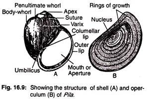
The body of Pila globosa is enclosed by a thick spirally-coiled globular shell. It has the form of an elongated cone coiled round a central axis in a spiral manner. A single revolution of the shell is called the whorl. The extreme top of the shell is designated as apex (Fig. 16.9A).
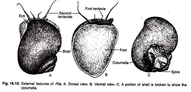
The apex of the shell is regarded as the oldest part of the shell. Starting from the apex the other whorls—the penultimate whorl and body-whorl are large to enclose the greater part of the body. The first whorl is smallest and the last one is the largest (Fig. 16.10A).
(ii) Operculum:
The last whorl contains a large aperture, which can be closed by a lid called operculum which is attached to the posterior side of the foot. The operculum is a flat calcareous plate. It is formed as a cuticular secretion of a group of cells from the foot. It has a lunate-oblong outline which corresponds to the aperture of the shell.
The operculum shows numerous concentric rings of growth around a well-marked nucleus. The inner surface of the operculum shows a distinct elliptical area, called boss, for the insertion of opercular muscle (Fig. 16.9B). The margin of the aperture (mouth) is smooth and is called peristome.
A spiral column arising from the centre of the shell is present on the inner side. It is called columella (Fig, 16.10C). The type of coiling is right-handed and is called dextral. In rare or abnormal cases left- handed coiling (sinistral) is also observed. Detailed account of coiling in gastropods is discussed in general notes on mollusca.
(iii) Microscopic structure of the shell:
The microscopic picture of shell of Pila globosa exhibits three layers. The outer layer is chitinous and known as periostracum. The two underlying layers, the ostracum and hypostracum, are composed of calcareous material. The periostracum is thin and consists of a large number of parallel bands in young stage.
These bands are separated from one another by wavy lines. Each band is made up of rectangular blocks. But in an adult, the periostracum appears as a homogeneous membrane without showing the bands or blocks. The ostracum and hypostracum are essentially similar. The plates constituting these calcareous layers are disposed differently.
(iv) Body:
The whole body is located within the whorls of the shell and is attached to the columella of the shell by columellar muscle. The columellar muscle arises from the foot and is attached with the columella. The columellar muscle plays a vital role.
It prevents the animal from extending out of the shell beyond certain limit and also helps to withdraw it into the shell. The body is divisible into the head, foot and visceral mass. The head along with the foot can be protruded to a limited extent through the aperture of the shell (Fig. 16.10A).
(v) Head:
The head is well-marked. It is prolonged into a partly contractile snout. It carries two pairs of tentacles. The longer pair are thread-like and contain stalked eye at the base, called ommatophores.
The shorter pair are called labial palps or first tentacles and are regarded as the antero-lateral prolongations of snout. Two fleshy and highly contractile projections, called nuchal lobes or pseudepipodia, are seen on the two sides of the head.
The lobes, although projected anteriorly over the foot, are the prolongations of the mantle and are innervated by nerves from the pleural ganglia. The left nuchal lobe is highly developed and forms the respiratory siphon, used for aerial respiration. The nuchal lobes of Pila are not homologous with the epipodia of other gastropods.
(vi) Foot:
The foot is more or less triangular when seen from ventral side (Fig. 16.10B). The anterior part of the foot is round and the posterior part of the foot holds a hard disc-like structure called the operculum. The foot is highly muscular and contains pedal glands, Pila is adapted to creeping movement. The visceral mass is a spiral coiled hump that includes all of the internal organs of the body.
(vii) Mantle:
The skin covering the visceral mass forms the pallium or mantle. It forms a cloak over the anterior part of the body including the head and its appendages in retracted state.
The mantle sub-serves three functions in the life of Pila globosa:
(i) Protects the visceral mass and head,
(ii) Serves as an additional respiratory organ and
(iii) Secretes the shell by the shell-secreting glands at the free margin of the mantle.
The mantle is free anteriorly and encloses a spacious cavity, known as pallial or mantle cavity.
This cavity contains visceral organs of the animal which are known as pallial complex. The mantle cavity is imperfectly divided into left and right chambers by a longitudinal muscular ridge, known as epitaenium. The right chamber contains the ctenidium, rectum and the genital duct. The left chamber contains the pulmonary sac.
There is a comb-like organ of taste known as osphradium close to left nuchal lobe. The mouth and anus are closely situated on the same side of the body. The anal and the genital apertures are located on the right mantle opening.
3. Coelom of Pila Globosa:
In adult, the general body cavity is a haemocoel as seen in other molluscs. The true coelom is represented by the pericardial cavity and the cavities round the kidney.
4. Digestive System of Pila Globosa:
Pila lives primarily on aquatic vegetation. The digestive system is composed of digestive canal and digestive glands (Fig. 16.11).
The digestive canal is distinguishable into:
(i) Foregut,
(ii) Midgut and
(iii) Hindgut.
The foregut and the hindgut possibly develop from the ectodermal layer, while the midgut is endodermal in origin. The foregut includes the buccal mass and the oesophagus, the midgut consists of the stomach and the intestine and the hindgut includes the rectum.
(i) Foregut or Stomodaeum:
The mouth is a vertical slit which leads into the anterior end of the digestive tract which becomes greatly swelled to form an oval buccal cavity. The buccal cavity is enclosed by a strong thick-walled muscular structure called buccal mass. The buccal mass is regarded as the pharynx by many workers.
The entrance of the mouth is guarded by a pair of chitinous jaws projecting from the roof of the buccal cavity (Fig. 16.12A). Covering the floor of the buccal cavity, is present a chitinous ribbon like structure. This structure is known as radula or lingual ribbon (Fig. 16.12B) and is produced from a radular sac. The radula is an elongated structure bearing transverse rows of serrations.
Each transverse row contains about seven teeth—two marginals, a lateral on either side of a median rachidian tooth, giving the formula as: 2, 1, 1, 1, 2 = 7. The radula is movably placed by muscles upon a large outgrowth of the floor of the buccal cavity, called tongue mass or odontophore. It is made up of muscles with cartilaginous support. It has an anteriorly placed subradular organ.
The subradular organ is a more or less rounded structure. It is divided into two by a median furrow. A small pouch like sublingual cavity is present beneath the subradular-organ. The radula at the posterior end enters into a radular sac which supplies new teeth to the radula.
The radula is pushed forward by muscles from behind and it works as a file by rasping food materials. Pila is a vegetable feeder and takes leaves of aquatic weeds by cutting with the jaws. The buccal cavity receives two salivary glands on the posterior side.
The buccal cavity leads into oesophagus. The oesophagus is a long tube and just after its origin from the buccal mass it gives out on each side, a small out-pushing called oesophageal pouch. The oesophagus ends in stomach.
(ii) Midgut:
The stomach is red in colour and is situated on the lower part of the visceral mass just below the pericardium. It is a large sac and bent on itself to form a ‘U’- tube, one limb of which received the oesophagus and the other leads into the intestine. The end which receives the oesophagus is called the posterior or cardiac chamber, while the other end is called the pyloric chamber.
The cardiac chamber actually constitutes the main part of the stomach. The pyloric chamber exhibits transverse folds at its inner wall, while that of cardiac chamber appears corrugated. A caecum or blind pouch opens at the junction of stomach and intestine. The caecum does not contain any crystalline style as observed in other gastropods. It is merely a blind diverticulum of the pyloric chamber of the stomach.
(iii) Hindgut:
The intestine is long and forms 2-½-3 coils. The posterior part of the intestine is nearly straight and turns to the anterior direction and continues as the rectum. The rectum lies on the floor of the right side of the mantle cavity and terminates in anus which is situated near the mouth within the right mantle opening.
Digestive Glands of Pila Globosa:
The digestive glands include the salivary glands and the liver or hepatopancreas. There are two salivary glands situated one on each side of the oesophagus. The liver or digestive gland is black in colour and constitutes the main bulk of the visceral hump. It gives out two ducts which unite to form a common duct and opens into the stomach.
Food and Feeding:
Usually the food of Pila globosa includes succulent aquatic plants and sometimes they are found to feed on dead animal’s tissue. The food is taken into the buccal cavity by the chain-saw mechanism of radula and make the leaves of plants into pieces by the jaws. Then the food is masticated in the buccal cavity.
Digestion:
The masticated food in the buccal cavity is digested partly by the action of secretion that is secreted by the salivary glands. This secretion helps to convert the starch into sugar.
The undigested part of food is digested in the stomach by the secretion of digestive gland. The secretion contains enzymes comparable to those of vertebrate enzyme. But the cellulose part of the food is digested within the resorptive cells of the digestive gland.
Both extracellular and intracellular types of digestion take place in Pila. Extracellular digestion takes place within the stomach of Pila but intracellular digestion occurs in the digestive gland. The major part of absorption takes place in the digestive gland and rest in the intestine. The undigested food passes out through the anus.
5. Locomotion of Pila Globosa:
Pila moves very slowly by creeping on the substratum by its foot. During movement the foot is protruded through the opening of the shell and its flat sole helps in the process. The extension of the foot is caused by sudden influx of blood into the foot. The glands present in the foot produce slimy secretion that helps the animal to glide on dry surface. The foot is provided with vertical, longitudinal and transverse muscles.
During locomotion the wave-like contractions on its surface are produced by the contractions of the vertical muscles. The contraction of the transverse muscles drives the blood forward which causes the extension of the foot in front. During this process the longitudinal muscles contract to pull the posterior end of the foot forward.
6. Respiratory System of Pila Globosa:
Pila exhibits double mode of respiration, i.e., it can absorb oxygen dissolved in water by ctenidium and can also utilise atmospheric oxygen by the pulmonary sac.
The mantle cavity is incompletely divided into right chamber (branchial) and left chamber (pulmonary) by the presence of epitaenia.
Aquatic respiration is performed by the single ctenidium or gill situated on the dorsolateral wall of the right portion of the mantle cavity (Fig. 16.13A).
The ctenidium is made up of numerous flattened, triangular leaflets or lamellae. These lamellae are arranged in a single row running only one side along the ctcnidial axis of the gill. This type of the ctenidium is known as monopectinate type (Fig. 16.14A). The basal end of each lamella is attached to the pallial epithelium and the other end hangs freely.
The ctenidial lamellae are not of same size. The lamellae are large in the middle of the ctenidium, while the lamellae decrease in size towards the two ends. Each branchial (ctenidial) lamella is composed of two layers of epithelia supported by muscle fibres and connective tissue. Two epithelial layers enclose a narrow space.
Each epithelial layers consists of three types of cells:
(i) Ciliated columnar cells,
(ii) Non-ciliated columnar cells and
(iii) Few glandular cells.
The detailed structures of a gill lamella is shown in Fig. 16.13B. The ctenidium is supplied with blood vessels. The right side of lamella is smaller called afferent side and the left side is longer called efferent side.
The ctenidial axis of the afferent side carries the afferent blood vessel that collects deoxygenated blood and left side of the ctenidial axis carries efferent blood vessel that supplies oxygenated blood. Each lamella is provided with many transverse ridges or pleats (Fig. 16.14B).
Mechanism of Respiration:
A. Aquatic respiration:
In aquatic respiration, a current of water containing oxygen is drawn in by the extended left siphon (left nuchal lobe). The water reaches the osphradium which tests the nature of water. Ultimately the respiratory water current reaches the pulmonary chamber and flows into the right branchial cavity crossing the epitaenia.
Here the water bathes the ctenidium and passes out through the right nuchal lobe. The gaseous exchange takes place between oxygen dissolved in water and carbon dioxide which is produced during respiration, diffuses into water.
Nuchal lobes:
The two fleshy, muscular nuchal lobes or pseudepipodia are situated, one on either side of the head. The mantle is prolonged into highly contractile nuchal lobes. The left lobe is highly developed while the right lobe is less developed and both form as extended funnels or siphons. Both the lobes help in respiration.
The course of water current in aquatic respiration in Pila is being represented here:
Pulmonary sac:
The pulmonary sac is a closed cavity which hangs from the dorsal wall of the mantle in the pulmonary chamber. The pulmonary sac in Pila globosa is a new attainment in response to its aerial respiration.
The pulmonary sac has one opening into the pulmonary chamber known as pneumostome which is guarded by two valves. The wall, specially the dorsal wall (Fig. 16.13C) of the pulmonary sac, is highly vascular and helps directly in gaseous exchange.
B. Aerial respiration:
On land, the pulmonary sac becomes filled up with atmospheric air and carries on the process of respiration. Pila can also respire through the pulmonary sac while it remains in water.
To inhale atomospheric air, Pila comes to the surface of the water. Before reaching the surface, Pila begins to expand the size of the left nuchal lobe (left siphon). It increases its size both in length and breadth and rolls up to form an elongated respiratory tube.
The outer end of the tube extends beyond the level of water and sucks in air from atmosphere. The inner end of the tubes comes in an immediate contact with the opening of the pulmonary sac. The alternate contraction and dilatation of the mantle wall as well as of the pulmonary sac help in the process of respiration.
After gaseous exchange the expelled air goes out of the pulmonary chamber by the same route. During this process the branchial chamber remains completely shut off from the pulmonary chamber by the epitaenia which comes in contact with the roof of the mantle.
Aquatic respiration takes place when Pila globosa remains either submerged in water or remains attached to the aquatic weeds. When the water of the pond becomes foul, Pila comes to the surface of water, or on land they perform aerial respiration.
7. Circulatory System of Pila Globosa:
The circulatory system of Pila globosa is well- developed owing to aquatic as well as aerial modes of respiration.
The heart is situated in the left-hand side of the visceral whorl very near to the posterior end of the ctenidium. The pericardial chamber encloses the heart and the aortic ampulla. As the ctenidium lies in front of the heart the animals are included under Prosobranchia. The heart consists of a single auricle and a single ventricle (Fig. 16.15).
The auricle is thin-walled and lies in the dorsal part of the pericardium. It communicates with the thick-walled musciilar ventricle through the auriculo-ventricular aperture. The ventricle is situated just below the auricle in the same axis.
The auriculo-ventricular aperture is guarded by semilunar valves which prevent regurgitation of blood from the ventricle to the auricle. The auricle receives oxygenated blood from the ctenidium and pulmonary sac through efferent ctenidial and pulmonary veins respectively.
The lower end of the ventricle gives rise to an aorta. The root of the aorta is provided with two semilunar valves which do not allow the backflow of blood into the ventricle. The aorta immediately bifurcates into two arteries, the anterior one is called cephalic aorta supplying blood to the head region and the posterior or visceral aorta which supplies blood to the posterior part of the body.
The cephalic aorta, just after its origin, gives a dilated sac-like outgrowth, known as aortic ampulla. Both the aortae supply arteries to different parts of the body.
The cephalic aorta gives off three arteries along its outer side:
(i) An artery to the skin,
(ii) An artery to the oesophagus and
(iii) An artery to the left part of the mantle, the osphradium and left siphon.
The cephalic artery gives off a pericardial artery on its inner side. This artery supplies the pericardium and enters into posterior renal chamber.
The main trunk of the cephalic artery enters into the perivisceral sinus (space surrounding the buccal mass and oesophagus) and then crosses beneath the oesophagus. It then gives off many arteries to the buccal mass, oesophageal wall, right side of the mantle, right siphon and the copulatory organ, eyes, tentacles, etc. (Fig. 16.16).
The visceral aorta, immediately after its origin, gives off an artery to supply the pericardium, digestive gland and skin. A little further, the visceral aorta gives origin to a stout gastric artery to the stomach.
The main aorta runs along the left margin of the posterior renal chamber and sends branches to the intestine and the posterior renal chamber. It then sends an artery to the digestive gland, the gonad and terminates in the wall of the rectum.
The blood, after being distributed to the various parts of the body by the arteries and their tributaries, passes into small spaces (lacunae). These lacunae unite to form large sinuses.
There are four main sinuses:
(i) Peri-visceral sinus,
(ii) Peri-intestinal sinus,
(iii) Branchio-renal sinus and
(iv) Pulmonary sinus.
The peri-visceral sinus sends blood to the ctenidium and pulmonary sac. The peri- intestinal sinus passes blood to the kidney for eliminating metabolic wastes. From the kidney, blood returns to the auricle by efferent renal veins.
Course of circulation of blood:
The type of circulation is of open type. The cephalic and visceral aortae supply blood to the different parts of the body. The cephalic aorta supplies blood to the head, mantle, buccal mass, oesophagus, copulatory organ, columellar muscle and associated structures. The visceral aorta supplies blood to the visceral mass.
Although there are four main sinuses, the blood is collected into the peri-visceral and peri-intestinal sinuses. From these sinuses, the blood is conveyed either into the pulmonary sac, ctenidium or into the kidney.
During aerial respiration, the blood flows into the pulmonary sac, while in aquatic respiration most of the blood from the perivisceral sinus goes to the ctenidium. After aeration, the blood comes to the auricle by the pulmonary vein or by the efferent ctenidial vein. The blood from the peri-intestinal sinus passes either into the anterior or posterior chamber of the kidney.
On its way through the anterior renal chamber, the blood gets rid of nitrogenous wastes and flows either into the ctenidium or into the posterior renal chamber. The posterior renal chamber gets blood either from the peri-intestinal sinus or from the anterior renal chamber. The blood gets rid of its excretory product but without being aerated. Thus mixed blood goes to the auricle for distribution via the ventricle.
The circulation of blood through the heart and the different parts of the body is shown below:
8. Excretory System of Pila Globosa:
The excretory organ is the kidney. It consists of two renal chambers—one anterior and another posterior (Fig. 16.17). The anterior renal chamber is more or less oval in shape and is situated anterior to the pericardium. It communicates to the posterior renal chamber by one end and the other end opens into the mantle cavity through a slit-like aperture near the epitaenia.
It is reddish in colour and its internal cavity presents numerous lamellated processes which reduce the internal cavity. The lamellae are arranged on the floor on either side of the afferent renal sinus, and on the roof they are similarly arranged on either side of the efferent renal sinus.
The posterior renal chamber is broad and the colour varies from brownish to gray. It is situated behind the anterior renal chamber. This chamber is separated from the pericardium by a vertical partition (renopericardial septum) and opens into it through a slit-like renopericardial aperture.
The roof has profuse branching of the afferent and efferent renal vessels. The renal chambers are provided with network of blood vessels and take up nitrogenous waste products from the blood. The wastes are discharged into the mantle cavity through the renal duct. From the mantle cavity the waste products are eliminated outside the body.
9. Nervous System of Pila Globosa:
The nervous system consists of ganglia, commissures, connectives and the nerves to different organs.
(i) Ganglia:
A small compact mass of nerve cells and connective tissue is called ganglion.
The main ganglia are:
(1) One pair of roughly traingular cerebral ganglia situated on the dorsolateral sides of the buccal mass, one on each side of the head.
(2) One pair of pleuropedal ganglia placed below the buccal mass on the lateral side. Each pleuropedal ganglionic mass is more or less rectangular in outline and is formed by the fusion of pleural and pedal ganglia. The infraintestinal ganglion is also fused with the right pleuropedal mass.
(3) Visceral ganglion is very large and appears to be unpaired. It is a bilobed structure and is formed by the fusion of two separate ganglia. The visceral ganglion is placed posteriorly very close to the heart.
(4) A pair of buccal ganglia are situated on the buccal mass on the two sides of the oesophagus.
(5) A single supraintestinal ganglion is located near the middle of the left pleurovisceral connectives (Fig. 16.18).
(ii) Commissures:
The nerve connections between two similar ganglia are generally called commissures. The ganglia are placed on the opposite sides of the body. Two cerebral ganglia are connected by a thick nerve cord, called the cerebral commissure. The buccal ganglia are also connected by a delicate buccal commissure. The inner sides of the pleuropedal ganglia are connected by a broad nerve, called the pedal commissure.
(iii) Connectives:
The nerve connections between two dissimilar ganglia are usually called connectives. The ganglia may be situated on the same or opposite sides of the body. The cerebral ganglia and the buccal ganglia are connected by cerebrobuccal connectives.
The pleuropedal ganglia are connected on each side with the cerebral ganglion by cerebropedal and cerebropleural connectives. That the pleuropedal ganglia are formed by the fusion of separate pleural, and pedal ganglia is indicated by the presence of an indistinct constriction and the existence of two separate connectives joining the cerebral ganglia.
The pleural ganglion is connected with the visceral ganglion by a pleurovisceral connective on each side. The right pleurovisceral connective lies below the level of the oesophagus and is generally designated as infraintestinal visceral connective and the left pleurovisceral connective is situated above the level of the oesophagus and is termed as supra-intestinal visceral connective.
The supra-intestinal ganglion is connected with the right pleuropedal ganglion by an oblique nerve placed above the oesophagus, called supraintestinal nerve. A very slender nerve, called the infraintestinal nerve, is present connecting the two pleural ganglia of two sides.
(iv) Nerves to the different parts of the body:
Each cerebral ganglion innervates the eye, the snout and the tentacles on its side. The statocyst is also innervated by a slender nerve arising from the cerebral ganglion. The pedal ganglion gives out numerous nerves to the foot and the pleural ganglion supplies the mantle.
The supraintestinal ganglion supplies nerves to the ctenidium and the pulmonary sac. The visceral ganglion sends nerves to kidney, genital organ, pericardium and intestine. The buccal ganglion innervates the buccal mass.
(v) Chiastoneury in Pila:
The nervous system of Pila Globosa exhibits streptoneurous chiastoneury condition. This is the result of torsion of visceral mass which has made the whole of the nervous system asymmetrical. The complexities in the nervous system in Pila are due to complete migration of the anal and genital openings to the oral end.
The chiastoneury is not so clear in Pila Globosa and typical figure-of-8-like arrangement is not produced between the supra and infraintestinal nerves. No crossing is possible on the right side because of the shifting and fusion of infraintestinal ganglion with the right pleural ganglion.
The zygoneury (a secondary connection between pleural and supraintestinal ganglia) is present only on the left side. So the typical chiastoneurous condition with double zygoneury as seen in many gastropods is not clear in Pila.
Sense Organs of Pila Globosa:
The sense organs are quite well developed.
(i) Osphradium:
The osphradium is the organ for taste. It helps to taste the chemical and physical qualifies of the incurrent water and also assists in the selection of food. There is only one osphradium which remains suspended from the roof of the pallial cavity on the left side.
It is small in size and is roughly oval in shape (Fig. 16.19A). Fig. 16.19B shows the detailed structures of osphradium in sectional view. It has a bipectinate arrangement of its leaflets on the two sides of a central axis. It consists of a single epithelial layer enclosing nervous tissue, connective tissue and blood spaces.
(ii) Tentacles:
The labial palps and the true tentacles are the tactile organs.
(iii) Statocyst:
The statocysts are two in number and are situated one on each side of the pleuropedal ganglionic mass. Each has a round sac-like body (Fig. 16.19C) and is kept in position by muscles. The centre of the sac is occupied by a solid mass of calcareous particles, known as statolith. It is the organ of balance and gets its nerve supply from the cerebral ganglion.
(iv) Eyes:
Two eyes are located one at the base of each longer pair of tentacles. Each eye has a closed vesicle (Fig. 16.19D) with the inner wall lined by photosensitive or retinal cells. The opening of the vesicle is covered by transparent outer and inner corneas.
The cavity between the cornea and the retina is filled up with an oval body constituting the lens. Although, in Pila, the eyes have all the essential components for photoreception, it is very poor in sight.
10. Reproductive System of Pila Globosa:
The sexes are separate but sexual dimorphism is almost absent.
(i) Male reproductive system:
The male reproductive system comprises of testis which lies in close contact with the digestive gland and occupies the upper two or three whorls (Fig. 16.20A). The testis is a flat plate- like structure and is more or less triangular in outline. It is cream-coloured. Many fine ducts—the vasa efferentia, originating from the testis unite together to open into the vas deferens.
The vas deferens or male gonoduct is differentiated into:
(i) An upper tubular part;
(ii) A terminal glandular part and
(iii) A curved blind tube, called vesicula seminalis, in between the junction of the two parts of the vas deferens.
The penis is present in the form of a whip-like flagellum which is partially ensheathed by the penis sheath and situated on the right side of the body near the mantle opening. The penis sheath is a simple outgrowth from the inner surface of the mantle. The penis is a long flagellar structure and is capable of great extension.
Two types of spermatozoa are encountered in Pila, one is pear-shaped—Eupyrene type (Fig. 16.20B) and the other is worm-like Oligopyrene type (Fig. 16.20C). The eupyrene types are functional and are capable of fertilizing the eggs.
(ii) Female reproductive system:
The female reproductive system consists of a much branched organge coloured ovary situated in the upper whorl of the visceral mass and remains embedded in the digestive gland (Fig. 16.20D). Female gonoduct or the oviduct which is differentiated into an upper tubular part comes down along the edge of the liver.
The lower glandular part remains on the floor of the mantle cavity parallel to the rectum. The glandular part is distinguishable into a yellow coloured albumen gland which secretes albumen and the posterior part is distinguished as the uterus.
At the junction of the tubular and glandular portions of the oviduct there is the receptaculum seminalis or the spermatheca where spermatozoa remain stored. The terminal part of the uterus is differentiated as vagina. The male intromittent organ, the penis, is rudimentary and useless in female. The eggs are fertilized by spermatozoa coming from the spermatheca.
Fertilization is internal and oviposition begins a day or two after copulation. The zygote, receives albumen and a coating of shell in the uterus. Few hundred eggs are laid at a time and they adhere together to form a mass (Fig. 16.20E). Existence of rudimentary penis in female is a very remarkable occurrence and is suggestive of the fact that this group had hermaphroditic ancestor.
11. Development of Pila Globosa:
The development occurs outside the body of the female. The development of Pila globosa is direct, and young snail develops from the fertilized egg. The embryo floats in a central core of liquid albumen which is surrounded by a thick layer of whitish solid albumen. The outer part of the egg consists of a white egg shell and a double layered shell membrane beneath the egg shell.


