In this article we will discuss about Cycadeoidea. After reading this article you will learn about: 1. History of Cycadeoidea 2. Morphological Features of Cycadeoidea 3. Anatomy 4. Reproductive Organs 5. Microsporophyll 6. Gynoecium.
Contents:
- History of Cycadeoidea
- Morphological Features of Cycadeoidea
- Anatomy of Cycadeoidea
- Reproductive Organs of Cycadeoidea
- Microsporophyll in Cycadeoidea
- Gynoecium of Cycadeoidea
Contents
1. History of Cycadeoidea:
Cycadeoidea, also called Bennettites by several European palaeobotanists is represented by about 30 species. The name Cycadeoidea was put forward in 1827 for petrified trunks from Isle of Portland. Though Bennettites is still employed for plant fossils from the Isle of Wight, Cycadeoidea is now the valid name of the genus.
It has been reported from Upper Jurassic to Upper Cretaceous rocks of America, India, Russia and several European countries. It occurs in the form of a large number of petrifactions in different parts of the world.
2. Morphological Features of Cycadeoidea:
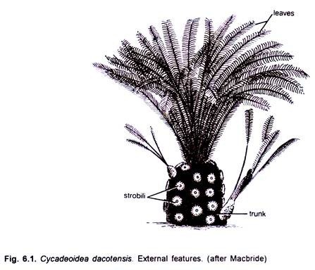
The Cycadeoid trunks were short, stout, spherical to sub-spherical (Figs. 6.1,6.2) and un-branched or branched. The trunks and leaves of many of its species show remarkable resemblance with those of living Cycads. Some of the species were short while others (Cycadeoidea jenneyana) attained a height of 3 to 3 .6 metres.
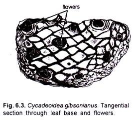
The trunk generally attained a diameter of about 50 cm, and had many, persistent, rhomboidal leaf bases (Figs. 6.2, 6.3). A compact crown of Cycad-like, large, pinnately compound leaves was present at the apex. The leaflets had many parallel veins.
3. Anatomy of Cycadeoidea:
The stem was roughly circular or oval in outline. It remained covered by heavy armour of leaf bases. The epidermis was not very distinct. The cortex was parenchymatous and possessed many mucilage canals and leaf traces. Many conjoint, collateral, open and endarch vascular bundles constituted the primary vasculature of the stem (Fig. 6.4). A large centrally located pith was present.
The xylem and the pholem have been studied in detail by Wieland (1906) (Fig. 6.5 A, C) Most of the tracheids were rectangular in shape. They were scalariform. The tracheids of protoxylem were spiral. The secondary xylem and the secondary phloem were traversed by secondary medullary rays, which were either uniseriate or bi-seriate. Cambium was clearly visible.
A leaf trace developed singly from the primary vascular strand. It divided into many mesarch strands upon entering into the cortex. At the place of its origin the leaf trace was C-shaped.
4. Reproductive Organs of Cycadeoidea:
The Bennettitalean reproductive organs are designated as “flowers “. The flower buds in the plants were present in the axil of leaf bases. As many on 500 flower buds were present on a single trunk in species such as Cycadeoidea dartonii (=Monanthesia dartonii).
In several species of Cycadeoidea all the flower buds were present on a trunk at almost the same stage of development. Some palaeobotanists believe that such a plant might have flowered only once during its lifetime. Except a few species (e.g. C. wielendii) the flowers in Cycadeoidea were bisexual.
Hermaphrodite flower developed on a short pedicel. They were surrounded by as many as one hundred bracts, which were hairy and protective (Fig. 6.6). Flowers in different species were of different size. In Cycadeoidea dartonii they attained a length of about 2 cm and a diameter of about 1.5 cm while in C. dacotensis each flower was about 8 cm long and 3 cm in diameter.
In C.dacotensis the lower two-third portion of the floral axis had about 100-150 bracts. A whorl of stamens was present above the bracts. Each stamen was pinnately branched (Fig. 6.7) and each pinna had a double row of purse-shaped sporangia. Each sporangium resembled with a synangium. A conical floral axis was present just above the whorl of stamens. The entire compact structure resembled with a strobilus.
5. Microsporophyll in Cycadeoidea:
According to Wieland (1906, 1916), the androecium or pollen-bearing region consisted of about 20 pinnate, microsporophyll’s. These were somewhat fixed or united at the base. Bean-shaped pollen capsules were arranged in two rows on each pinna of the sporophyll.
These microsporophyll’s remained folded round the gynoecium when young, but probably at maturity they expanded. Delevoryas (1963), however, opined that the microsporophyll’s never expanded.
He further concludes that synangia-bearing structures, described as pinnae by Wieland (1906), were similar to the trabeculae. These trabeculae established a connection between outer and inner walls of the androecium. Pollen capsules or synangia were borne along these trabeculae.
Several (20-30) pollen sacs or microsporangia were present in a pollen capsule or synangium. The wall of a synangium consisted of outer palisade-like, thick-walled cells followed by thin-walled layer and then a tapetum.
The tapetum was not clearly demarcated (Figs. 6.8, 6.9). The pollen grains were oval in shape and measured up to 68fi in length. Multicellular pollen grains in Cycadeoidea have been reported by Taylor (1973).
6. Gynoecium of Cycadeoidea:
The gynoecium receptacle was spherical or conical in shape. Hundreds of the stalked ovule along with an approximately equal number of inter-seminal scales were present on the receptacle (Fig. 6.10). Each ovules was about 1 mm in length. The integument of the ovule was fused with the nucellus, except at the apex.
The ovule was orthotropous with a long micropylar beak. A pollen chamber and a nucellar beak was present in each ovule (Fig. 6.11) . The seed was somewhat elongated or oval in shape and possessed two cotyledons (Fig. 6.12) . Crepet and Delevoryas (1972) reported a linear tetrad in the nucellar region of Cycadeoidea.


