In this article we will discuss about:- 1. Habit and Habitat of Equisetum 2. Structure of Equisetum 3. Reproduction 4. Life Cycle.
Habit and Habitat of Equisetum:
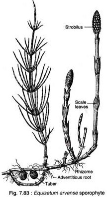
The plant body of Equisetum has an aerial part and an underground rhizome part (Fig. 7.83). The rhizome is perennial, horizontal, branched and creeping in nature. The aerial part is herbaceous and usually annual. Majority of the species are small with a size range in between 15 and 60 cm in height and 2.0 cm in diameter.
However some species grow up in higher heights [e.g., E. giganteum (13 m), E. telmateia (2 m); E. ramosissimum (4 m), though their stem are relatively thin (0.5-2.0 cm in diam.)] showing vine-like habit and climb over adjacent forest trees.
Equisetums generally grow in wet or damp habitats and are particularly common along the banks of streams or irrigation canals (E. debile, E. palustre). However, some species are adapted to xeric condition (e.g., Equisetum arvense). Some common Indian species are : E. arvense, E. debile, E. diffusum, E. ramosissimum.
Some species of Equisetum are indicators of the mineral content of the soil in which they grow. Some species accumulate gold (about 4.5 ounce per ton of dry wt.), thus they are considered as ‘gold indicator plants.
Hence these plants help in prospection/exploration for new ore deposits. In Equisetum, silica is deposited on the outer wall of the epidermal cells giving the characteristic rough feeling, thus it provides a protective covering against predators and pathogens.
Structure of Equisetum:
The Sporophyte:
The sporophytic plant body of Equisetum is differentiated into stem, roots and leaves (Fig. 7.83).
Stem:
The stem of Equisetum has two parts: perennial, underground, much-branched rhizome and an erect, usually annual aerial shoot. The branching is monopodial, shoots are differentiated into nodes and internodes.
In majority of the species, all the shoots are alike and chlorophyllous and some of them bear strobili at their apices (e.g., E. ramosissimum, E. debile). Sometimes shoot shows dimorphism (two types of shoots i.e., vegetative and fertile) e.g., E. arvense.
Some shoots are profusely branched, green (chlorophyllous) and purely vegetative. The others are fertile, unbranched, brownish in colour (achlorophyllous) and have terminal strobili.
The underground rhizome and the aerial axis appear to be articulated or jointed due to the presence of distinct nodes and internodes. Externally, the internodes have longitudinal ridges and furrows and, internally, they are hollow, tube-like structures. The ridges of the successive internodes alternate with each other and the leaves are normally of the same number as the ridges on the stem.
Internal Features of Stem:
In T.S., the stem of Equisetum appears wavy in outline with ridges and furrows (Fig. 7.84). The epidermal cell walls are thick, cuticularised and have a deposition of siliceous material.
Stomata are distributed only in the furrows between the ridges. A hypodermal sclerenchymatous zone is present below each ridge which may extend up to stele in E. giganteum. The cortex is differentiated into outer and inner regions.
The outer cortex is chlorenchymatous, while the inner cortex is made up of thin-walled parenchymatous cells. There is a large air cavity in the inner cortex corresponding to each furrow and alternating with the ridges, known as vallecular canal. These are schizolysigenous canals extending the entire length of internodes and form a distinct aerating system.
New leaves and branches of Equisetum are produced by the apical meristem, however, most of the length of the stem are due to the activity of intercalary meristem located just above each node. The activity of intercalary meristem causes rapid elongation of the inter- nodal region.
The stele is ectophloic siphonestele which is surrounded by an outer endodermal layer. An inner endodermis is also present in some species of Equisetum (e.g., E. sylvaticum). The endodermis is followed by a single-layered pericycle.
The vascular bundles are arranged in a ring which lies opposite to the ridges in position and alternate with the vallecular canals of the cortex. Vascular bundles are conjoint, collateral and closed. In the mature vascular bundle, protoxylum is disorganised to form a carinal cavity which lies opposite to the ridges.
The metaxylem tracheids (scalariform or reticulate) are present on both sides of the phloem. In some species vessels with reticulate perforations are reported. The central part of the internode of aerial shoot is occupied by a large pith cavity which is formed due to rapid elongation of the internodal region.
The vascular bundles remain unbranched until they reach the level of node. At the nodal region, each vascular bandle trifurcates (divided into three parts).
The middle branch of the trifurcation enters the leaf. Each lateral branch of the trifurcate bundle joins a lateral strand of an adjacent trifurcate bundle to form a vascuiar bundle of internode (Fig. 7.85). Thus the vascular bundles of internode alternate with those of internodes above and below.
In the nodal region, the xylem is extensively developed as a conspicuous circular ring. There are no vallecular or carinal canals at this level. In addition, a plate of pith tissue occurs at the node which separates one internode from another.
The internal structures of the shoot of Equisetum is peculiar because it shows xerophytic as well as hydrophytic features.
The xerophytic features are:
(i) Ridges and furrows in the stem,
(ii) Deposition of silica in the epidermal cells,
(iii) Sunken stomata,
(iv) sclerenchymatous hypodermis,
(v) Reduced and scaly leaves, and
(vi) photosynthetic tissue in the stem.
The hydrophytic characteristics on the other hand are (i) we 11-developed aerating system like carinal canal, vallecular canal and central pith cavity, and (ii) reduced vascular elements.
Root:
The primary root is ephemeral. The slender adventitious roots arise endogenously at the nodes of the stems. In T.S., the root shows epidermis, cortex and stele from periphery to the centre. The epidermis consists of elongated cells, with or without root hairs.
The cortex is extensive; cells of the outer cortex often have thick walls (sclerenchymatous) and those of the inner cortex are thinner parenchymatous. The stele is protostelic where the xylem is triarch or tetrarch, or, in smaller roots, may be diarch.
A large metaxylem element is present in the centre of the stele and the protoxylem strands lie around it. The space between the protoxylem groups is filled with phloem. There is no pith.
Leaves:
The leaves of Equisetum are small, simple, scale-like and isophyllous; they are attached at each node, united at least for a part of the length and thus form a sheath around the stem. The sheath has free and pointed teeth-like tips.
The number of leaves per node varies according to the species. The species with narrow stems have few leaves (e.g., 2-3 leaves in E. scirpoides) and those with thick stem have many leaves (e.g., up to 40 leaves in E. schaffneri).
The number of leaves at a node corresponds to the number of ridges on the internode below. The leaves do not perform any photosynthetic function and their main function is to provide protection to young buds at the node.
Reproduction in Equisetum:
Equisetum reproduces vegetatively and by means of spores.
i. Vegetative Reproduction:
The subterranean rhizomes of some species (e.g., E. telmateia, E. arvense) form tubers (Fig. 7.83) which, on separation from the parent plant, germinate to produce new sporophytic plants. The tubers develop due to irregular growth of some buds at the nodes of the rhizomes.
ii. Reproduction by Spores:
Spores are produced within the sporangia. The sporangia are borne on the sporangiophores which are aggregated into a compact structure termed strobilus or cone or sporangiferous spike.
Strobilus:
The strobilus are terminal in position and generally are borne terminally on the chlorophyllous vegetative shoot (Fig. 7.86A). However, they may be borne terminally on a strictly non- chlorophyllous axis (e.g., E. arvense).
The strobilus is composed of an axis with whorls of sporangiophores (Fig. 7.86B, C). Each sporangiophore is a stalked structure bearing a hexagonal peltate disc at its distal end (Fig. 7.86D). On the under surface of the sporangiophore disc 5-10 elongate, cylindrical hanging sporangia are borne near the periphery in a ring.
The flattened tips of the sporangiophores fit closely together which provide protection to the developing sporangia. The axis bears a ring-like outgrowth, the so-called annulus immediately below the whorls of sporangiophores which provide additional protection during early development.
The annulus has been interpreted as a rudimentary leaf sheath by some botanists, whereas others consider it to be sporangiophoric in nature as occasionally it bears small sporangia.
Development of Sporangium:
The mode of development of sporangium is eusporangiate, as it is not entirely formed from a single initial. Superficial cells adjacent to the original initial may also take part in the development of sporangium.
Sporangia are initiated in single superficial cell around the rim of the young sporangiophore. The periclvnal division of the sporangium initial forms an inner and an outer cell. The inner cell, by further divisions in various planes, gives rise to sporogenous tissue.
The outer cell, by periclinal and anticlinal divisions, gives rise to irregular tiers of cells, the inner tiers of which may transform into sporogenous tissue and the outer tiers become the future sporangial wall cells.
The innermost layer of the sporangial wall differentiates as the tapetum. The sporogenous cells separate from each other, round off and eventually transform into spore mother cell. All but the two outermost wall layers disorganise to form periplasmodial fluid.
However, not all of the sporogenous cells function as spore mother cells. Many of them degenerates to form a multinucleate nourishing substance for the spore mother cells. Each spore mother cell undergoes meiotic division (reductional division) and produces spore tetrad. All spores in a sporangium are of same size and shape i.e., homosporous.
Structure of Mature Sporangium:
The mature sporangium is an elongated saclike structure, attached to the inner side of the peltate disc of the sporangiophore (Fig. 7.86D). It is surrounded by a jacket layer which is composed of two layers of cells. The inner layer is generally compressed and the cells of the outer layer have helical thickenings which are involved in sporangial dehiscence.
Dehiscence of Sporangium:
At maturity, the strobilar axis elongates, as a result the sporangiophores become separated and exposed. Then the sporangium splits open by a longitudinal line due to the differential hygroscopic response of the wall cells.
Spores:
The spores are spherical and filled with densely packed chloroplasts. The spore wall is laminated and shows four concentrate layers. The innermost is the delicate intine, followed by thick exine, the middle cuticular layer and the outermost epispore or perispore. The intine (endospore) and exine (exospore) are the true walls of the spore.
The outer two layers i.e., cuticular layer and epispore are derived due to the disintegration of the nonfunctional spore mother cells and tapetal cells. At maturity, the epispore (the outermost layer) splits to produce four ribbon like bands or strips with flat spoon-like tips.
These bands are free from the spore wall except for a common point of attachment and remain tightly coiled around the spore wall until the sporangium is fully matured.
These are called elaters (Fig. 7.87A). The elaters are hygroscopic in nature. The spores remain moist at early stages of development, thus the elaters are spirally coiled round the spore. The spores dry out at maturity and consequently the elaters become uncoiled.
These uncoiled elaters become entangled with the elaters of other spores. Through these actions the elaters help in the dehiscence process and also the dispersal of spores in large groups from the sporangium.
The elaters of Equisetum are different from those of the bryophytes (Table 7.6).
Gametophyte Generation:
Equisetum is a homosporous pteridophyte. The haploid spores germinate to form gametophyte. The germination takes place immediately if the spores land on a suitable substratum. If the spores do not germinate immediately, their viability decrease significantly.
The spores swell up by absorbing water and shed their exine (Fig. 7.87B). The first division of the spore results in two unequal cells: a small and a large cell (Fig. 7.87C). The smaller cell elongates and forms the first rhizoid. The larger cell divides irregularly to produce the prothallus. The prevailing environmental conditions determine the size and shape of the prothallus.
If a large number of spores are developed together within a limited space, then the prothalli formed are of thin filamentous type. But a relatively thick and cushion-shaped prothalli are formed from sparsely germinating spores. Mature gametophytic plants may range in size from a few millimeters up to 3 centimeters e.g., E. debile) in diameter.
They are dorsiventral and consist of a basal non-chlorophyllous cushion-like portion from which a number of erect chlorophyllous muticellular lobes develop upwards. Unicellular rhizoids are formed from the basal cells of cushion (Fig. 7.87D).
The prothallus bears sex organs and reproduces by means of sexual method.
Sexuality in Equisetum:
The gametophytic plant body bears sex organs i.e., antheridium (male) and archegonium (female). The gametophyte are basically bisexual (homothallic) i.e., they bear both male and female sex organs (Fig. 7.87D). Although, some unisexual (dioecious) members are also reported (Fig. 7.87E, F). Some are initially unisexual and then become bisexual.
This early sex determination appears to be related to the environmental conditions viz., temperature, light, humidity and the supply of nutrients as well. Ducket (1977), in order to explore the sexuality in Equisetum, observed that some of the fragments of male gametophyte remained male throughout the successive subcultures under laboratory conditions.
Some other fragments produced archegonia, which subsequently bore antheridia in increasing numbers. This phenomenon supports the contention that Equisetum gametophytes are potentially bisexual. However, Hanke (1969) observed that gametophyte of Equisetum bogotense were unisexual (bearing antheridia) and never change to bisexual type.
However, the initial male gametophyte of E. ramosissimum, E. variegatum and E. bogotense never became bisexual.
Schratz (1928) observed that 50% spores germinate to produce male gametophytes, while the remaining 50% spores produce female gametophytes though they do not loose their male potentiality (i.e., antheridia develop later if fertilisation fails). He termed this as ‘incipient heterospory’.
A study of sexuality based on enzymatic analysis revealed the intragametophytic self- fertilisation in E. arvense.
Equisetum is homosporous and, therefore, definite sex-determining mechanism is absent. But, the sexuality demonstrated by some of the members appears to be related to environmental factors. Therefore, it is termed as environmental sex determination.
Sex Organs of Equisetum:
i. Antheridium:
In monoecious species, antheridia develop later than archegonia. They are of two types — projecting type and embedded type. Antheridia first appear on the lobes of the gametophyte (Fig. 7.87D). The periclinal division of the superficial antheridial initial gives rise to jacket initial and an androgonial cell (Fig. 7.88A, B).
The jacket initial divides anticlinally to form a single-layered jacket. The repeated divisions of androgonial cells form numerous cells which, on metamorphosis, produce spermatids/antherozoids (Fig. 7.88C-E). The antherozoids escape through a pore created by the separation of the apical jacket cell.
The apical part of the antherozoid is spirally coiled, whereas the lower part is, to some extent, expanded (Fig. 7.88F). Each antherozoid has about 120 flagella attached to the anterior end.
ii. Archegonium:
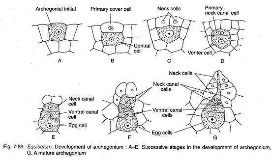
Any superficial cell in the marginal meristem acts as an archegonial initial which undergoes periclinal division to form a primary cover cell and an inner central cell (Fig. 7.89A, B). The cover cell, by two vertical divisions at right angle to each other, forms a neck (Fig. 7.89C). The central cell divides transversely to form a primary neck canal cell and a venter cell (Fig. 7.89D).
Two neck canal cells are produced from the primary neck canal cell. While, the venter cell, by a transverse division, forms the ventral canal cell and an egg (Fig. 7.89E).
At maturity, an archegonium has a projecting neck comprising of three to four tiers of neck cells arranged in four rows, two neck canal cells of unequal size, a ventral canal cell, and an egg at the base of the embedded venter (Fig. 7.89F-G). The archegonia are confined to cushion region in- between the aerial lobes (Fig. 7.87D).
Fertilisation:
Water is essential for fertilisation. The gametophyte must be covered with a thin layer of water in which the motile antherozoides swim to the archegonia. The neck canal cells and ventral canal cell of the archegonia disintegrate to form a passage for the entry of antherozoids.
Many antherozoids pass through the canal of the archegonium but only one of them fuses with the egg. Thus diploid zygote is formed. Generally more than one archegonia are fertilised in a prothallus.
Embryo (The New Sporophyte):
The embryo is the mother cell of the next sporophytic generation. Unlike most pteridophytes, several sporophytes develop on the same prothallus. The first division of the zygote is transverse. This results in an upper epibasal cell and lower hypobasal cell. The embryo is therefore exoscopic (where the apical cell is duacted outward i.e., towards the neck of the archegonium) in polarity.
No suspensor is formed in Equisetum. The epibasal and hypobasal cells then divide at right angles to the oogonial wall, and as a result a tour-celled quadrant stage is established (Fig. 7.90A). All the four cells of the quadrant are of different size and shape.
The four-celled embryo undergoes subsequent divisions and the future shoot apex originate from the largest cell and leaf initials from the remaining cells of one quadrant of the epibasal hemisphere.
One cell of the epibasal quadrant and a portion of the adjacent quadrant of the hypobasal region contribute to the development of root. The first root develops from one of the epibasal quadrants and a portion of the adjacent hypobasal quadrant. The shoot grows rapidly.
Later the root grows directly downward and penetrate the gametophytic tissue to reach the soil or substratum (Fig. 7.90B, C). A number of such sporophytes may develop from a large mature gametophyte if more than one egg is fertilised (Fig. 7.90D).


