In this article we will discuss about:- 1. Salient Features of Psilotum 2. Reproduction in Psilotum 3. Phylogeny.
Salient Features of Psilotum:
i. The sporophytes are dichotomously branched with an underground rhizome and upright branches.
ii. The upright branches are leafless.
iii. Rhizoids present instead of roots.
iv. Stem have a relatively simple vascular cylinder.
v. The sporangia are borne in groups (trilocular) and form synangia.
vi. Spores produced are all alike (homosporous).
vii. The development of gametophyte is exosporic and form monoecious subterranian gametophyte.
viii. The development of embryo is exoscopic.
Sporophyte:
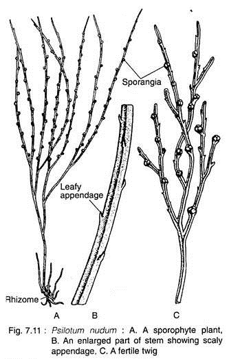
The plant body of Psilotum is sporophytic branched rhizome system and dichotomously branched, slender, upright, green aerial systems (Fig. 7.11 A) that bears small appendages and synangia (singular: synangium).
Aerial Stem:
Any one of the rhizome tips may turn upward and undergo several dichotomies to give rise to a green aerial shoot (Fig. 7.11 A). The aerial shoots are slender, generally erect but may be pendent in epiphytes (P. flaccidum). They are perennial and become shrubby by repeated dichotomies and sometimes attain a height up to one meter.
The aerial axis may be cylindrical at base, furrowed in the upper parts (wavy in C.S.), but somewhat flattened with three longitudinal ridges (triangular in C.S.) at the top. The basal part of the aerial axes is smooth but the distal part bears small, scaly appendages and synangia (Fig. 7.1 IB, C).
The aerial stems are photosynthetic and the general appearance is xerophytic although the plants grow in very moist environment.
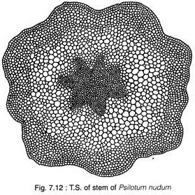
In T.S., the aerial system is covered by an epidermis, followed by extensive cortical areas, single-layered endodermis and stele. The stele is siphonostelic in the basal part which becomes actinostelic in the younger branches (Fig. 7.12 & 7.13).
The epidermis is single-layered, in which the outer tangential cell walls are heavily cutinised and covered by a layer of cuticle. The epidermis is broken regularly by stomata (Fig. 7.12). The stomata of Psilotum do not have subsidiary cells.
The cortex is extensive and is divisible into three regions (Fig. 7.13). The outer portion (chlorenchymatous) directly beneath the epidermis consists of elongated, lobed chlorophyllous cells with intercellular spaces. This region is two- to five-layered thick and the cells are full of chloroplastids and starch grains.
Internal to this zone, there is a middle cortex of 4 to 5 layers of vertically elongated thick-walled sclerenchymatous cells without intercellular spaces. The walls of these cells apparently become lignified in the lower portion of the aerial stem.
These tissues provide mechanical support to the plant. Further inwards, there is a broader zone of parenchymatous cells (the inner cortex), the cell walls of which become thinner and thinner towards the centre. These cells are without intercellular space but contain more starch grains.
The cortical tissue is bounded inwardly by a single-layered endodermis whose vertically elongated cells have a conspicuous casparian bands on the radial and end walls.
The centre of the stem is occupied by a ridged or flattened cylinder of primary xylem with protoxylem elements at the tip of each ridge. This cylinder may have as many as ten ridges near the transition region from rhizome to aerial stem. Fewer ridges are present in the more distal parts of the aerial stem.
A single-layered parenchymatous pericycle is present just below the endodermal layer. The phloem is internal to the pericycle and located between the ridges of the xylem.
At the extreme base, the stem is protostelic (actinostelic). In the middle portion the stele is siphonostelic as the centre of the xylem is occupied by a patch of elongated sclerenchymatous cells (sclerotic pith).
Rhizome:
The basal subterranean branched rhizome is generally hidden beneath the soil or humus. It bears numerous rhizoids, instead of roots, which perform the functions of absorption and ancho- rage.
In T. S., the rhizome reveals an outermost epidermis, cortex, endodermis, pericycle and stele (Fig. 7.14). The epidermis is indistinct and gives rise to 2-celled rhizoid. The cortex is extensive, parenchymatous and differentiated into outer, middle and inner layers.
The outer cortical layer is characterised by the presence of intracellular endophytic mycorrhiza. The cells of middle cortex are large with starch grains, while the cells of the inner cortex are often dark brown in colour because of the presence of phlobaphene (an oxidative product of tannins).
The stele is protostelic (haplostele or actinostele) and is surrounded by a well-defined endodermal layer with conspicuous casparian bands on the radial walls.
Appendages:
These are small scale-like structures helically arranged on the upper part of the aerialsystem. Internally, the appendage is composed of parenchymatous photosynthetic cells, bounded by a single-layered cutinised epidermis. There is no stomata in the appendages. There is no vascular trace in the appendages of P. nudum, although minute leaf traces originate from stele in P. flaccidum which fade out in cortex.
Reproduction in Psilotum:
The Psilotum reproduces vegetatively as well as by spores.
i. Vegetative Reproduction:
The sporophyte as well as gametophyte of Psilotum (e.g., P. nudum) propagate vegetatively through the production of Gemmae (Fig. 7.15). They are small, multicellular and ovoid structures developing on surface of rhizome (in sporophytic plant body) or prothallus (in the gametophyte).
After detachment from the parent body, gemmae of sporophyte may germinate to form a subterranean shoot, while the gemmae of prothallus, on germination, form a new prothallus.
ii. Reproduction by Spores:
Spore-Producing Structure:
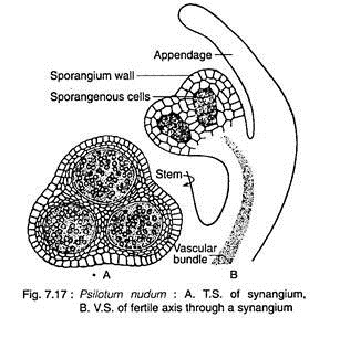
At maturity, many of the dichotomously branched aerial shoots become fertile and produce trilocular sporangia known as synangia (Fig. 7.11C & 7.16A, B). The mature synangium is generally a three-lobed structure (Fig. 7.17A) and each lobe of the synangium corresponds to a sporangium.
The synangia located at the tip of very short axis, measuring 1-2 mm in diameter and closely associated with a forked, foliar appendage (Fig. 7.1 7B). At maturity, the synangium exhibits loculicidal dehiscence.
Development of Sporangium:
The mode of development of each sporangium of the synangium of Psilotum nudum is of eusporangiate type. Each sporangium develops from a group of superficial initial cells which divide periclinally to produce primary wall initials and primary sporogenous cells.
A sporangial wall of four or five layers is produced through repeated periclinal and anticlinal divisions of the primary wall initials. The sporogenous tissue is produced from repeated divisions of primary sporogenous cells in various planes.
Further development of sporangenous tissue gives rise to spore mother cells and numerous spores are produced as a result of meiosis of the spore mother cells. The spores are of equal size and shape (i.e., homosporous), bilaterally symmetrical, colourless and kidney-shaped with monolete aperture.
Gametophyte:
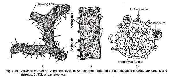
The mature gametophyte shows a striking similarity with a piece of sporophytic rhizome (Fig. 7.18A). It grows as saprophyte with an associated fungus.
Spores germinate exosporically to form the gametophyte. The mature gametophytes are brown, cylindrical, subterranean, radially symmetrical and usually dichotomously branched, but may sometimes become irregular. The surface of the gametophyte is covered by long unicellular, brownish rhizoids. The gametophyte grows by means of apical meristem (Fig. 7.18A).
In T.S., the gametophyte reveals cutinised peripheral cells which encloses many-layered thin-walled parenchymatous cells (Fig. 7.1 8C). Many of the internal parenchyma cells are filled with the hyphi of a symbiotic fungus. It has been observed that in the gametophyte of tetraploid P. nudum, the centre is occupied by xylem with annular, scalariform and reticulate tracheids, surrounded by phloem and an endo- dermis.
Thus, Psilotum is the only plant in the plant kingdom where the vascular tissues develop in the gametophytic generation. The external resemblance of the sporophytic rhizome and gametophyte, coupled with the presence of vascular tissue in the gametophyte, supported the homologous theory on the origin of alternation of generations.
Sex Organs:
The gametophytes of Psilotum are monoecious (i.e., homothallic). Sex organs i.e., antheridia and archegania, are superficial and scattered over the surface of the gametophyte (Fig. 7.18A-C). Generally, antheridia are more in number than archegonia.
Antheridium:
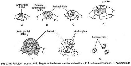
The antheridium develops from a single superficial cell (antheridial initial) of the prothallus. The periclinal division of the superficial cell produces an outer jacket initial and an inner primary androgonial cell (Fig. 7.19A, B).
The outer jacket initial undergoes repeated anticlinal divisions and forms a single-layered jacket. The inner primary androgonial cell divides in various planes and produces a mass of developing androgonial cells, the last generation of which are the androcytes (Fig. 7.1 9C- F).
At this stage, the antheridium projects above surface of the prothallus as a minute protuberance. Each androcyte eventually becomes a spirally-coiled, multiflagellate antherozoid (Fig. 7.19G) and escapes by the disintegration of the opercular cell.
Archegonium:
The archegonium is also developed from a single superficial cell (archegonial initial) of the prothallus. The initial cell undergoes periclinal division to form an outer primary cover cell and an inner central cell (Fig. 7.20A, B). The anticlinal divisions followed by periclinal divisions of the outer cover cell produces a long projecting neck arranged in four vertical rows of cells with four to six cells in each row.
The central cell divides transversely to form an upper primary neck canal cell and a lower primary venter cell. The nucleus of the primary neck canal cell divides to form two neck canal nuclei and generally this division is not accompanied by wall formation resulting into a binucleate neck canal cell. The primary venter cell divides transversely to produce a large egg and a small ventral canal cell (Fig. 7.20C-F).
Fertilisation:
At maturity, the cell wall of the lower tier of neck cells becomes thick walled and cutinized. The apical tier, however, breaks away in presence of water and the mucilagenous contents of the neck cells are released. Thus, a free passage is formed for the entry of the antherozoids.
Fertilisation is accomplished by the union of a multiflagellate sperm and egg, resulting in the formation of a diploid zygoie.
Embryo (New Sporophyte):
The diploid zygote is the mother cell of the sporophytic generation. The first division of the zygote is transverse, forming an outer epibasai cell (directed towards the neck of the archegonium) and an inner hypobasal cell (directed towards the base of the archegonium) (Fig. 6.21 A-D).
The apical epibasai cell ultimately gives rise to the sporophytic branch system (aerial and underground), while the lower hypobasal cell produces the foot. This type of embryogeny where the shoot forming apical cell is directed outward (towards the neck of the archegonium) is called exoscopic mode of embryo development.
The foot anchors the young sporophyte securely to the gametophyte and absorbs nutrients until the sporophyte becomes pysiologically independent.
Fig. 7.22 depicts the life-cycle of Psilotum.
Phylogeny of Psilotum:
Affinity with Ferns:
Bierhorst (1971) placed Psilotum along with Tmesipteris within Filicopsida primarily on the basis of similarities with some ferns like Gleichenia, Stromatopteris, etc., by the following characteristics:
1. Axial nature of gametophytes.
2. Superficial position of antheridia on the prothallus.
3. Exoscopic type of embryogeny.
4. Development of sporangial structure.
5. Mutiflagellated spermatozoids.
However, this has been highly criticised by several pteridologists. Except for the above characteristics Psitolates (Psilotum and Tmesipteris) differ from ferns in almost all principal characteristic features.
Moreover, Psilotales are root-less, leaf-less plants (Psilotum) or with microphyllous leaves (Tmesipteris) showing eusporangiate mode of sporangial development, while Gleichenia, Stromatopteris have well-developed sporophyte with megaphyllous leaves and roots, showing lepto- sporangiate mode of sporangial development.
Affinity with Rhyniopsida:
Psilotum and Tmesipteris resemble the earliest known Silurian-Devonian vascular plants — Rhyniopsida — in the following characteristic features:
1. The sporophytes are dichotomously branched with subterranean rhizome and upright branches.
2. Absence of roots and sporophytic generation bears rhizoids.
3. The branches are leafless e.g. Psilotum.
4. Sporangia multilayered, in rare instance are terminal.
5. Terminal branch shows protostelic configuration.
The above similarities suggest that Psilotales (Psilotum and Tmesipteris) is the most primitive extant group among the vascular plants. Moreover, biochemical characteristics of Psilotum and Tmesipteris have strengthened the above consideration. Tse and Towers (1967) isolated an unique phenolic compound, Psilotin, from the two genera which is not found in other groups of pteridophytes.
Hence Psilotum and Tmesipteris constitute a natural group in Pteridophyta and should be placed at the beginning of pteridophyte classification next to early vascular plants and before Lycopsida.
It has been observed through Gel electrophoresis that histone protein profiles of Psilotum are more or less identical with the histones of mosses. Thus it suggests the primitiveness of Psilotum. Hence Psilotum must be treated as a primitive vascular plant.
