In this article we will discuss about:- 1. Habit and Habitat of Pteridium 2. External Features of Pteridium 3. Internal Structure 4. Reproduction.
Habit and Habitat of Pteridium:
Pteridium a monotypic genus (represented by a single species P. aquilinum; Reimers, 1954), is one of the oldest known fern (fossil records over 55 million years old have been found). It is cosmopolitan in distribution and occurs in both temperate and tropical regions of the world except the Arctic zone and the temperate South America.
In India it is commonly found at an elevation of 2000 to 3000 feet. It is one of the primary coloniser of the land and once it is established, it does not allow other ferns to grow in that area.
External Features of Pteridium:
The mature plant body is sporophytic and can be differentiated into rhizome, roots and leaves (Fig. 1A).
1. Rhizome:
It grows horizontally, 8-12 cm. beneath the surface of the soil. It is long, slender and branched. The branching is first dichotomous but lateral unequal dichotomies are seen. Watt (1940) reported three types of branches (long shoots, intermediate shoots and short shoots) developing from the rhizome. Rhizome is covered with fine, multicellular, pale brown hairs called ramenta.
2. Roots:
The primary root of the young sporophyte is short lived. In mature sporophyte, adventitious roots arise in acropetal succession at irregular intervals from the lower surface of the rhizome. These roots are slender, spraingly branched and endogenous in origin.
3. Leaves:
The rhizome is differentiated into nodes and internodes. On nodes, the young leaves appear alternately on the dorsal surface. Young leaves are circinately coiled and are separated by long internodes. In older plants, the leaves seem to be restricted on the short and thick branches.
Leaves (also called fronds) are very long (0.5 to 3.5 meter) and their petiole is as long as the lamina. The petiole portion which extends into the lamina is called rachis. The lower branches of the rachis are longer and gradually decrease towards the apex of the lamina (deltoid lamina).
Each leaf is tripinnately compound. Each pinna is sessile. A nectary is present at its base. It has a distinct mid rib that gives out lateral branches. A prominent mid rib is present in each pinnule. The vein of the pinnule runs upward and branches dichotomously.
In mature leaves many sori are present on the abaxial (lower) surface of the pinnule. These sori are continuous for a considerable distance along the ventral surface of the margins of the pinnules. This type of sorus (a group of sporangia) is called coenosorus or continuous linear sorus. Sorus bearing leaves are called sporophylls. In each sorus many sporangia are present.
Internal Structure of Pteridium:
1. Transverse Section (T.S.) of Rhizome:
It can be differentiated into three zones: epidermis, cortex and stele.
(i) Epidermis:
It is the outermost layer. Its cells are narrow, thin or thick with thickened brown outer walls.
(ii) Cortex:
Epidermis is followed by cortex. Many layers just below the epidermis are thick walled (scelerenchymatous) and are called hypodermis. Rest of the space inside the hypodermis is occupied by thin walled (parenchymatous) ground tissue in which are embedded many meristeles. In between the meristeles two patches of sclerenchymatous tissue are present. One patch lies above and one lies below the inner meristeles (fig- 2A).
(iii) Stele:
In mature rhizome of the Pteridium, the stele is polycyclic [according to Webster (1970) the stele is perforated, amphiphloic, siphonostele with medullary bundles]. It consists of two concentric cylinders of vascular bundles. The outer and inner cylinders of vascular tissues are separated by two patches of sclerenchymatous tissue. The outer ring of vascular tissue is much dissected dictyostele and consists of many small meristeles.
The inner ring consists of medullary or accessory meristeles which are usually two in number (Fig. 2A). Each meristele of the outer and inner ring is surrounded by a single layer of endodermis and one or two layers of pericycle (Fig. 2B). Just below the pericycle is first formed phloem known as protophloem.
The inner portion of the phloem is called metaphloem. It surrounds the xylem and consists of large sieve tubes. Companion cells are absent. The xylem is mesarch, the protoxylem is surrounded on all the sides by metaxylem. In between the protoxylem and metaxylem small parenchymatous cells are present.
2. Transverse Section of Petiole:
It can be differentiated epidermis, cortex and stele.
(i) Epidermis:
It is the outermost, protective single layer of cells.
(ii) Cortex:
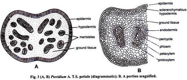
Epidermis is followed by a few layered thick sclerenchymatous hypodermis, which, in turn, encircles the parenchymatous ground tissue.
(iii) Stele:
Embedded in the ground tissue are haphazardly embedded leaf traces or vascular strands or meristeles. The number of leaf traces varies within the size and age of the leaf. The larger petioles have several haphazardly arranged vascular strands while smaller petioles have vascular strands arranged in the shape of a much convoluted horse-shoe open towards the upper side of the petiole. The structure of vascular strands resembles with that of meristeles of rhizome.
3. Anatomy of the Root:
It is circular in outline and can be differentiated into piliferous layer or epidermis, cortex and stele (Fig. 4).
(i) Epidermis:
It is the outer most single layer. Some of its cells grow out to form unicellular root hairs.
(ii) Cortex:
It is differentiated into two zones: The outer zone consists of thin walled parenchymatous cells and the inner zone consists of thick walled, sclerenchymatous, lignified cells. Inner zone completely surrounds the stele. It provides mechanical support to the root (fig. 4). The innermost layer of the cortex is endodermis. It has the casparian strips on its radial walls. It is followed by unilayered or bilayeredpericycle.
(iii) Stele:
A protostele is present in the centre of the root. It is diarch and exarch. It has a xylem plate in the centre with two protoxylem groups, one at each end. Outside the xylem lie two groups of phloem arranged on either side of the xylem plate.
4. Transverse Section of Pinnule or Lamina:
It can be differentiated into epidermis, mesophyll tissue and vascular bundles (Fig. 5).
(i) Epidermis:
The pinnule is bounded on both sides by single layered upper and lower epidermis. Stomata are confined to lower epidermis.
(ii) Mesophyll tissue:
It is differentiated into palisade tissue and spongy parenchyma. The palisade tissue is one to three cells in thickness and is present just below the upper epidermis. The space between the palisade tissue and lower epidermis is filled with spongy parenchyma with well-developed intercellular spaces. All the cells of mesophyll tissue contain abundant chloroplast.
(iii) Vascular Bundles:
The vascular bundles are embedded in the mesophyll tissue. These are spherical in shape, concentric collateral and are surrounded by a distinct endodermal layer.
5. Transverse Section (T.S.) of Fertile Leaflet or Pinnule:
Transverse section of fertile pinnule is similar to that of sterile pinnule with the exception that fertile pinnule bears many stalked sporangia (coenosorus) along the lower margins. The coenosorus is surrounded by two well-formed indusial lips between which lies the receptacle.
The outer indusial lip is well developed. It is formed by the reflexed margin of the pinnule. It overlaps the coenocorus and its sporangia. It is called false indusium. The inner indusial lip is ill developed. It consists of a sheet one cell in thickness and lies adjacent to receptacle. It is the outgrowth of placenta and is called true indusium (Fig. 6 A, B).
 Reproduction in Pteridium:
Pteridium reproduces both vegetatively and sexually.
A. Vegetative reproduction:
It takes places by death and decay of the older portion of the underground rhizome. The rhizome is dichotomously branched and grows indefinitely. When the death and decay process reaches a point of dichotomy, the two branches separate and behave as an independent individual. This is the most common method of multiplication. Due to this region the plants are found in gregarious (glowing close together, but not matted) habit.
B. Sexual Reproduction:
1. Sporophytic Phase:
Spore producing organs:
Pteridium is homosporous i.e. It produces only one type of spores. These spores are produced inside sporangia, which in turn are grouped together to form sorus (pi. sori). Many sporangia are present in each sorus. The sorus is continuous along the under margin of the pinnules and this type of sorus is known as continuous linear sorus (coenosorus).
Structure of Mature Sporangium:
Sporangium is oval, biconvex lens shaped and can be differentiated into stalk and capsule. The stalk is long slender and mode up of three vertical rows of cells. The capsule wall is composed single layer of thin walled cells called jacket.
Few thin walled cells form the stomium (Fig. 7A, B). It provides an easy cleavage when the sporangiums dehisce. Few cells modify to form annulus. Capsule wall encloses 8-16 spore mother cells which change into 32-64 spores after the reduction division.
Development of Sporangium:
The development of sporangium is leptosporangiate type. A marginal cell functions as sporangial initial. It projects above the receptacular surface in the form of papilla. It divides by a transverse or oblique transverse wall into two cells the outer cell and inner or lower basal cell (Fig. 8 A, B). The basal cell takes no further part in sporangial development.
The entire sporangium develops from the outer cell. It divides by three intersecting diagonal walls in such a way that a three sided pyramidal apical cell is formed (Fig. 8 C-E). It cuts off segments along its three sides. The lower segments form the stalk while the upper three segments form a portion of the sporangial wall.
The pyramidal apical cell divides by periclinal division to form outer jacket initial or cap cell and inner central cell or primary sporogenous cell or archesporial cell (Fig. 8F). The jacket initial divides anticlinally to form single layered sporangial wall (Fig. 8 G). Archesporial cell cuts four tapetal initials which divide both periclinally and anticlinally to form a two layered tapetum.
The sporogenous cell later divides to form 8-16 spore mother cells (Fig. 8H) which undergo meiosis to produce 32-64 spores. The tapetal cells break down and form a nutritious fluid. The spore mother cells float in it.
On maturity the indusium dries exposing the sorus. In dry weather, the cells of the annulus loose water from their outer walls. The thin outer wall bends inwards and becomes concave. It brings about the unequal tension of the cell walls. Due to these changes the annulus tends to straighten and in doing so it tears the sporangial wall from the stomium.
Some of the spores are blown away by wind but the greater mass of the spores is held in the upper open end of the sporangium. Sudden release of tension in the annular cells results in the snap back of annulus again in its original position ejecting the spores forcibly from capsule with a jerk (Fig. 9 A-C).
2. Gametophytic Phase:
Structure of spore:
Spores are the first cells of the gametophytic generation. They are haploid, uninucleate, bluntly or roughly triangular, about 0.3 mm in diameter and have a distinct triradiate mark. All the spores are of one kind (homosporous). They are double layered. The outer wall is known as exine and inner intine. Exine is much thicker than intine and may be variously sculptured. The spore germinates to form a prothallus (gametophyte).
Development of Prothallus:
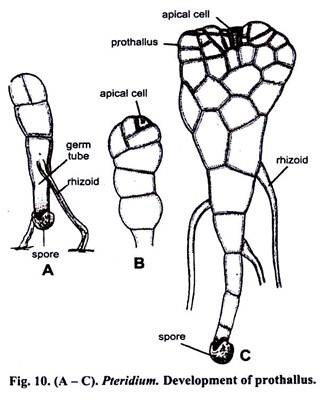
Under suitable conditions the spore germinates. Exine ruptures and initine comes out in the form of a germ tube (Fig. 10A). The germ tube elongates and divides by a transverse wall into a lower smaller cell and upper larger cell.
The smaller cell grows into the soil forming the first rhizoid. The larger cell which contains many chloroplasts divides by a few transverse divisions and ultimately results in 3 to 6 called filament. After the transverse divisions are over, two oblique walls are laid down in the terminal cell of the filament and thus a two sided apical cell is formed (Fig. 10B).
There is no development in the other cells of the filament. The apical cell cuts off segments on its right and left alternately and ultimately forms a green, multicellular one celled thick young prothallus. The apical cell soon gives rise to many marginal initials which help in further growth of gametophyte.
Mature prothallus:
On maturity the prothallus becomes heart shaped and shows dorsiventral symmetry. It is 3 mm to 8 mm in diameter, many celled thick in the centre but only one cell thick at the margins. A deep notch is present at the anterior side in which growing point is situated.
It has a distinct anterior and posterior side. Posterior side is narrower and many unicellular rhizoids arise from its ventral surface. Main functions of the rhizoids are to attach the prothallus on the soil and to absorb water and mineral nutrients.
In mature prothallus, sex organs develop on the ventral surface. Prothalli are monoecious and protandrous i.e., antheridia develop earlier than archegonia. Antheridia are produced towards the lower side near the rhizoids while the archegonia are produced towards the notch. Sex organs are sessile and remain partly embedded in the tissue of the prothallus (Fig. 11).
Structure of mature antheridium:
Sessile antheridium is a globular structure. It consists of a wall which is made up of three tubular cells: opercular or cap cell, first ring cell and second ring cell (Fig. 12 G). The cap cell allows the release of antherozoids during dehiscence of antheridium.
The first ring cell or funnel cell forms the base of the antheridium. The second ring cell or circular cell forms the middle portion of the antheridial wall. The cap cell forms a lid which allows the antherozoids to escape at the time of dehiscence of antheridium.
A fourth cell or the basal cell is also present at the base of antheridium. It is regarded as the single celled stalk of the antheridium. The wall of the antheridium encloses 32 uninucleate, multiflagellate antherozoids.
Development of antheridium:
It develops from a superficial cell on the lower surface of prothallus (near the rhizoids). It divides by a transverse division to form a basal cell and upper antheridial initial (Fig. 12 A, B). The antheridial initial divides by a transverse wall.
This wall undergoes curvature. It looks like a funnel and almost touches the wall of the basal cell (Fig. 12 C, D). It results in the formation of upper dome cell or central cell and a lower first ring cell. The dome cell or central cell again divides by a curved wall to form an outer jacket cell and a central primary androgonial cell (Fig. 12 E).
The jacket cell divides periclinally to form upper cap or cover cell and second ring cell. Primary androgonial cell divides and redivides to form 16 sperm mother cells which divide again to form 32 androcytes. The protoplast of each androcyte metamorphosis into spirally coiled multiflagellate antherozoid or spermatozoid (Fig. 12 F-I).
Structure of Mature Archegonium:
Mature archegonium can be differentiated into elongated neck and basal swollen venter. Neck is slightly curved and projected from the surface of the prothallus towards the surface moisture of the soil and points towards the mature antheridia.
It is made up of four vertical rows of sterile neck cells (Fig. 13 E). Each row is 4 cells in height. Neck cells enclose a binucleate neck canal cell (Fig. 13E). There is no venter wall. The egg and single ventral canal cell remain surrounded by the cells of the prothallus.
Development of archegonium:
It develops near the notch of the prothallus from an archegonial initial (Fig. 13 A). It divides by a transverse division to form an upper primary cover cell and a lower cell (Fig. 13B). The lower cell again divides by a transverse division.
In this way three cells are formed (Fig. 13C) Upper primary cover cell, middle central cell and lower basal cell. Upper primary cover cell divides by two vertical walls at right
angles to each other, it results in the formation of four neck initials. The neck initials divide by transverse division to form four celled high neck of the archegonium.
The central cell divides transversely and forms three cells-an egg, a venter canal cell and a neck canal cell. The nucleus of the neck canal cell further divides and a binucleate neck canal cell is formed (wall formation is not accompanied. Fig. 13 D, E).
Fertilization:
Water is essential for fertilization. When the mature antheridium comes in contact with external water, jacket layer swells. The walls of the androgonial cells also disorganise to form a mucilaginous mass.
It results in an increase in the pressure inside the antheridium which pushes apart the cover cell of the antherdium and antherozoids are liberated within the thin membrane (this membrane is a portion of the wall of the androcyte). The membrane soon dissolves in water and multiflagellate antherozoids are liberated in water.
Simultaneously, in mature archegonium the neck canal cell and the venter canal cell also disintegrate and get converted into mucilage mass. It swells due to absorption of water, exerts a pressure on the neck and causes it open. The mucilage fills the neck canal and is partially extruded at the mouth of archegonium (Fig. 13F).
It contains chemical substances like malic acid which attract antherozoids. Many antherozoids are attracted chemotactically towards the archegonium and enter the neck (Fig. 13G) but only one fertilizes the egg. The female and male nuclei fuse to form a diploid structure called zygote.
Development of Embryo:
After fertilization zygote increases in size and completely occupies the cavity of the venter (Fig. 14A). It divides by a vertical wall parallel to long axis of archegonium forming two cells. The cell which is towards the apex of the prothallus is known as epibasal cell and the cell towards the base of prothallus is called hypobasal cell.
The wall separating the two halves are known as basal wall. The second division is vertical (at right angle to the basal wall) and it results in the formation of 4-celled embryo (quadrant stage). The third division is transverse (to the basal wall) and it results in the formation of 8-celled embryo (octant stage).
The anterior quadrant of the octant forms the stem and leaf (superior forming stem and interior, the leaf), the posterior quadrant gives rise to foot and root (Fig. 14A-G). The young sporophyte is dependent upon the gametophyte for its nutrition.
It draws its nutrition with the help of foot. But as soon as two or three leaves develop, the prothallus begins to exhaust and the young sporophyte becomes an independent plant, attached to the soil by its own roots (Figs. 14G, 15).


