In this article we will discuss about:- 1. Occurrence of Sargassum 2. Plant Body of Sargassum 3. Internal Structure 4. Features 5. Features 6. Reproduction 7. Life Cycle Pattern.
Occurrence of Sargassum:
The genus Sargassum (Spanish sargazo, seaweed) is represented by about 150 species, out of which 1 6 species are found in India. It is found in temperate, subtropical and tropical regions of both northern and southern hemispheres. It is very common in Africa, South America, Australia etc.
In West Africa, a part of Atlantic Ocean becomes densely occupied by Sargassum and the region is called as ‘Sargasso sea’. Sargassum filipendula, a free-floating large kelp found in the Sargasso Sea, was discovered by Columbus in 1492, as the ships were held fast by the sea weeds. In India it is found in Porbandar, Bombay, Okha, Lakshadweep Island etc.
Plant Body of Sargassum:
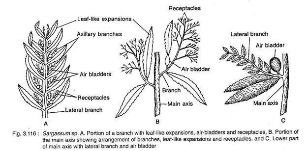
The plant body is diploid (2n), erect and branched thallus (Fig. 3.116). The thallus is differentiated into a basal holdfast and an expanded, leafy, cylindrical main axis.
The holdfast is discoid and serves the function of anchorage with the substratum. The main axis is generally of 10 to 50 cm in length. It is erect, flattened or cylindrical structure. The main axis bears many primary laterals arranged spirally in a phyllotaxy of 2/5.
Due to its unlimited growth, the primary laterals are also called long shoots. The main axis and primary laterals (long shoots) bear flat’ expanded structures, called secondary laterals or leaves.
The leaves are flat, simple structures with distinct midrib and dentate, serrate or entire margins, with an acute apex.
Sometimes, the leaves growing towards sunlight show many dots on both the surfaces. These dots are the ostioles i.e., openings of the sterile conceptactes. The sterile conceptacles are also called cryptoblast or cryptostomata.
On the main axis as well as on the primary laterals, the secondary laterals i.e., the leaves are replaced by many spherical, hollow bodies, called air bladders. The air bladders help to float them in water (Fig. 3.116).
The axils of leaves develop long much branched flattened or cylindrical structures called receptacles. The receptacle bears many fertile flask-shaped structures, the conceptacles. The conceptacles bear sex organs.
Internal Structure of Sargassum:
Axis:
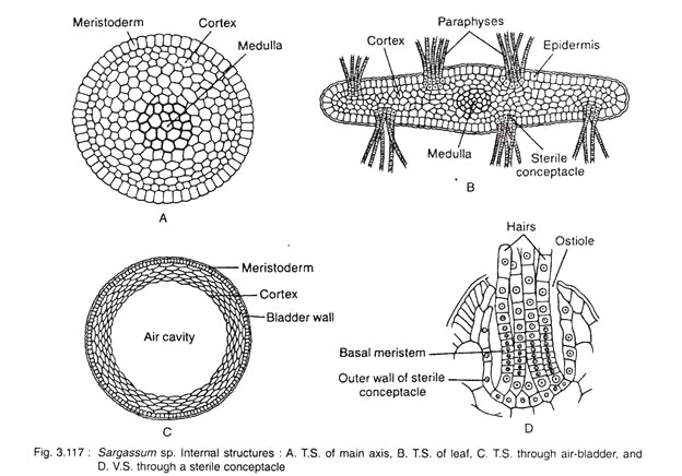
It is generally of circular in outline and differentiated into three regions: outer meristoderm, middle cortex and innermost medulla (Fig. 3.117A).
The meristoderm is made up of single layer of closely packed cells. The cells are meristema- tic in nature. The cells contain chromatophores and perform photosynthesis. This layer can store food material.
The cortex is situated next to meristoderm and occupies major part of the axis. It consists of compactly arranged parenchyma cells of polygonal shape, rarely with intercellular spaces. The cells are smaller in size than meristoderm. Like the outer layer this layer also stores food material.
The medulla i.e., the inner layer consists of narrow, thick walled elongated cells. This layer possibly helps in conduction.
Leaf:
It is flat and differentiated into outer meristoderm, middle cortex and inner medulla like the axis (Fig. 3.117B). The medulla is round and present in the middle region. On both surfaces of the leaf there are many sterile conceptacles, the cryptostomata or cryptoblasts (Fig. 3.117D).
These are flask-shaped with many sterile unbranched filaments, the paraphyses developed from the base. The paraphyses protrude out through the opening present on the outer side, the ostiole. The cells of the wall have many chromatophores.
Air Bladder:
Internally it is almost alike with the axis but without medulla (Fig. 3.117C). The central region is occupied by a large hollow cavity filled with air and gases. Outer to the cavity, cortex is present; which consists of a few layers and thinner cells than axis and finally it ends with a single layered outer meristoderm.
Thus the thallus shows division of labour along with differentiation of tissues. It serves the function of anchorage, photosynthesis, storage, conduction and support.
Features of Sargassum:
1. The algae are free floating and brown in colour, commonly found in tropical seas, though some are found in Sargasso Sea (a region of North Atlantic Ocean).
2. The plant body is diploid and differentiated into root, stem and leaf-like structures.
3. The main axis i.e., stem is vertically elongated and differentiated into nodes and internodes.
4. It bears long shoots of unlimited growth (primary laterals), leaves (secondary laterals), air bladders and receptacles.
5. Both stem and leaves are differentiated into epidermis, cortex and medulla.
6. Apical growth takes place by a three-sided apical cell.
7. Reproduction takes place by vegetative and sexual means. Asexual reproduction is absent.
8. Vegetative reproduction takes place by fragmentation.
9. Sexual reproduction is oogamous.
10. Oogonia and antheridia are borne in unisexual conceptacles, those remain embedded in receptacles.
11. Oogonium produces one egg and the antheridium produces 64 biflagellate sperms.
12. Fertilization is internal, as the egg is not come out from the oogonium.
13. Zygote germinates directly and produces a new sporophytic (2n) plant.
Reproduction in Sargassum:
It reproduces by both vegetative and sexual means. Asexual reproduction is absent.
1. Vegetative Reproduction:
It takes place by fragmentation. Due to death and decay of the older part, the younger region gets separated. The separated region grows and finally develops into a new individual like the mother. The free floating members like S. hystrix and 5. natans, multiply only by this method.
2. Sexual Reproduction:
It is of oogamous type and takes place by the union of antherozoid and egg, developed in antheridia and oogonia respectively. The sex organs develop in separate flask-shaped bodies the conceptacles, developed on branched receptacles. The conceptales with antheridia or oogonia are called male or female conceptacles.
Development of conceptacle. The fertile and sterile conceptacle are almost similar. The difference lies in the activity of basal cells of the linear wall of conceptacle. In sterile conceptacle it only develops sterile hairs, the paraphyses, but in fertile conceptacle it develops either antheridia or oogonia and also paraphyses in some regions.
During development (Fig. 3.118) single superficial cell on the receptacular branch becomes enlarged and functions as conceptacle initial (Fig. 3.118A). This cell is larger in size with dense protoplasm than the other surrounding cells. The conceptacle initial becomes flask- shaped.
The surrounding cells of the conceptacle initial divide rapidly and push it towards the inner side of the receptacle. The conceptacle initial then undergoes mitotic division and by oblique septation it forms upper elongated tongue cell and lower broad basal cell (Fig. 3.118B).
The tongue cell elongates and gradually disappears. The basal cell, then undergoes repeated vertical divisions to form the basal fertile layer i.e., the inner layer of the conceptacle. The reproductive organs are developed from this inner layer.
Development of Antheridium:
The antheridia are developed from the inner fertile layer of the antheridial conceptacle (Fig. 3.119). The lower basal cells of the conceptacle are the antheridial initials forming papilla like outgrowths (Fig. 3.119A). Each one divides by a transverse wall into two cells. The lower one remains as conceptacle Wall, whereas the upper one (Fig. 3.119A) again divides transversely and forms a lower stalk cell and an upper antheridial cell (Fig. 3.119B).
The antheridial cell develops into an antheridium (Fig. 3.119C). Due to rapid growth of the stalk cell, the antheridium becomes pushed at one side (Fig. 3.119D). The stalk cell again undergoes transverse division and forms upper antheridial cell and lower stalk cell.
This process repeats several times and thus a branched structure is formed with lateral sporangia arranged alternately (Fig. 3.119D, E). The apical cell of the stalk remains sterile and behaves as paraphysis. Many thread like filaments also develop from the basal cells of the conceptacle which are also called paraphyses (Fig. 3.119H).
The diploid nucleus of the antheridial initial undergoes meiosis followed by repeated mitotic divisions forming 32-64 haploid nuclei. The nuclei then accumulate some cytoplasm and form many uninucleate bodies. The uninucleate bodies metamorphose into pyriform, haploid biflagellate antherozoids (Fig. 3.119C).
The mature antheridium (Fig. 3.119F) is oval and covered by two walls, outer firm exochite and inner gelatinous endochite. At maturity the antheridium is detached from the stalk and comes out from the conceptacle through ostiole. After coming out, the wall of sporangium gets gelatinised and the antherozoids are liberated.
Development of Oogonium:
Like antheridium, oogonium also develops from the basal fertile layer of the conceptacle (Fig. 3.120). Some of the cells of this layer function as an oogonial initials (Fig. 3.120A). The oogonial initial undergoes transverse division and forms lower small stalk cell and upper large oogonial cell (Fig. 3.120B).
The oogonial cell becomes enlarged and forms a spherical structure. The diploid (2n) nucleus undergoes first meiotic (Fig. 3.120C), then mitotic divisions and 8 nuclei are formed. Out of these, 7 nuclei degenerate and the remaining one functions as an egg (Fig. 3.120D) which remains in the centres.
The wall of the oogonium consists of three layers, the outer exochite, the middle mesochite and inner endochite (Fig. 3.120C). The mature oogonia come out of the conceptacle through the ostiole, but still they remain attached with the conceptacle base by a long gelatinous stalk formed by the exochite.
Some of the basal cells of the inner layer of conceptacle instead of forming oogonia remain sterile and form sterile, long, hair-like structures, the paraphyses (Fig. 3.120E). Thus conceptacle contains oogonia intermingled with paraphyses.
Fertilisation:
Fertilisation takes place when the eggs remain outside but still attached with the conceptacle by gelatinous stalks (Fig. 3.120F). Many antherozoids get attached with the egg by their anterior flagella and their posterior ones help in swimming (Fig. 3.121A).
Later on only one penetrates the oogonial wall. The remaining antherozoids get separated and gradually degenerate, Initially after fertilisation both the nuclei remain side by side (Fig. 3.121 B), but later they fuse together and form the zygote (Fig. 3.121C).
Germination of Zygote:
Just after fertilization the zygote undergoes germination (Fig. 3.121D-H), while the oogonium still remains attached with the conceptacle. After some time it comes out of the gelatinous wall. After liberation, the zygote gets attached with any solid substratum.
The zygote then divides transversely and forms lower and upper cell. The lower cell develops into rhizoid and the upper cell undergoes repeated periclinal and anticlinal divisions, thus forming a thalloid sporophyte (2n) of Sargassum.
Life Cycle Pattern of Sargassum:
Sargassum shows diplontic life cycle without any alternation of generations (Fig. 3.122). The plant body is diploid except the antherozoids and eggs. The antherozoids and eggs i.e., the gametes, represent only the haploid (n) stage. After fusion zygote is formed. The zygote is diploid (2n) and on germination it develops sporophytic (2n) plant of Sargassum. Thus it shows a typical example of diplontic life cycle.
Common Indian species:
Sargassum ilicifolium, S. tenerrium, S. wightii, S. duplicatum, S. myriocystum, S. christifolium, S. carpophyllum, S. cinereum and S. plagiophyllum.


