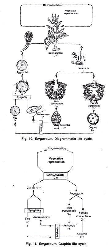
In this article we will discuss about the vegetative and sexual reproduction that occur in the life cycle of sargassum.
(A) Vegetative Reproduction in Sargassum:
Sargassum multiplies profusely by vegetative fragmentation. The thallus breaks into fragments due to mechanical injury or death and decay of older parts. The species like S. hystrix and S. natans growing in Sargasso sea are completely sterile as they do not form any reproductive structures. In these species the fragmentation is the only method of multiplication.
(B) Sexual Reproduction in Sargassum:
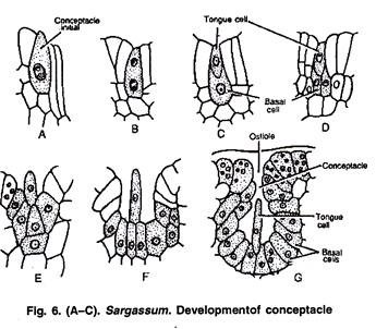
Sexual reproduction in Sargassum is oogamous. The male sex organs are called antheridia and the female oogonia. The sex organs develop in special flask shaped cavity called conceptacle. These conceptacles are present is specially modified laterals called receptacles (Fig. 6 A-C). The male and female sex organs develop in separate conceptacles.
The conceptacles bearing antheridia are called male conceptacles and those bearing oogonia are called female conceptacles.
In homothallic or monoecious species the male conceptacle and female conceptacles are produced on same receptacle, but antheridia and oogonia are not produced in same conceptacles. In dioecious plants the male and female conceptacles are produced on separate male and female plants. Sargassum species are mostly monoecious.
Development of Conceptacles:
The conceptacle develops from a single superficial cell on the receptacular branch. This cell called conceptacle initial is flask shaped and differs from the adjacent cells due to its larger size and prominent nucleus (Fig. 6A). The initial cell divides slower than other cells. As a result it gets lower in position than adjacent cells.
The initial cell divides by transverse division; the two cells formed are separated by a curved septum. The lower cell is called basal cell and the upper is called tongue cell (Fig. 6 B, C).
The tongue cell divides transversely to make small filament which later disintegrates. The basal cell undergoes many vertical divisions to make fertile layer of the conceptacles. The cells of fertile layer later form antheridia and oogonia (Fig. 6 D-G).
In fertile conceptacles the cells of basal layers do not spread in upper part, this forms narrow opening called ostiole.
Development of Antheridium:
Any cell of the fertile layer can function as antheridial initial. This cell is dense cytoplasmic and develops a papilla like outgrowth. It divides by transverse division to make lower stalk cell and upper antheridial cell (Fig. 7 A-B). The antheridial cell rounds off to make antheridium.
The stalk cell elongates and pushes the antheridium to one side. The growing stalk cell divides again to make basal cell and the antheridial cell. This process repeated many times and results in formation of many antheridia and a sterile paraphysis (Fig. 71).
The antheridia are oval structures with two layered cell walls. The outer wall is called exochite and the inner is called endochite (Fig. 7 G). At young stage the antheridia are inside conceptacles and on maturity the antheridia are detached from stalk and come out of ostiole.
The antheridium has one diploid nucleus which divides first by meiotic division and later by mitotic divisions. This results in formation of 32-64 haploid nuclei. The protoplast of antheridium also divides in equal number of segments. Each protoplast segment with haploid nucleus develops into an antherozoid (Fig. 7 H). The antherozoid is pear shaped structure with two lateral flagella.
The flagella are heterokontic, one being acronematic and the other pantonematic.
The antherozoids are liberated in water after gelatinization of the antheridial wall.
Development of Oogonium:
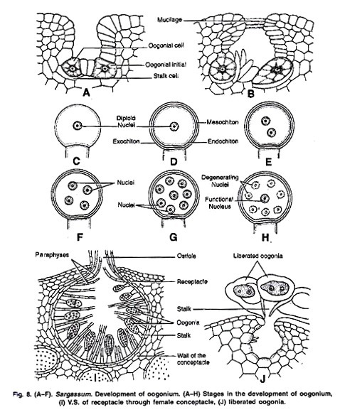
Any cell of the fertile layer of the female conceptacle can function as oogonial initial (Fig. 8 A). The oogonial initial divides by transverse division to make small, lower stalk cell and the large, upper oogonial cell (Fig. 8 B). The stalk cell further does not divide or elongate, so the oogonial cells are almost sessile.
The oogonial cell enlarges and makes spherical oogonium. The oogonia wall has three layers—the outer exochite, middle mesochite and the inner endochite. On maturity of the oogonium the exochite ruptures, the mesochite forms the gelatinous stalk and the oogonial nuclei- and protoplast remains surrounded by endochite.
The diploid oogonial nucleus undergoes meiotic and mitotic divisions to form 8 nuclei. The seven of these eight nuclei degenerate and only one remains functional. This nucleus with protoplasm forms single ovum or oosphere (Fig. 8 C-H).
The cells of female conceptacle which do not form oogonia develop into long hair like paraphyses.
Fertilization:
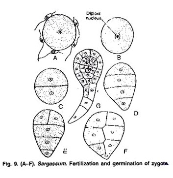
The antherozoids are released in water and the oogonia remain attached to the conceptacle base by mucilaginous stalk. The oogonia protrude out of the ostiole (Fig. 8 J). A large number of antherozoids surround the oogonium and attach to oogonial wall with the help of anterior flagellum (Fig. 9 A). Only one antherozoid penetrates the oogonial wall. The male and female nuclei fuse to form a diploid zygote (Fig. 9 B).
Germination of Zygote:
The zygote germinates immediately after fertilization when the oogonium still remains attached to the wall of conceptacle by a mucilaginous stalk. After some time the zygote is liberated by gelatinization of the oogonial wall. After liberation the zygote gets attached to any substratum in sea water. The zygote first divides by transverse division to make a lower cell and upper cell (Fig. 9 C-F).
The lower cell forms the rhizoids. The upper cell first divides by transverse division and later by anticlinal and periclinal divisions. It results in the differentiation of three layers—the meristoderm, cortex and medulla. The divisions of upper cell result in formation of a diploid, sporophytic Sargassum plant.
The life cycle of Sargassum is diplontic type and there is no alternation of generation. The thallus is diploid sporophytic. It forms diploid antheridia and oogonia. The reduction division in antheridia and oogonia forms haploid antherozoid and oognial nuclecus. The gametes only are haploid structure in the life cycle. After fertilization a diploid zygote is formed which divides to make a diploid sporophytic thallus (Fig. 10, 11).
