In this article we will discuss about:- 1. Occurrence of Polysiphonia 2. Plant Body of Polysiphonia 3. Cell Structure 4. Features 5. Reproduction 6. Life Cycle.
Occurrence of Polysiphonia:
The genus Polysiphonia (Gr. poly — many; siphon — tube) is represented by more than 150 species, out of which about 16 species are reported from India. They grow in marine habitat and are cosmopolitan in distribution.
Commonly they are found in littoral and sublittoral zones. In India they are found in western and southern coasts. Commonly, they grow as lithophytes i.e., on rocks or stones (e.g., P. elongata), some species like P. urceola, P. terulacea grow as epiphytes on Laminaria. P. fastigiata grows as semiparasite on Ascophyllum nodosum.
Plant Body of Polysiphonia:
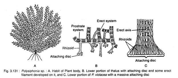
Plant body is multiaxial well branched thallus of dark brown, reddish or bluish red colouration appearing as a very small bush (Fig. 3.131 A). The height of the bush varies from a few to several centimeters.
Most of the species are heterotrichous in habit, consisting of prostrate and erect systems.
Prostrate System:
It may be multiaxial and well developed. From the lower side of the prostrate system many unicellular rhizoids are developed (Fig. 3.131B, C). The rhizoids are much lobed at the apex and form definite attachment discs (e.g., P. urceolata, P. nigrescens).
[In P. elongata and P. violacea, the prostrate system is absent and many rhizoids develop from the lowermost cells of the erect system and by aggregation they form massive attachment disc (Fig. 3.131C).]
Erect System:
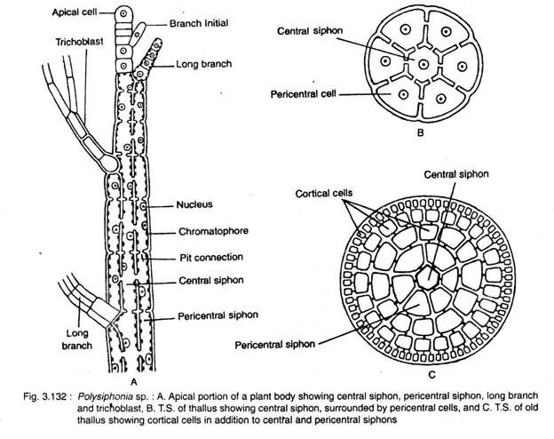
The erect filaments develop from the prostrate system. The erect system consists of main axis and many branches (Fig. 3.132A). The branches are of two types: long branch and short branch
The long branches are called branches of unlimited growth or long lateral branches and the short branches i.e., branches of limited growth are called trichoblasts. The long branches develop in a spiral or radial symmetry. The trichoblasts are spirally arranged, dichotomously branched, colourless and mostly annual structures bearing sex organs. The trichoblasts may develop both from main axis and long branches.
The main axis and long branches consist of a central siphon of many elongated cylindrical cells situated in vertical row (Fig. 3.132B, C). It is surrounded by 4-20 peripheral siphons. So the plant body is polysiphonous and named Polysiphonia. Only the central siphon is present at the apical region of both main axis and the long branches.
In most of the species, pericentral siphon is covered by 3 layers of cortical cells formed due to periclinal and anticlinal divisions of the cells of pericentral siphon. All cells of the plant body are connected with each other by pit connections (cytoplasmic connections). The short branches or trichoblasts are monosiphonous.
Cell Structure of Polysiphonia:
The cells have thick wall, differentiated into outer pectic and inner cellulosic layers. The cells are uninucleate with many discoid chromatophores without pyrenoids. Neighbouring cells are connected by pit connections. The cells contain large central vacuole. Reserve food is floridean starch.
Important Features of Polysiphonia:
1. Plant body is polysiphonous.
2. Apical growth takes place by single dome- shaped apical cell.
3. Sexual reproduction is of advanced oogamous type.
4. Post-fertilisation stage is much elaborate.
5. Cystocarp is well-developed.
Reproduction in Polysiphonia:
Polysiphonia reproduces both asexually and sexually. Sexual reproduction is of oogamous type. In the life cycle of Polysiphonia three kinds of plants are recognised.
These are:
1. Diploid tetrasporophyte,
2. Haploid gametophyte, and
3. Diploid carposporophyte (Fig. 3.138).
1. Diploid Tetrasporophyte:
It develops on direct germination of carpospore (2n = 40), thus the plant is diplsid (2n). It is an independent plant which, instead of developing sex organs develops tetrasporangia. The diploid nucleus of tetrasporangia undergoes meiosis and develops four (4) haploid (n = 20) tetraspores.
2. Haploid Gametophyte:
It develops on direct germination of tetraspore (n); thus the independent plant is haploid (n). Most of the species are heterothallic, thus the spermatangia (male sex organ) and carpogonia (female sex organ) are developed on different plants.
3. Diploid Carposporophyte:
This stage is diploid (2n) and dependent on haploid gametophytic plants. The union between haploid (n) spermatium (developed inside spermatangium) and haploid female gamete (developed inside carpogonium) forms diploid (2n) nucleus inside the carpogonium.
Further development of diploid nucleus forms diploid carposporophyte. Later carpospores are formed by mitotic division of carposporangium. The carpospore on direct germination forms diploid tetrasporophyte plant.
Asexual Reproduction:
Asexual reproduction takes place by haploid non-motile tetraspores.
The carpospores (2n) on direct germination develop diploid tetrasporophytic plants. The plants are independent and polysiphonous. Some pericentral cells of the thallus near apical region develop sac-like tetrasporangia. The diploid nucleus of tetrasporangium undergoes meiosis and forms four tetraspores. The spores are arranged tetrahedrally (Fig. 3.133A).
Development of Tetraspores:
Tetraspores are produced in tetrasporangia. Single pericentral cell of each tier, towards apical region functions as tetrasporangial initial (Fig. 3.133B). This initial cell is smaller than other pericentral cells of any particular tier. This initial cell divides vertically into inner and outer cells.
The inner cell functions directly into sporangial mother cell and the outer cell further divides and forms two or more cover cells. The sporangial mother cell divides transversely into lower stalk cell and upper tetra- sporangial cell.
The latter undergoes further enlargement and develops into a tetrasporangium. The diploid nucleus of tetrasporangium undergoes meiosis and forms 4 tetraspores or meiospores. The tetraspores are arranged tetrahedrally inside the tetrasporangium.
The mature tetraspores are liberated by rupturing the wall of the sporangium. On germination they develop gametophytic polysiphonous plant. Being heterothallic, out of four tetraspores, two produce male and the remaining two produce female gametophytic plants.
Sexual Reproduction:
Sexual reproduction is of oogamous type. Plants are commonly dioecious. The male sex organs i.e., spermatangia and female sex organs i.e., carpogonia, are developed on male and female plants, respectively.
1. Male Reproductive Organ:
It is called spermatangium or antheridium.
Initially male trichoblast develops as side branch on the plant body (Fig. 3.134A). It becomes branched. In some species both the branches become fertile, but in others only one remains fertile and the rest undergo repeated dichotomy to form dichotomous sterile structure.
The monosiphonous fertile branch(es) of male trichome bears many unicellular and spherical spermatangia. Each spermatangium is a uninucleate structure which produces single spermatium, the male gamete.
During development of spermatangium (Fig. 3.134B-D), all cells except a few basal cells, divide periclinally and form pericentral cells on both the sides. Each pericentral cell undergoes several divisions and forms spermatangial mother cells. Each one cuts off 2-4 unicellular bodies, the spermatangia. Each spermatangium develops into a single non-motile male gamete, the spermatium.
The spermatia are liberated from the spermatangium, through a narrow apical slit on the wall. The spermatia are dispersed through water.
2. Female Reproductive Organ:
The female reproductive organ is called carpogonium.
The carpogonium develops at the top of 2-5 celled carpogonial filament (Fig. 3.135). The carpogonial filament develops on the female trichoblast. The carpogonium is a flask-shaped body, with a basal swollen region containing an egg and an upper elongated neck region, the trichogyne.
During development of carpogonium, initially a female trichoblast initial is developed on central siphon, a few cells (3-4) below the apical cell. The female trichoblast initial, then undergoes repeated divisions and forms a female trichoblast of 5-7 cells. The lowermost three cells of the female trichoblast divide vertically and form three tiers of pericentral cells.
Any one of the pericentral cells of the middle tier towards the mother axis becomes the supporting cell. The supporting cell cuts off a small initial at its outside, the procarp initial (Fig. 3.135A). The procarp initially undergoes repeated divisions and forms a 4-celled branch, the procarp or carpogonial filament (branch) (Fig. 3.135B).
The apical cell of the carpogonial filament functions as carpogonium mother cell. The cell further develops into a carpogonium. The carpogonium has a swollen basal region containing egg and an elongated tubular region, the trichogyne (Fig. 3.135C).
At the later stage, the carpogonium develops two initials from the supporting cell, one at the base, the basal sterile filament initial (Fig. 3.135D) and another at the lateral side, the lateral sterile filament initial. The lateral sterile initial divides transversely and forms two-celled lateral sterile filament (Fig. 3.135E).
The carpogonium is ready for fertilisation at this stage. The pericentral cell adjacent to the supporting cell starts growing to cover the fertilised carpogonium. Later on they form sheath (the protective covering) around the fruit body, called as pericarp.
Fertilisation:
The spermatia are dispersed with the help of water. A few spermatia become attached at the tip of the. receptive trichogyne. Out of many, only one becomes successful. The common wall of successful spermatium and trichogyne dissolves at the point of contact and the male nucleus passes to the female nucleus present at the base of the carpogonium. The fusion between the nuclei results in the formation of zygote.
Post-Fertilisation Changes:
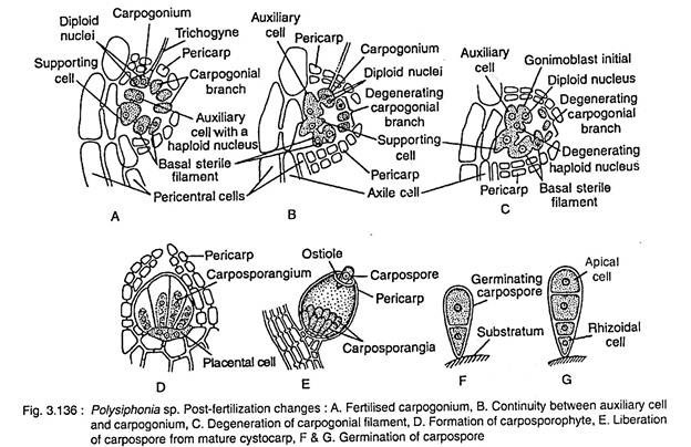
At the starting of this phase, an auxiliary cell is developed from the supporting cell situated just below the basal region of the carpogonium (Fig. 3.136A). Simultaneously, the lateral,.sterile filament increases in length (4-10 celled) by cell division as well as elongation and the basal sterile initial divides to form a two (2)-celled filament. The auxiliary cell has a single haploid nucleus.
A tubular connection is then developed between the auxiliary cell and carpogonium (Fig. 3.136B). The carpogonial nucleus (2n) divides mitotically into two nuclei, of which one is transported to the auxiliary cell and the other one remains in the carpogonium. Thus the auxiliary cell contains one haploid and one migrated diploid nuclei. The haploid nucleus (n) is degenerated. Gradually the trichogyne shrivels (Fig. 3.136B).
Many vegetative filaments then develop from the adjacent vegetative pericentral cells, which gradually develop the total covering. The diploid nucleus of auxiliary cell then divides mitotically and forms two nuclei. One of them then migrates into the outgrowth developed on the auxiliary cell.
This outgrowth after separating by a partition wall forms gonimoblast initial (Fig. 3.136C). In this way many gonimoblast initials can develop on auxiliary cell. Each initial by repeated mitotic divisions forms gonimoblast filament. The terminal cell of the gonimoblast filament develops into carposporangium, which forms single diploid carpospore inside (Fig. 3.136D, E).
During this development the auxiliary cell, supporting cell, carpogonium and some cells of basal and sterile filaments fuse together and form an irregular cell, the placental cell (Fig. 3.136D). The haploid nuclei (n) of the placental cell gradually degenerate and have simply a nutritive function.
The placental cell, gonimoblast filament and carpogonia are covered by many vegetative filaments and form an urn-shaped structure, the cystocarp (Fig. 3.136E, 3.137). The outer covering of cystocarp is called pericarp. The diploid part of the cystocarp represents the carposporophyte. Some cells of basal and sterile filament along with some cells of carpogonial filament gradually degenerate.
The carposporangium develops single diploid carpospore. After liberating from the carpogonium they come out through the ostiole of cysto- carp (Fig. 3.137).
Germination of Carpospore:
Coming in contact with any solid surface, the diploid carpospore gets attached and then undergoes first mitotic division and forms large upper and small lower cells (Fig. 3.136F, G). Both the cells undergo mitotic division and form 4 celled stage.
The lower most cell forms the rhizoid, the upper one functions as apical cell and the rest cells undergo further development and form the polysiphonous body. This plant body is diploid i.e., the tetrasporophytic plant, which later develops the tetraspores and complete the cycle.
Life Cycle of Polysiphonia:
Life cycle of Polysiphonia consists of three distinct phases: diploid tetrasporophyte, haploid gametophytes and diploid carposporophyte.
Out of 4 tetraspores produced in tetrasporangia on diploid tetrasporophytic plant, two tetraspores develop haploid (gametophytic) male and other two haploid (gametophytic) female plants. The male gametophytic plants develop male gametes inside spermatangia and female gametophytic plants develop female gametes inside carpogonigia.
Zygote develops inside carpogonium after gametic fusion. With gradual development gonimoblast filament, carposporangia and carpospores are developed inside a composite structure, the cystocarp. It is the carposporophytic stage. Diploid carpospore on germination produces the diploid tetrasporophytic plant again.
Thus the life cycle is triphasic and haplo- diplobiontic type (Fig. 3.138).
Indian Species:
Polysiphonia tuticorinensis, P. sertularioides, P. platicarpa, P. suotilissima, P. unguiformis etc.


