
The following points highlight the three main parts of toad’s nervous system (With Diagrams). The parts are: 1. The Central Nervous System 2. The Peripheral Nervous System 3. The Autonomic Nervous System.
Part # 1. The Central Nervous System:
This is composed of a hollow median tube, the anterior enlarged part of which is known as the brain and the posterior narrow part as the spinal cord. The cavity of the brain is continuous with that of the spinal cord and is filled up with a fluid called cerebrospinal fluid.
The solid parts of the central nervous system are composed of neurones or nerve cells, together with their processes, the nerve fibres. The part of the neurone in which the nucleus lies is the cell-body. The extended processes of the neurones which run together within a connective tissue sheath form a nerve.
Aggregated masses of nerve cells with their contained nuclei has a greyish appearance and are, therefore, recognised as the grey matter. Bundles of nerve fibres, on the other hand, appear to be white and are, therefore, called white matter.
The grey matter lies on the outer surface of the brain and covers up the white matter. In the spinal cord, however, the condition is reversed; the white matter is on the outer side and the grey matter is on the inner side.
The brain and the spinal cord are enclosed in two protective coverings or meninges:
(i) An outer and thicker dura mater, and
(ii) An inner and thinner Pia mater.
Brain:
The brain is housed within the cranium of skull. It is composed of three primary regions: the forebrain, the midbrain, and the hindbrain. Subsequent changes in forebrain and hind- brain result in a further subdivision of these two parts, so that ultimately, the brain consists of five portions which may be tabulated anteroposteriorly in the following manner:

I. The forebrain is composed of telencephalon and diencephalon. At the anterior end of the telencephalon there is a pair of small olfactory lobes, which are concerned with the sense of smell. The rest of the telencephalon consists of two elongated lobes, the cerebral hemispheres.
The floor of each hemisphere is thickened to form a mass called corpus striatum. The thin roof and sides of the hemispheres are smooth and constitute the pallium. The cerebral hemispheres are the seat of intelligence, consciousness, and voluntary motion.
The posterior part of the forebrain is the diencephalon or thalamencephalon. It is a depressed region behind the cerebral hemispheres which gives out dorsally a thin projection called epiphysis. To this is attached a small pineal body of unknown function. In front of the epiphysis is a highly vascular membrane, the anterior choroid plexus.
On the ventral surface of the diencephalon is an X-shaped structure, the optic chiasma, formed by the crossing of the two optic nerves. A little behind the chiasma is a projection called hypophysis or infundibulum, and to this is attached a rounded glandular structure, the pituitary body. Each side of the diencephalon is thickened to form the optic thalamus.
II. The midbrain or mesencephalon is the widest part of the brain. A pair of hollow oval bodies grow out from its dorsal surface. These are the optic lobes or corpora bigemina. Ventral to the optic lobes, there are two longitudinal bands or peduncles, the crura cerebri, which connect the forebrain with the hindbrain.
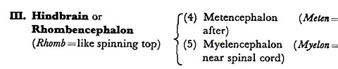
III. The hindbrain is composed of metencephalon and myelencephalon. The metencephalon is represented dorsally by a thin transverse shelf called cerebellum, which lies behind the optic lobes. It is the centre for the co-ordination of movements.
The myelencephalon connects the hindbrain with the spinal cord. Its floor and sides are greatly thickened to form the medulla oblongata. Dorsally, on the roof of the myelencephalon, is a thin membrane richly supplied with blood vessels. This is known as the posterior choroid plexus.
The medulla contains the centres for the control of important vital processes such as regulation of heart beat, respiration, and metabolism. It has been proved by experiment that if all the parts of the brain be destroyed and only the medulla is retained the toad can hop, swim, and breathe. Removal of medulla, however, is at once followed by death.

Cavities of the Brain:
The brain is not a solid mass. It has cavities within, which are known as the ventricles. The hollow of the cerebral hemispheres constitute the first and the second ventricles which are also recognised as the lateral ventricles. The cavity of the diencephalon is the third ventricle. Each lateral ventricle communicates with the third ventricle by a small opening called foramen of Monro.
The fourth ventricle is the shallow cavity of the medulla oblongata. Its thin roof is composed of the highly vascular posterior choroid plexus. The third ventricle communicates with the fourth ventricle by means of a narrow passage called iter or aqueduct of Sylvius which extends throughout the length of the midbrain. Posteriorly, the fourth ventricle communicates with the central canal of the spinal cord.

Spinal Cord:
It is a hollow tube encased within the neural canal of the vertebral column. Extending backwards from the medulla oblongata, the spinal cord ends within the urostyle as a slender filament, the filum terminale. Two median grooves run along the whole length of the cord. The one running in the mid- ventral line is the ventral fissure and the other in the mid-dorsal line is the dorsal fissure.
The cavity of the cord, the central canal or neurocoel, is continuous with the fourth ventricle of the brain. The spinal cord is covered with two protective meninges, outer dura mater and inner pia mater, which are continuous with those of the brain.
Surrounding the central canal is a zone of nerve cells forming the grey matter and arranged like a butterfly with extended wings. The grey matter is enclosed in a cylinder of nerve fibres forming the white matter.
Part # 2. The Peripheral Nervous System:
This includes the paired cranial and spinal nerves originating from the cerebrospinal axis. A nerve is a white thread-like structure composed of a bundle of microscopic nerve fibres.
The nerve fibres originate from the cell bodies of the neurones which are situated within the grey matter of the central nervous system. Sometimes the cell bodies may aggregate to form a small swelling in the course of a nerve trunk. Such a swelling is known as a ganglion.
The nerve fibres, are of two kinds:
(i) Afferent or sensory fibres, and
(ii) Efferent or motor fibres.
The sensory fibres convey messages from receptor organs, such as eyes, ears, nose, tongue, and skin, to the central nervous system. Motor fibres are those which carry impulses from the central nervous system to the effector organs, such as glands and voluntary muscles.
A sensory nerve is composed entirely of sensory fibres, and similarly a motor nerve is composed of motor fibres. A nerve may contain both sensory and motor fibres and in that case it is known as a mixed nerve.
Cranial Nerves:
Ten pairs of cranial nerves originate from the brain. Some of the cranial nerves are entirely sensory, some are motor and some are mixed. The cranial nerves are numbered in sequence to their origin. They are known by their respective names as well as by number. The following is a detailed account of the origin, course, distribution, and function of the cranial nerves.
I. Olfactory Nerve:
The olfactory nerves originate from the olfactory lobes of the telencephalon. Extending forward into the nasal capsule, each nerve is distributed to special sensory cells on the mucous membrane of the nose of the same side. It is a purely sensory nerve concerned with the sense of smell.
II. Optic Nerve:
Arising from the optic lobes, the two optic nerves run across the ventral surface of the diencephalon in front of the pituitary body. At this point they form the X-shaped optic chiasma in which the two nerves cross each other.
Thus the left optic nerve crosses over to the right side and vice versa. Emerging from the chiasma, the optic nerve enters the orbit and is distributed to the retina of the opposite eye. It is a purely sensory nerve concerned with vision.
III. Oculomotor Nerve:
The oculomotor nerves arise from the ventral surface of the mesencephalon. Proceeding forward, it enters the orbit and supplies four of the extrinsic muscles which move the eye, namely, superior rectus, inferior rectus, internal rectus and inferior oblique. As the name implies oculomotor is a purely motor nerve.
IV. Trochlear or Pathetic Nerve:
It is a slender motor nerve which arises from the dorsal surface of the mesencephalon just behind the optic; lobe. It follows the course of the third nerve and innervates another extrinsic eye muscle, called superior oblique.
V. Trigeminal Nerve:
The trigeminal is a stout mixed nerve arising from the lateral side of the medulla oblongata. It bears a Gasserian or pro-otic ganglion near its origin and comes out of the skull through a small foramen in front of the auditory capsule. Outside the skull the nerve is characteristically divided into three main branches: the opthalmic, the maxillary, and the mandibular nerves.
The opthalmic nerve is the smallest of the three branches. It proceeds forward through the orbit and supplies sensory fibres to the skin round the anterior part of the head, the upper eyelid and the mucous membrane of the nose.
The maxillary nerve is the main branch to the upper jaw, supplying sensory fibres to the upper lip, lower eyelid and mucous membrane of the upper jaw.
The mandibular nerve curves downward and backward to the corner of the moudi. It now proceeds along the outer side of the lower jaw supplying sensory fibres to the skin and motor fibres to the muscles on the floor of the buccal cavity. Thus the mandibular branch is a mixed nerve.
VI. Abducens Nerve:
The abducens is a slender motor nerve arising from the ventral surface of the medulla oblongata. It runs through the orbit and supplies the external rectus muscle of the eye.
VII. Facial Nerve:
The facial is a mixed nerve arising from the medulla oblongata just behind the V nerve. It runs into the Gasserian ganglion and leaves the skull through the same foramen along with the trigeminal. Outside the skull the facial nerve divides into two branches: the palatine, and the hyomandibular.
The palatine nerve proceeds forward beneath the mucous membrane of the roof of the buccal cavity and is distributed mainly to the roof of the mouth. It is joined anteriorly by a small branch from the maxillary division of the V nerve.
The hyomandibular nerve turns outward and then proceeds forward along the floor of the buccal cavity. It receives an anastomosing branch from the glossopharyngeal nerve and then breaks up into branches which supply the tympanum, the mandibular joint, and the mucous membrane and muscles on the floor of the buccal cavity.
VIII. Auditory Nerve:
It is a short and stout nerve which arises from the lateral side of the medulla oblongata posterior to the facial nerve. It runs outward to supply the internal ear. The auditory is a purely sensory nerve concerned with hearing and balancing.
IX. Glossopharyngeal Nerve:
This is a mixed nerve originating from the medulla posterior to the auditor nerve. It immediately runs into the vagus ganglion at the base of the X nerve. Separating from the ganglion, the glossopharyngeal gives off an anastomosing twig which joins the hyomandibular branch of the facial.
The main trunk of the nerve curves round the pharynx and then turns forward, pursuing a tortuous wavy course along the floor of the buccal cavity. It supplies sensory fibres to the floor of the buccal cavity and the tongue. Motor fibres innervate muscles.
X. Vagus or Pneumogastric Nerve:
The vagus is a large mixed nerve arising from the medulla oblongata by a number of roots. It bears a vagus ganglion near its origin. Coming out of the cranium through a small foramen in the ex-occipital bone, the vagus nerve gives off a few twigs to the pharyngeal wall.
It then turns back and divides into four main branches:
(i) Laryngeal to the voice-box,
(ii) Cardiac to the heart,
(iii) Pulmonary to the lung of the same side, and
(iv) Gastric to the alimentary canal.
Because of its long course, the vagus is recognised as the wandering nerve. The X nerve is perhaps the most important cranial nerve for it controls heartbeat, respiratory movements and peristalsis of the alimentary canal.
Spinal Nerves:
There are ten pairs of spinal nerves. Each spinal nerve is connected with the spinal cord by two roots: a dorsal sensory root, and a ventral motor root. The dorsal root bears a ganglion near its origin. The two roots soon unite to form a common trunk, which comes out through the intervertebral foramen. Soft chalky masses, called calcareous bodies, surround the openings by which the spinal nerves come out.
Coming out, each spinal nerve divides into three branches:
(i) A small dorsal branch supplying the skin and muscles of the back;
(ii) A large ventral branch forming the main trunk of the nerve and supplying the skin and muscles on the ventral surface of the body; in some spinal nerves, the ventral branch runs into the limb;
(iii) A very short ramus communicans which at once joins the nearest sympathetic ganglion. All the spinal nerves are mixed nerves.
The first spinal nerve is the hypoglossal nerve. It leaves the vertebral column behind the atlas and accompanies the glossopharyngeal nerve to supply the muscles of the tongue.
The second and third spinal nerves join to form a network, called brachial plexus. From this arises the brachial nerve for supplying the forelimb of the side.
The fourth, fifth and sixth spinal nerves supply the skin and muscles of the trunk region.
The seventh, eighth, ninth and tenth spinal nerves join with one another to form the sciatic plexus, from which arises the large sciatic nerve for supplying the hind limb. The tenth spinal nerve is very small and is often absent at one side.
Part # 3. The Autonomic Nervous System:
The autonomic (auto = self; nemo = distributing) nervous system consists of two minute cords, the sympathetic trunks, one on each side of the dorsal aorta. The sympathetic trunks begin posteriorly near the point of bifurcation of the dorsal aorta. They diverge anteriorly to accompany the systemic arches.
Further forward, each splits to encircle the subclavian artery of its own side. It now enters the cranium, supplies a communicating twig to the vagus ganglion, and ends by joining the Gasserian ganglion of the V nerve. Each sympathetic trunk bears ten small ganglia in its course and each sympathetic ganglion is connected to the corresponding spinal nerve by a short ramus communicans.
")
The sympathetic trunks send out slender branches for the innervation of involuntary muscles on the wall of heart, blood vessels, digestive tube, etc. These branches often unite to form ganglionated nerve plexuses.
Thus there is a cardiac plexus supplying the heart, bearing Bidder’s ganglion in the inter-auricular septum and Remak’s ganglion on the sinus venosus. Similarly, there is a solar plexus surrounding the coeliacomesenteric artery, which innervates the stomach and the intestines.
The autonomic nervous system is mainly concerned with the regulation of involuntary actions such as beating of the heart, peristaltic movements of the alimentary canal, and the activities of the viscera in general.
It is not completely independent of the central nervous system, because most of the sympathetic ganglia are connected with peripheral nerves. It is autonomic in the sense that its actions are not disturbed by the central nervous system.