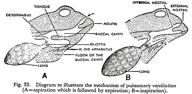
In this article we will discuss about the respiratory system in toad. This will also help you to draw the structure and diagram of respiratory system in toad.
Respiration is the exchange of oxygen and carbon dioxide between the toad and its environment. Oxygen is taken into the body and carbon dioxide is given off. Every cell of the body consumes oxygen. With the help of oxygen, the assimilated food which is stored within the cells is slowly oxidised.
The result is the liberation of heat energy, and formation of carbon dioxide and water. Thus when glucose is oxidised, one molecule of glucose combines with six molecules of oxygen to form six molecules of carbon dioxide and six molecules of water.
This is not all; because one gramme of glucose, when completely oxidised in the body, yields about four calories of heat, which is utilised by the animal for carrying out its other activities. Respiration, therefore, is not merely exchange of oxygen for carbon dioxide, but it also involves transformation of assimilated food into energy.
When no reserve food is available, the animal transforms its own protoplasm into energy and thereby loses weight. It is to be noted that the source of the oxygen is the air and the carbon dioxide is formed in the tissues.
The transport of the two gases occurs through the agency of the blood. Acting as a ‘middleman’ the blood carries oxygen from the air to the tissues, and carbon dioxide from the tissues to the outside air.
The exchange of oxygen of the air for the carbon dioxide of the blood takes place in the respiratory organs and is known as external respiration. The true respiration, resulting in the release of energy, occurs in the tissues by the exchange of oxygen of blood for the carbon dioxide of the tissues. This is known as internal or tissue respiration.
While in blood, the oxygen is actually carried by the haemoglobin of the erythrocytes. Haemoglobin combines with oxygen to form an unstable compound called oxyhaemoglobin. In this form, oxygen is carried to all the body cells. As the blood is circulating through the capillary networks of deoxygenated tissue, the unstable oxyhaemoglobin releases the bulk of its oxygen and is reduced to its former state.
It is then sent back to the respiratory surface for reloading with fresh oxygen. The oxygen that is set free diffuses out through the capillaries into the lymph, which comes into direct contact with the body cells. Tissue respiration now takes place.
Oxygen combines with food materials which are stored in the tissues for this purpose. Carbon dioxide is formed along with release of energy. The carbon dioxide is carried by the lymph and diffuses out into the blood. It is transported chiefly by the blood plasma in the form of bicarbonates and finally released and expelled through the capillary networks on the respiratory surface.
Types of Respiration:
As the toad is an amphibian, it is able to receive oxygen and given out carbon dioxide from three kinds of respiratory organs.
These are:
(1) The lungs,
(2) The skin, and
(3) The mucous membrane of the buccal cavity.
The moist epithelium of these organs are richly supplied with blood capillaries. Atmospheric oxygen dissolves in the moisture of the respiratory surface and diffuses into the blood, through the thin wall of the capillary networks. Carbon dioxide of the blood diffuses out in the opposite direction.
Pulmonary Respiration:
The organs concerned in the pulmonary respiration are:
(1) The external nares,
(2) The internal nares,
(3) The buccal cavity,
(4) The glottis,
(5) The laryngotracheal chamber,
(6) The bronchi, and
(7) The lungs.
The lungs are thin-walled spongy structures which extend backward into the anterior part of the body cavity. There are two lungs, one on each side of the heart.
Each lung is a pink-coloured elastic sac which is connected to the base of the laryngo-tracheal chamber by a short tube called bronchus. The laryngo-tracheal chamber or voice-box communicates with the floor of the buccal cavity by a slit-like glottis. The buccal cavity opens into the exterior by the internal and external nares.
The outer wall of each lung is hollowed out internally to produce innumerable simple chambers which are called air-sacs or alveoli. Each alveolus receives its own blood supply from a minute branch of the pulmonary artery. The alveolar epithelium, presenting a moist surface and being richly supplied with capillary network, carries out O2—CO2 exchange.
Mechanism of Respiration:
Ventilation of the lungs is effected by two processes, namely, breathing in of air or inspiration and breathing out of air or expiration.
The two processes occur alternately in three stages:
(1) Aspiration,
(2) Expiration, and
(3) Inspiration.
(1) The toad closes its mouth and lowers the floor of the buccal cavity, thus drawing in air through the external nares. This air is sucked into the buccal cavity through the internal nares. As the glottis remains closed, the air cannot pass into the lungs. This is known as aspiration.
(2) Aspiration is followed by expiration. The trunk muscles forcibly contract upon the elastic lungs. This expels air rich in CO2 through the glottis, which opens at this stage. The buccal cavity, which is already full of inhaled atmospheric air, becomes very much distended with the exhaled air from the lungs. A mixture of the two kinds of air now rushes out through the open nostrils.
(3) Expiration is quickly followed by inspiration. The external nares are tightly closed and the floor of the distended buccal cavity is forcibly raised. The mixed air is thus pumped into the lungs through the glottis.
In a living toad, the floor of the buccal cavity is constantly moved up and down, and the mouth is kept closed. This means that the animal is breathing. The respiratory movements are caused by the protraction and retraction of the hyoid apparatus which lies on the floor of the buccal cavity.
The animal appears to use its lungs only when oxygen is urgently needed. Otherwise it is satisfied with buccal respiration. The buccal cavity is always distended with air and its mucous membrane is very richly supplied with blood vessels. Hence, O2—CO2 exchange can occur freely in this part.
Since the skin is exposed to air, no movement is necessary to carry on cutaneous respiration. This goes on at all times but is specially useful in the winter when the animal hibernates. The tadpoles breathe through gills. These are slender epithelial outgrowths containing capillary networks. As the gills are constantly bathed in water, they can absorb oxygen and give out carbon dioxide.

