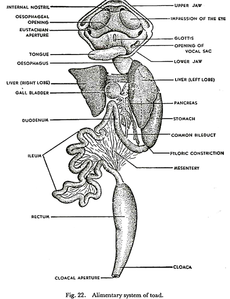
The following points highlight the two main types of alimentary system of toad. The types are: 1. Alimentary Canal 2. Digestive Glands.
Type # 1. Alimentary Canal:
The alimentary canal is a long muscular tube running through the body from end to end. It begins anteriorly in the mouth and ends posteriorly at the cloacal aperture.
The tube is swollen in some places and at others it is narrowed and coiled, so that one can easily recognise different distinct parts. The various parts of the alimentary canal are mouth, buccal cavity, pharynx, oesophagus, stomach, small intestine, large intestine, cloaca and cloacal aperture.
The mouth is a wide opening at the anterior end of the snout. It extends from corner to corner as far back as the tympanic membrane. Mouth leads into a spacious buccal cavity which is bounded by two jaws. The upper jaw is immovable but the lower jaw can be moved up and down, closing and opening the mouth. The jaws are toothless and covered by hard sheaths.
The buccal cavity is lined by mucous membrane. On the roof of the buccal cavity, and situated anteriorly, are two small openings called internal nares which communicate through the nose with the external nares. Just behind the internal nares are the impressions of the eyeballs. These are increased by pushing the eyes into the orbits.
Still further behind, near the angle of the jaws, are the eustachian apertures, one on each side. These are large openings leading into the tympanum of the same side. Attached to the floor of the buccal cavity is a fleshy tongue. It is a broad mucous-covered flap which is fixed by the anterior margin.
The free posterior margin of the tongue may be flicked out of the mouth and quickly retracted in, so as to form an efficient apparatus for catching insects. Behind the tongue is a raised area with a median longitudinal slit. This is the glottis which communicates with the lungs. Beyond this, the buccal cavity narrows down to form an indistinct pharynx which leads directly into the gullet or oesophagus.
The oesophagus is a short wide tube which runs into the body cavity and connects with the stomach. Internally its mucous membrane is thrown into numerous longitudinal folds, so that it may be expanded during the passage of a big bolus of food. The stomach is a thick-walled, slightly bent, muscular bag which lies a little to the left of the middle line. Its broader anterior end is connected with the oesophagus.
This is the cardiac end (kardia — heart), because it lies nearest to the heart. The opposite end of the stomach is the pyloric end which opens into the small intestine through the pylorus (pylorus—gate-keeper). The opening is guarded by a circular band of constricted sphincter muscle, the pyloric valve, which regulates the departure of food from the stomach.
Embedded in the internal mucous membrane of the stomach there are branched and tubular gastric glands secreting gastric juice for the digestion of food. Following the stomach is the small intestine. The first part of the small intestine is known as the duodenum and the second part as the ileum.
The duodenum is a relatively short tube which runs forward from the pylorus and lies nearly parallel to the body of the stomach to which it is attached by a fold of peritoneum. It receives the ducts from the liver and the pancreas and is continued backwards as the much-coiled ileum.
The ileum is the longest part of the alimentary canal. The coils of the ileum are held together and fastened to the body wall by a fold of mesentery. The mucous membrane of the small intestine contains a large number of intestinal glands which secrete intestinal juice.
Moreover, the mucous membrane is thrown into folds which hang freely within the lumen of the gut for absorbing the digested food. The small intestine is abruptly enlarged to form the large intestine which consists of the rectum and the cloaca. The rectum is a wide flask-shaped tube about 2 “5 cm. in length.
Posteriorly it is joined to the cloaca. The cloaca receives the ducts from the kidneys, the urinary bladder and the reproductive organs and finally opens to the exterior through the vent. The vent or cloacal aperture is a small opening at the posterior end of the trunk.
Histologically, the wall of the alimentary canal is composed of five layers:
(1) A thin outer layer of peritoneum forming the serous coat;
(2) An outer layer of longitudinal muscles;
(3) An inner layer of circular muscles, the two together forming the muscular coat;
(4) A connective tissue layer containing blood vessels and forming the sub-mucous coat;
(5) The innermost mucous coat or mucous membrane composed of epithelial cells. These layers are best seen in a cross-section of the ileum (Fig. 23). It is to be noted that by the contraction of the muscular coat, the alimentary canal drives the food onwards in peristaltic waves (peri=around; stalsis—constriction).
Type # 2. Digestive Glands:
Besides the minute gastric and intestinal glands already mentioned, there are two important digestive glands which are associated intimately with the gut. These are the liver and the pancreas [pan—all; kreas—flesh).
The liver is a large, dark-red organ suspended by a mesentery in the anterior part of the body cavity. It consists of two main lobes, right and left, connected by means of a narrow portion. The left lobe of the liver is incompletely subdivided into two masses.
The liver secretes bile which comes out by tubes called hepatic ducts (hepar—liver). Between the right and left lobes of the liver is a dark-green, spherical sac, the gall-bladder, which acts as reservoir of the bile. The hepatic ducts from the liver join the cystic duct from the gall-bladder to form the common bile duct which runs backward towards the duodenum.
On the way, it has to pass through the substance of the pancreas where it receives numerous small pancreatic ducts. Coming out from posterior end of the pancreas, the common bile duct, which may now be called the hepato-pancreatic duct, finally opens into the duodenum near the pylorus of the stomach.
The pancreas is a pale yellow, elongated structure of irregular shape lying in the angle between the stomach and the duodenum. It secretes pancreatic juice which is carried, along with bile, to the duodenum through the common bile duct.
Events during feeding and digestion:
The toad is a carnivorous animal fond of ingesting living insects, caterpillars, earthworms and snails. It is attracted by the movement of the prey. Insects are snapped in by the tongue, and earthworms are crushed by the jaws till they become quiet, and then swallowed as whole.
The body of the prey is composed of:
(i) Proteins,
(ii) Fats,
(iii) Carbohydrates,
(iv) Mineral salts,
(v) Vitamins, and
(vi) Water.
These are known as the proximate principles of the food. Vitamins, mineral salts and water are directly absorbed into the body and need not undergo digestion. Carbohydrates, fats and proteins, however, are very complex organic compounds which cannot be absorbed unless they are broken down and prepared into simpler substances. The process of preparing the food for absorption into the body is known as digestion.
The ingested food cannot be considered as a part of the body unless it is digested and absorbed by the cells. Digestion of the food is effected by the digestive juices which contain among other things chemical substances called enzymes or ferments. The enzymes are responsible for the digestion of proteins, fats and carbohydrates.
Each enzyme is specific, that is, it can act upon only one particular kind of food. An enzyme which splits proteins will not break down carbohydrates or fats. Moreover, the digestive enzymes act by causing hydrolysis.
This means that a molecule of water is chemically added to a molecule of the substrate upon which the enzyme is acting. Lastly, the enzymes are catalysts and hence they are not spent up in the course of the chemical reaction.
Considering these properties, the digestive enzymes are classified into three main groups:
(i) Proteolytic enzymes such as pepsin of the gastric juice, trypsin of the pancreatic juice, and erepsin of the intestinal juice;
(ii) Lipolytic enzyme such as lipase of the pancreatic juice;
(iii) Amylolytic or diastatic enzyme such as amylase of the pancreatic juice. The proteolytic enzyme pepsin breaks down proteins into peptones which are further broken down to amino acids by trypsin and erepsin.
The lipolytic enzyme lipase splits fat into fatty acids and glycerine. The amylolytic enzyme amylase acts upon carbohydrates like starch and breaks them down to complex sugars like maltose. The maltose is then further broken into simple sugars like glucose by the action of maltase of the intestinal juice.
No digestion takes place in the mouth of the toad. The food is mixed with mucus and propelled into the stomach by the peristaltic contraction of the oesophagus. The food is churned and thoroughly mixed with gastric juice in the stomach.
The gastric juice contains hydrochloric acid which helps the pepsin to convert proteins into peptones. The food is temporarily stored in the stomach for a short time and then partially digested food passes by peristalsis through the pylorus into the duodenum.
Here the acid chyme is treated with bile, pancreatic juice and intestinal juice. The bile is alkaline in reaction. It neutralises the acid and helps in the digestion and absorption of fats. There is, however, no digestive enzyme in the bile.
The pancreatic trypsin then converts peptones into amino acids. The process is hastened by the erepsin of the intestinal juice. The proteins are thus converted completely into soluble amino acids. The pancreatic lipase digests fat into soluble fatty acids and glycerine.
The amylase and maltase together convert starchy matter into glucose. The soluble food is then absorbed into the blood stream through the folds of the mucous membrane of the ileum.
The indigestible residue accumulates in the rectum and then egested as faeces through the cloacal aperture. The absorbed food is assimilated, that is, changed into the protoplasm of the toad. It is then utilised by the animal for maintaining its growth and repairing its tissues. A part of the assimilated food is spent as a readily available source of energy for carrying out vital activities.
The surplus is stored in the body for future use. The glucose is converted into glycogen or animal starch and stored as such in the liver and skeletal muscles. The fatty acids are reconverted into fat and stored in the fat bodies. The amino acids are not stored, the surplus being converted into urea and excreted as such in the urine.

