Let us learn about Monocotyledonous Stems. After reading this article you will learn about: 1. Regions of Monocotyledonous Stems 2. Features of Monocotyledonous Stems 3. Types.
Contents
Regions of Monocotyledonous Stems:
Stele:
The vascular bundles of monocotyledonous stems, instead of being arranged in a cylinder as in dicotyledonous stems, are usually scattered throughout the stele, including the pith, so that there is no distinction between pith and pith rays.
Sometimes the centre of the stele is free from vascular bundles and is occupied by parenchyma cells, which dry up and disappear at an early stage, resulting in a hollow stem, as in most grasses.
Vascular Bundles:
The vascular bundles of monocotyledonous stems are like those of dicotyledonous stems in consisting of xylem towards the centre of the stele and phloem towards the periphery. The vascular bundles of monocotyledonous stems do not possess a cambium layer which is found in dicotyledonous stems. This means that monocotyledonous stems usually do not have secondary thickening.
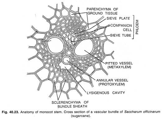
Each bundle remains more or less completely surrounded by a sheath of sclerenchyma cells, the bundle sheath, which is particularly well developed on the sides toward the centre and toward the periphery of the stem. The phloem is made up mostly of sieve tubes and companion cells, and the xylem of vessels and wood parenchyma.
Features of Monocotyledonous Stems:
The most distinctive and characteristic anatomical features of the monocotyledonous stem are as follows:
1. The vascular bundles are many.
2. The stele is broken up into bundles. The vascular bundles are lying scattered in the ground tissue of the axis.
3. The endodermis is not found. The cortex, pericycle and pith are not differentiated because of the presence of scattered bundles throughout the axis.
4. The vascular bundles are collateral and closed. The secondary growth of usual type is lacking, but vestiges of cambial activity in bundles may present in the plant body.
5. Leaf trace bundles are numerous. The leaf traces when enter the stem, penetrate deeply. The median traces penetrate more deeply than lateral. The bundles are common. Each common bundle somehow or other fuses with other bundle in the due course of time. The anastamoses occur at the nodes.
6. Each vascular bundle remains surrounded by a well developed sclerenchymatous sheath.
7. The vascular bundles are commonly oval shaped.
8. The phloem is represented by sieve tubes and companion cells only. The phloem parenchyma is not found.
9. The pith is not marked out.
10. Usually sclerenchymatous hypodermis is present.
11. Usually epidermal hairs are not present.
Types of Monocotyledonous Stems:
Anatomy of the Stem of Zea Mays:
Epidermis:
The epidermis consists of a single layer of compact cells having no intercellular spaces among them. It is covered with thick cuticle. The epidermal hairs are altogether absent.
Hypodermis:
Below the epidermis, usually two or three layers of sclerenchyma cells represent hypodermis.
Ground tissue system:
It consists of thin walled parenchyma cells having well-defined intercellular spaces among them. This tissue extends from below the sclerenchyma (hypodermis) to the centre. It is not differentiated into cortex, endodermis, pericycle and pith.
Vascular system:
It is composed of many collateral and closed vascular bundles scattered in the ground tissue. The vascular bundles lie toward periphery in greater number than the centre. Comparatively the peripheral bundles are smaller in size than the central ones.
Each bundle is more or less surrounded by a sheath, which is more conspicuous towards upper and lower sides of the bundle. The bundle consists of two parts, i.e., xylem and phloem.
Usually the xylem is Y-shaped and consists of pitted and bigger vessels of metaxylem and smaller vessels (annular and spiral) of protoxylem. In between metaxylem vessels, small pitted tracheids are also found. Around the lysigenous or water cavity wood parenchyma is present. The lysigenous cavity is formed by the breaking down of the inner protoxylem vessel.
Phloem consists of sieve tubes and companion cells. Phloem parenchyma is altogether absent in most of monocotyledonous stems. The outer phloem which is broken mass may be called as protophloem and the inner portion is metaphloem. Sieve tubes and companion cells are quite conspicuous.
Anatomy of the Stem of Asparagus:
Epidermis:
It is outermost uniseriate layer composed of approximately rounded cells with cuticularized outer walls.
Ground tissue system:
Just beneath the epidermis a few layers of parenchyma are found which contain chloroplasts in them. This may be called cortex. The innermost layer of the cortex consists of compact cells and called the starch sheath.
Below the starch sheath a multi-layered complete band of sclerenchyma occurs, which gives mechanical support to the stem. The rest of the portion is ground tissue which consists of thin walled parenchyma cells having well developed intercellular spaces among them. The vascular bundles remain scattered in the ground tissue.
Vascular system:
The vascular bundles remain scattered in the ground tissue. The central bundles are comparatively larger than the peripheral ones. They are always collateral and closed. Each vascular bundle consists of xylem and phloem. The xylem is Y-shaped. The metaxylem vessels form arms of Y and protoxylem, the base. Phloem consists of sieve tubes and companion cells. Bundle sheath is not found.
Anatomy of the Scape of Canna:
Epidermis:
It is the outermost uniseriate layer consisting of small, polygonal cells with cuticularized outer walls.
Ground tissue system:
Just beneath the epidermis a few layers of parenchyma occur forming small cortical region. The cells of cortex are sufficiently large and polygonal. Immediately below the cortex, a single-layered chlorophyllous tissue is found consisting of chloroplast bearing cells.
The sclerenchyma patches also remain attached to the chlorophyllous tissue here and there. The rest of the portion consists of a continuous mass of large, thin walled, parenchymatous cells having sufficiently developed intercellular spaces among them. It is called the ground tissue.
Vascular bundles:
They are many and of various sizes, lying scattered in the ground tissue. The bundles are closed and collateral. Each bundle is incompletely surrounded by a sheath of sclerenchyma called bundle sheath. The outer sclerenchyma patch of the bundle is more distinct and cap like whereas inner patch is not so developed.
Each bundle consists of xylem and phloem. The xylem is situated on the inner side and the phloem towards outer side. The xylem consists of a large spiral vessel with one or two smaller vessels of same nature. The phloem consists of sieve tubes and campanion cells.
Anatomy of the Stem of Wheat (Triticum Aestivum):
Epidermis:
It is the outermost uniseriate layer, usually composed of compact tabular cells with cuticularized outer walls. The stomata are also seen here and there on the epidermis.
Ground tissue system:
Just beneath the epidermis the sclerenchyma cells occur in small patches which are not arranged in a continuous band, but are interrupted by chlorenchyma tissue here and there. The stomata are confined on the epidermis only in chlorenchymatous regions. The rest of the ground tissue consists of thin walled rounded or oval parenchyma cells having sufficiently developed intercellular spaces among them. The central region of the stem is hollow.
Vascular bundles:
The closed and collateral vascular bundles occur in two series. The peripheral series consists of smaller bundles, whereas the inner series is of bigger bundles. The vascular bundles of outer series are lying embedded in sclerenchyma band.
The bundles of inner series are also surrounded by sclerenchymatous bundle sheath like that of maize stem. The bundle sheath of peripheral bundles actually touches the epidermis.


