The below mentioned article provides an overview on the epidermal tissue system of plants.
Epidermis:
This system solely consists of the outermost skin or epidermis of all the plant organs beginning from the underground roots to the fruits and seeds.
This layer represents the point of contact between the plants and the outer environment and, as such exhibits diversities in structure.
It is primarily a protective tissue, which protects the internal tissues against excessive loss of water by transpiration and mechanical injury. Subsidiary functions like storage of water, mucilage, secretion and, though rarely, even photosynthesis, may also be carried on.
But for stomatal and lenticular openings the epidermis is a continuous layer. Normally it is uniseriate—typically consisting of one layer of cells. It derives its origin from the protoderm of the meristematic region.
The protoderm cells divide anticlinally and in course of time uniseriate epidermis is formed. Many-layered or multiseriate epidermis, usually called multiple epidermis, is found in some organs like roots of orchids, leaves of Ficus spp. (Fig. 507A), Nerium, Peperomia, etc.
Normally it may be assumed that these layers have originated from the protoderm by periclinai divisions. The outermost layer of multiple epidermis is similar to ordinary uniseriate one. The inner layers are different from other tissues in absence of chlorophyll.
There is room for doubts if all these layers belong to epidermis from ontogenetic point of view. They may be outer layers of cortex originating from the ground meristem, but resemble the epidermis both in structure and function.
The epidermal cells are living with lining layer of protoplast around large central vacuole. The plastids are normally small and colourless. Chloroplasts are present only in the guard cells of the stomata in case of organs exposed to sunshine, but they occur in the epidermal cells of aquatic plants and plants growing in moist and shady situations. Mucilage, tannins and crystals may occasionally be present.
Anthocyanins may occur in the cell-sap of the vacuoles. Epidermal cells retain the potentiality of cell division. During normal course of development or due to external stimuli they may divide and produce new cells.
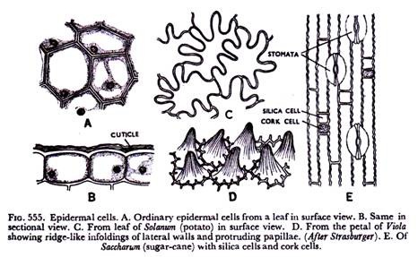
Epidermal cells exhibit wide diversities as regards their size, shape and arrangement. But they may be said to be essentially tabular in shape (Fig. 555 A & B) compactly set, so that a continuous layer without intercellular spaces is formed. Only in the petals of some flowers intercellular spaces are found, but they remain covered by outer cuticle. In surface view they are more or less isodiametric in shape.
In the leaves and petals of flowers they may have irregular shapes, often with teeth and flanges (Fig. 555 C & D) which remain peculiarly interlocked with one another. In monocotyledonous stems and leaves with parallel venation the epidermal cells are rather elongated in the direction of the long axis (Fig. 555 E), so much so that in extreme cases they may be fibre-like in appearance.
Epidermal cells have unevenly thickened walls, the outer and radial walls being much more thick than the inner walls. In some cases they may be so massive that the central lumen is almost obliterated. The walls are strongly cutinised, what is very important for protection against mechanical injuries and prevention of loss of water.
The fatty substance cutin is found in the wall—in interfibriller and intermicellar spaces of the cellulose and forms the cuticle occurring all over the outer wall of the epidermal cells (Fig. 556 A&B).
It remains as a separate layer and in some cases it may be removed as a whole. The cuticle is often found to project into the radial walls as peg-like bodies (Fig. 556C). Cuticle is absent only in the epidermis of roots and some submerged aquatic plants.
The thickness of the outer walls of the epidermal cells depends on the environmental conditions of the plants. It is quite thin in plants with adequate water supply, and it is unusually thick in plants growing in dry situations.
The surface of the cuticle may be smooth or may possess ridges and cracks. The cutinised portion of the walls, the portion lying beneath the cuticle, has been found to consist of alternating layers of cutin and pectic materials.
Waxy matters are often deposited on the cuticle in form of rods and grarules (Fig. 556E). The so-called ‘bloom’ of many fruits and glaucous characters of many stems and leaves are due to these deposits. Lignification is rather rare in epidermal cells.
It occurs in the pine needles (Fig. 556D), in cycad, in grass leaves outside the sclerenchyma patches and in a few dicotyledons. Deposition of silica is common in the epidermal cells of horse-tails (Equisetum) and grasses.
It is really interesting to find long epidermal cells having corrugated margin (Fig. 555E) associated with two kinds of short cells—the silica cells and cork cells in grasses. The silica cells contain silicon oxide and cork cells with suberised walls contain organic materials.
In some dicotyledonous families like Malvaceae, Rutaceae, etc., the epidermal cells individually or in groups undergo mucilaginous changes, particularly in the seeds. Special sac-like cells remain scattered in the epidermis of some members of family Cruciferae.
These are idicblastic cells resembling the lati ciffers, but they contain an enzyme, myrosin, and so they are called myrosin cells. It has been stated in a preceding chapter that many dicotyledonous families like Urticaceae, Moraceae, possess cystoliths.
The cystolith-containing cells of epidermis are referred to a lithocysts. The epidermis is often made up of a layer of sclereids, as found in the seed-coats of Pisum and Phaseolus of family Leguminosae (Fig. 537D) and in the scales of garlic—Allium sativum of family Liliaceae (Fig. 537E).
Radial and inner walls of epidermal cells possess pit-fields. Plasmodesmata have also been reported, those on the outer walls of leaves have also been called ectodesmata.
The origin of the shoot epidermis may be traced from the apical meristem. It arises from the outer layers-of tunica, according to tunica-corpus theory, or from the dermatogen of Haustein or protoderm, as suggested by Haberlandt, which may be called primordial epidermis.
But the root epidermis fundamentally differs from that of shoot in origin, structure as well as in function. It has been pointed out in the previous chapter that epidermis of root is related to the root-cap or the cortex from the developmental point of view.
The cells are tabular, lack in cutinisation of wall and their function is mainly absorption of water and solutes. So the terms epiblema, piliferous layer or rhizodermis have been applied to it.
Epidermis, as a rule, persists as uniseriate layer throughout its life in the organs where distinct secondary growth does not take place. In some monocotyledons, though secondary increase is absent, a kind of periderm is formed, and thus the epidermis is destroyed.
In organs with distinct secondary growth in thickness epidermis continues till cork cells are formed. In leaves, flowers and fruits, it persists as long as the organs do. In roots the epidermis with a part of cortex becomes dead, lignified or suberised after the root hairs are destroyed.
Bulliform Cells:
In the leaves of monocotyledons, excepting a few families, a peculiar type of comparatively larger, highly vacuolate and thin-walled cells occur in the epidermis. These are called bulliform (meaning, bubble-like) cells.
In transverse section they appear as a fan-like band because the median cell is usually the largest in size (Figs. 557 & 557A). They may be present on both sides of a leaf, but are more common on the upper side running parallel to the veins.
They either cover large areas or remain restricted to the grooves. These are mainly water-containing cells with no chlorophyll. The walls are usually thin, but the outer walls may be thick and cutinised like other epidermal cells, often filled with silica.
There are three views as regards the functions of bulliform cells. According to the first view they are concerned with the unrolling of the developing leaves. It is suggested that these cells undergo sudden and rapid expansion at a certain stage of leaf development and consequently bring about unfolding of the leaves.
The second view is that they have a role to play in the hygroscopic opening and closing movements of mature leaves, due to changes in turgor. They have also been called motor cells by workers holding the above view. The third view is that they are simply concerned with water-storage and have no other function.
Stomata:
The continuity of the epidermis of aerial organs is interrupted by the presence of some minute pores or openings on it. These pores are called the stomata, through which exchange of gases takes place between the internal tissues and the outer atmosphere. A stoma has a small slit or pore and two specialised epidermal cells, called guard cells, on the two sides. Often other epidermal cells adjacent to the stoma undergo modifications.
They differ from other epidermal cells and become associated with the stoma functionally. These are referred to as subsidiary or accessory cells (Figs 559 & 561). Though gaseous interchange actually occurs through the pore, called stomatal aperture or opening, the term stoma includes the whole thing, the pore, guard cells and subsidiary cells, when present.
In surface view the guard cells look cresent or kidney-shaped in appearance, being attached to each other at the margin of the concave side with the aperture lying in between them (Fig. 558A). They may be easily distinguished from ordinary epidermal cells, because they possess dense cytoplasm, prominent nuclei, chloroplasts, and even starch grains.
A cavity is present just beneath the stoma, what is called sub-stomatal chamber or cavity (Fig. 558B). It is in communication with the intercellular space system of the internal tissues.
The walls of the guard cells are unevenly thickened, the wall along the aperture being strongly built and that away from the aperture being thin and extensible. The guard cells have cutinised outer walls with a layer of cuticle which extends through the aperture and joins the inner wall.
Due to strong cutinisation often ledges of wall materials are noticed on the upper and lower sides of the ventral wall, so that in sectional view they appear like horns or beaks. The ledges project above and below and overarch the two chambers, referred to as the front cavity and back cavity, which communicate with each other through the pore (Figs. 558B & 560A).
The guard cells, due to uneven thickening of the wall, what is really an outstanding character, can regulate the opening and closing of the stomatal aperture. Normally stomata remain open in daytime and close up with nightfall.
The opening is influenced by the changes in the turgor of the guard cells. With increase of turgor the thinner walls of the guard cells get stretched and the thicker walls become more concave, thus the gap becomes wide.
In the grass and sedge families the guard cells of the stomata are peculiarly dumb-bell- shaped where the middle portion is straight and strongly thickened and the two ends are swollen or bulbuous (Fig. 559) and thin-walled.
Here increase in turgor causes further swelling of the bulbuous ends and, as a result, the straight median portions get separated from each other. Decrease in turgor brings about reverse changes. The physiological factors influencing detailed mechanism of the opening and closing of stomatal aperture will be taken up in the portion on plant physiology.
Stomata occur in all aerial parts of the plants, most abundantly in the foliage leaves. Those present on the floral parts and in the aquatic plants are normally functionless. In leaves they may occur on both upper and lower surfaces. In woody plants with dorsiventral leaves they are located on the lower epidermis. In herbaceous plants with isobilateral or centric leaves they occur on both the surfaces. Even in that case stomata are more abundant on the lower side than on the upper.
In floating leaves they occur only on the upper epidermis. In an individual leaf stomata are more numerous near the apex and minimum near the base, the middle portion having a distribution, which is an average of the apex and base. In leaves with parallel venation, as in the monocotyledons, and the needles of conifers stomata remain arranged in parallel rows (Figs. 555E & 559), whereas in reticulately-veined leaves they lie scattered (Fig. 563).
The number of stomata occurring on the epidermis of leaves is fairly large, which may range between a few thousand to over a hundred thousand per square cm. It has been estimated that a maize plant may have more than two hundred million stomata. So one can hardly estimate the number in a large tree.
The guard cells may be at the same level with adjacent epidermal cells or they may be placed above or lie sunken below the surface of the epidermis. Sunken stomata (Fig. 560) are characteristic of the plants of dry situations, where they often appear to be located at the bottom of a cup-shaped depression, which is called the external cavity or outer chamber. This is an effective mechanism for reducing transpiration.
The subsidiary cells are highly thickened here. In the leaves of Nerium a groove or depression is formed, what is called stomatal pit (Fig. 560B) and stomata remain very much sunken.
Stomata raised above the surface of epidermis (Fig. 560D) are found in the peduncle of Cucurbita where they appear
to be placed at the summit of a conical papilla. Stomata also occur on the sporophytes of bryophytes like Anthoceros and mosses.
The stomata of mosses representing really the simplest types, show departure from other types in the nature of thickening of the wall—ventral walls being thin and dorsal thick (Fig. 561) and in the mechanism of opening and closing of the aperture.
Ontogeny of the Stomata:
Stomata arise from the protoderm cells. Normally a protoderm cell undergoes anticlinal division, one of them serves as the stoma mother cell. It eventually divides into two cells leaving a small slit between them (Fig. 562).
The two cells develop into two kidney-shaped guard cells and the slit into the stomatal aperture. In many families the protoderm undergoes several divisions before the stoma mother cell differentiates.
Commonly subsidiary cells arise from protoderm cells lying adjacent to the stoma mother cell. They may be sister cells of the mother cell or may arise by division of the cells lying adjacent to the mother cells. A number of types of stomata have been recognised on the basis of their modes of development, relation with neighbouring cells and occurrence and number of subsidiary cells.
Without going into detail the following types may be cited as common ones:
In Allium, Iris, etc., the protoderm cell divides anticlinally into two unequal cells; the smaller one serves as the stoma mother cell which gives rise to the stoma. The subsidiary cells are absent. It is a very common type of stoma.
In Zea, bamboos and other members of grass and sedge families the guard cells are peculiarly dumb-bell-shaped in appearance. Two subsidiary cells arise by division of the protoderm cells lying adjacent to stoma mother cell and they occur on two sides of the guard cells. This is referred to as Zea type.
In Tradescantia four subsidiary cells are formed which originate from four protoderm cells surrounding the stoma mother cell. In Bryophyllum the protoderm cells have been found to produce a series of spirally arranged subsidiary cells, and finally they give rise to the guard cells.
The stomata occurring in bryophytes as found in the sporophyte of Mnium, are the simplest where wall ledges are absent and, unlike other types, the ventral wall is thin and the dorsal wall thickened. In recent years intensive investigations have been in progress regarding the mode of development of stomata, their relation to the neighbouring cells and the occurrence of the subsidiary cells.
In fact, these characters have been used in problems of classification and phylogeny. The stomata on the basis of investigations particularly in the gymnosperms (Florin and others) have been put into two types: viz., (1) Haplocheilic, where the guard cells originate by a single division of the stomatal initial, and some of the neighbouring cells become modified into subsidiary cells.
(2) Syndetocheilic type, when the guard cells and subsidiary cells originate from the same mother cell. It was thought that haplocheilic type is more primitive than the syndetocheilic one, but actual studies on a large number of plants do not support that contention.
Both the types have been noticed in gymnosperms and many families of angiosperms. In fact, different types have been found in the different genera of the same family, and even in different species of the same genus.
Modern workers (Cf. Metcalfe and Chalk) have suggested the following types of stomata in the dicotyledons on the basis of the characters stated above.
A. Anomocytic or irregular-celled type (Fig. 563A):
Stoma remains surrounded by a limited number of cells which cannot be distinguished from other epidermal cells. Thus the subsidiary cells are absent. This is also called ranunculous type, common in the families Ranunculaceae, Capparidaceae and others.
B. Anisocytic or unequal-celled type (Fig. 563B):
Here the stoma remains surrounded by three subsidiary cells of which one is distinctly smaller than the other two. It is otherwise known as cruciferous type common in Cruciferae.
C. Diacytic or cross-celled type (Fig. 563C):
Here the stoma remain enclosed by a pair of subsidiary cells whose common wall is at right angles to the guard cells. This is also called caryophyllaceous type, common in Caryophyllaceae, Acanthaceae and others.
D. Paracytic or parallel- celled type (Fig. 563D):
The stoma is accompanied on either side by one or more subsidiary cells which lie parallel to the long axis of the pore of guard cells. This is also referred to as rubiaceous type common in Rubiaceae, Magnoliaceae and others.
In view of the fact that diversities occur as regards the nature of the stomata the terms ranunculous, etc., are rather confusing, and anomocytic, etc., suggested by Metcalffe and Chalk appear to be more appropriate.
Another classification on the basis of development was devised (Pant, 1965), and stomata have been put in three categories:
(1) Mesogenous type—guard cells and subsidiary cells derived by consecutive division of a mother cell, e.g., Rubiaceae, Cruciferae.
(2) Mesoperigenous type—where the surrounding cells are of dual origin, some from the mother cell and some from the neighbouring cell, e.g, Ranunculaceae, Caryophyllaceae.
(3) Perigenous type—all neighbouring and subsidiary cells having independent origin, e.g., Cucurbitaceae, Nympheaceae.
In the monocotyledons the most common one is the graminaceous or grass type (Fig. 559). Here the two guard cells are dumb-boll-shaped having a narrow middle portion and bulbuous ends. Two distinct subsidiary cells lie parallel to the long axis of the pore.
In recent years intensive investigations have revealed a few other types as well (Fig. 563A). In Orchidaceae, Amaryllidaceae and others the guard cells are not associated with any subsidiary cells.
Guard cells surrounded by four to six subsidiary cells have been noticed in many species of Araceae, Commelinaceae, Musaceae and others. In Palmae, Pandaceae guard cells have four subsidiary cells—two of them are lateral and two polar ones.
The latter ones are smaller in size and round in shape. The term tetracytic has been used for this type.
It has been suggested that stomata with many subsidiary cells are primitive, and those with few or no subsidiary cells have been derived by reduction.
The stomata are very important from physiological point of view. It is through them that interchange of gases takes place between the intercellular space system of the internal tissues and the outer atmosphere and thus important physiological functions like photosynthesis, respiration and transpiration become possible.
Water-stomata or hydathodes are also epidermal openings through which liquids often with dissolved salts, are exuded from the plants. They have been discussed in the preceding chapter.
Epidermal Outgrowths:
Outgrowths of diverse forms, structures and functions develop from the epidermis. All these appendages which are epidermal in origin, are referred to as trichomes.
Thus they are different from the emergences like the prickles of roses, as the latter are formed by epidermis and a part of cortex. Trichomes may occur on all parts of the plant body.
Some of them persist throughout the life of the organs, whereas many of them are ephemeral bodies. They may remain alive or become dead and continue as such. Trichomes have been put into a number of groups on the basis of their morphological characters.
(a) Hairs:
Hairs constitute a very common type of trichome. They may be unicellular or multicellular. Unicellular hairs are often simple unbranched elongated bodies or they may be branched.
Some of them are very much elongated and twisted, so that they have woolly appearance (Fig. 564-C). Multicellular hairs may be formed of one row of cells (Fig. 564 A, D, E & F) or of many layers as found in the base of petiole of Portulaca.
Often these hairs branch in very peculiar fashions; some of them assume dendroid or tree-like appearance (Fig. 564 G & H), or the branches come out in one plane giving it stellate or star-like shape.
They are also called stellate hairs (Fig. 564 I), A multicellular hair has usually two parts, the basal part which remains embedded in the epidermis is the foot and the other which projects out is the body. An initial cell divides periclinally into two parts, of which the outer one forms the body and the inner one, the foot.
(b) Scales or Peltate hairs:
These hairs consist of disc-like plate of cell (Fig. 564 J) put on a short stalk or directly attached to the foot.
(c) Colleters:
These are glandular trichomes. Some hairs have multicellular stalk and head, the latter is composed of glandular cells (Fig. 565). Sticky exudations present on the surface of certain leaves and buds are secreted by colleters.
Salt-secreting glands as found in Tamaricaceae and calcium- secreting glands of Plumbaginaceae are really interesting (Fig. 565A).
(d) Water vesicles or bladders:
They form a very interesting type of trichome where some epidermal cells become greatly distended and serve as water reservoir.
They occur in
the so-called ‘ice-plant’ (Mesembryanthemum crystallinum of family Aizoaceae) where the surface of the leaves and young stems appear to be covered by ice-beads (Fig. 566).
Those occurring in Artiplex, also called vesiculate hairs, dry up with maturity and persist as a white layer on the leaf surface (Fig. 565A).
The walls of trichomes are commonly of cellulose covered by cuticle. They sometimes remain impregnated with silica and calcium carbonate. Trichomes other than glandular ones have highly vacuolated protoplast. The cotton fibres, which are really hairy outgrowths from the seeds, have secondary walls of almost pure cellulose. The stinging hairs of nettle (Urtica dioica) possess a peculiar type of wall structure for releasing the contents of the gland.
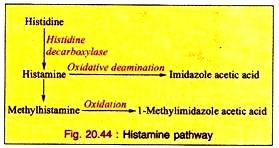
The hair (Fig. 244) resembles of fine capillary tube with silicified upper end and calcified lower end. The base remains embedded in the epidermal cells. Coming in contact with the skin the tip breaks at a predetermined point and the sharp edge penetrates into the skin when the contents (histamine and acetycholine) are injected, so to say, to the wound.
Root-hairs:
As already reported the root epidermis fundamentally differs from shoot epidermis in origin and in absence of cuticle and stomata. But it bears hairs at a particular zone. Unlike the hairs and trichomes discussed above, the root-hairs are not outgrowths or appendages, but they are prolongations of the epidermal cells. During the formation of root-hairs, growth in length of the epidermal cells is checked.
It has been found in some plants that root epidermis possesses two types of cells, short cells and long cells due to unequal division, and the hairs are formed from the short ones (Fig. 567) which are called trichoblasts.
It comes out as a protuberance, continues elongation and thus the hair is formed. It has vacuolate protoplast and the nucleus moves on to the tip. The wall is thin, composed of cellulose and pectic materials. Root-hairs are short-lived bodies.
During the growth of the root, old hairs are destroyed and replaced by new ones. They are responsible for the absorption of water and mineral solutes from the soil.
