The following points highlight the three major groups of Plant Tissues. The groups are: 1. Meristems Tissue 2. Permanent Tissue 3. Secretory Tissue.
Contents
Plant Tissue: Group # 1. Meristems Tissue:
A meristematic tissue consists of a group of cells which remain in a continuous state of division or they retain their power of division.
The characteristic features of meristematic tissue are as follows:
1. They are composed of immature cells which are in a state of division and growth.
2. Usually the intercellular spaces are not found among these cells.
3. The cells may be rounded, oval or polygonal in shape; they are always living and thin-walled.
4. Each cell of meristematic tissue possesses abundant cytoplasm and one or more nuclei in it.
5. The vacuoles in the cells may be quite small or altogether absent.
Meristems and Growth of Plant Body:
Beginning with the division of the oospore, the vascular plant generally produces new cells and forms new organs until it dies. In the beginning of the development of the plant embryo cell division occurs throughout the young organism.
But as soon as the embryo develops and converts into an independent plant the addition of new cells is gradually restricted to certain parts of the plant body, while the other parts of the remain concerned with activities other than growth.
This shows that the portions of embryonic tissue persists in the plant throughout its life, and the mature plant is a composite of adult and juvenile tissues. These juvenile tissues are known as the meristems. The presence of meristems remarkably differentiates the plant, from the animal.
In the growth resulting from meristematic activity is possible throughout the life of the organism, whereas in animal body the multiplication of the cells mostly ceases when the organism attains adult size and the number of organs is fixed.
The term meristem (Greek meristos, meaning divisible) emphasizes the cell-division activity characteristic of the tissue which bears this name. It is obvious that the synthesis of new living substance is a fundamental part of the process of the formation of new cells by division.
The living tissues other than the meristems may also produce new cells, but the meristems carry on such activity indefinitely, because they not only add cells to the plant body, but also perpetuate themselves, that is, some of the products of division in the meristems do not develop into adult cells but remain meristematic.
The meristems usually occur at the apices of all main and lateral shoots and roots and thus their number in a single plant becomes quite large. In addition, plants bearing secondary increase in thickness possess extensive meristems, the vascular and cork cambia, responsible for the secondary growth.
The combined activities of all these meristems give rise to a complex and large plant body. The primary growth, initiated in the apical meristems expands the plant body and produces the reproductive parts. On the other hand, the cambia, aid in maintenance of the expanding body by increasing the volume of the conducting system and forming supporting and protecting cells.
Classification of Meristems:
Various systems of classifying meristems have been proposed by many eminent workers which are based on the characteristics such as stage of development, position in plant body, origin, function and topography. No system is exclusive and rigid.
A few important types have been discussed here:
1. Meristems Based on Stage of Development:
Promeristem or primordial meristem:
Promeristem is the region of new growth in a plant body where the foundation of new organs or parts of organs is initiated. Sometimes, it is also called primordial meristem, urmeristem and embryonic meristem. From the view point of its structure, this region consists of the initials and their immediate derivatives.
The cells of this region are isodiametric, thin walled, vacuolate, with active cytoplasm and early stages of pits. Prominent nuclei and inconspicuous intercellular spaces may be seen. As soon as the cells of this region begin to change in size, shape, and character of wall and cytoplasm, setting off the beginning of tissue differentiation, they are no longer a part of typical meristem; they have passed beyond that earliest stage.
2. Meristems Based on Origin of Initiating Cells:
Primary and secondary meristems:
The meristems are classified as primary and secondary, on the basis of type of tissue in which origin occurs.
The primary meristems are those that build up the primary part of the plant and consist in part of promeristem. In primary meristems, promeristem is always the earliest stage. The possession of promeristem continuously from a early embryonic origin is characteristic of primary meristems. The main primary stems are the apices of roots, stems, leaves and similar appendages.
The secondary meristem appears later at a stage of development of an organ of a plant body. Secondary meristems always arise in permanent tissues and they are always found lying lateral along the side of the stem and root. Sometimes, some of the primary permanent tissues acquire the power of division and become meristematic.
These tissues build up the secondary meristem. Secondary meristems are so called because they arise as new meristems in tissue which is not meristematic. The most striking example of secondary meristem is phellogen or cork cambium. It is formed from mature cells cortical, epidermal or phloem cells.
The primary meristems build up the early and structurally and functionally complete plant body. The secondary meristems later add to that body forming supplementary tissues that functionally replace the early formed tissues or serve in protection and repair of wounded regions.
The cambium does not fall definitely in either group (primary and secondary). It arises from apical meristem of which it is late and specialized stage. However, the accessory cambia are secondary. The tissues formed by the cambium are secondary, whereas the primary meristems form only primary tissues.
3. Meristems Based on Position in Plant Body:
As regards their position in plant body, the meristems may be classified into three groups:
i. Apical meristem,
ii. Intercalary meristem and
iii. Lateral meristem.
(i) Apical Meristem:
The apical meristem lies at the apex of the stem and the root of vascular plants. Very often they are also found at the apices of the leaves, Due to the activity of these meristems, the organs increase in length. The initiation of growth takes place by one or more cells situated at the tip of the organ.
These cells always maintain their individuality and position and are called ‘apical cells’ or ‘apical initials’. Solitary apical cells occur in pteridophytes, whereas in higher vascular plants they occur in groups which may be terminal or terminal and sub-terminal in position.
(ii) Intercalary Meristems:
The intercalary meristems are merely portions of apical meristems that have become separated from the apex during development by layers of more mature or permanent tissues and left behind as the apical meristem moves on in growth. The intercalary meristems are inter-nodal in their position.
In early stages, the internode is wholly or partially meristematic, but later on some of its part becomes mature more rapidly than the rest and in the internode a definite continuous sequence of development is maintained. The intercalary meristems are found lying in between masses of permanent tissues either at the leaf base or at the base of internode.
Such meristems are commonly found in the stems of grasses and other monocotyledonous plants and horsetails, where they are basal. Leaves of many monocotyledons (grasses) and some other plants, such as Pinus, have basal meristematic regions. These meristematic regions are short living and ultimately disappear; ultimately, they become permanent tissues.
(iii) Lateral Meristems:
The lateral meristems are composed of such initials which divide mainly in one plane (periclinally) and increase the diameter of an organ. They add to the bulk of existing tissues or give rise to new tissues. These tissues are responsible for growth in thickness of plant body. The cambium and the cork cambium are the examples of this type.
4. Meristems Based on Function:
As regards their function a system of classification of meristems was proposed by Haberlandt in the end of nineteenth century. He suggested that the primary meristem at the apex of the stem and root is distinguished into three tissues — protoderm, procambium and ground or fundamental meristem.
The protoderm is the outermost tissue which develops into epidermis. The procambium develops into primary vascular tissue. It forms isolated strands of elongated cells very near to the central region; in cross-section each procambium appears as a small group of cells in the ground or fundamental meristem, but in longitudinal section the cells appear to be long and pointed.
The ground or fundamental meristem develops into ground tissue and pith; the cells of this region are large, thin walled, living and isodiametric. In later stages, they become differentiated into hypodermis, cortex, endodermis, pericycle, pith rays and pith.
Meristem and Meristematic:
The terms meristem and meristematic, as applied to developing cells and tissues, are somewhat loose. According to Eames and MacDaniels, the term ‘meristem’ is applied to regions of more or less continuous cell and tissue initiation; the adjective ‘meristematic’ is used to indicate resemblance in an important way to a meristem, but not necessarily as consisting of or constituting meristem, i.e., it is applied to those cells, tissues and regions that have characteristics of developing structures — especially cell division — but do not themselves strictly constitute meristems.
For example, the apices of the stems and the cambium are regions of tissue initiation, developing xylem and phloem are meristematic tissues, because they form some new cells and are immature but they are not permanent or semi-permanent initiating regions (meristems).
On the other hand, cells in mature tissue, such as the primary cortex of stems, may divide. Such cells are meristematic, but neither they nor the tissues of which they are a part constitute a meristem.
Meristems and Permanent Tissues:
In active meristems there occurs a continuous separation between cells that remain meristematic the initiating cells and those that develop into the various tissue elements the derivatives of the initiating cells. During this process of developing the derivatives gradually change physiologically and morphologically, and assume more or less specialized characteristics.
In other words, the derivatives differentiate into the specific elements of the various tissue-systems. As the cells of vascular plants vary in their physiologic and morphologic characteristics, they also vary in details of differentiation. Different types of cells attain different degrees of differentiation as compared with their common meristematic precursors.
For example, various parenchyma cells diverge relatively little from their meristematic precursors and retain the power of division to a high degree. On the other hand, such as sieve elements, fibres, tracheary elements are more thoroughly modified and lose most, or all of their former meristematic potentialities.
These variously differentiated cells are known as mature or permanent in the sense that they have reached the degree of specialization and physiological stability that normally characterizes them as components of certain tissues of an adult plant part. During the differentiation of tissues from meristems the derivatives of meristematic cells synthesize protoplasm, enlarge and divide.
It is difficult to delimit the meristem proper from its recent derivatives. The development of meristematic derivatives into mature cells also is gradual. In other words, the differentiation is a continuous process.
Plant Tissue: Group # 2. Permanent Tissues:
The permanent tissues are those in which growth has stopped either completely or for the time being. Sometimes, they again become meristematic partially or wholly. The cells of these tissues may be living or dead and thin-walled or thick-walled. The thin-walled permanent tissues are generally living whereas the thick-walled tissues may be living or dead.
The permanent tissues may be simple or complex. A simple tissue is made up of one type of cells forming a uniform of homogeneous system of cells. The common simple tissues are – parenchyma, collenchyma and sclerenchyma.
A complex tissue is made up of more than one type of cells working together as a unit. The complex tissues consist of parenchymatous and sclerenchymatous cells; collenchymatous cells are not present in such tissues.
The common examples are:
The xylem and the phloem.
Simple Tissues:
1. Parenchyma:
The parenchyma tissue is composed of living cells which are variable in their morphology and physiology, but generally having thin walls and a polyhedral shape, and concerned with vegetative activities of the plant. The individual cells are known as parenchyma cells.
The word parenchyma is derived from the Greek para, beside and enchein, to pour. This combination of words expresses the ancient concept of parenchyma as a semi- liquid substance poured beside other tissues which are formed earlier and are more solid.
Phylogenetically the parenchyma is a primitive tissue since the lower plants have given rise to the higher plants through specialization and since the single type or the few types of cells found in the lower plants have become by specialization the many and elaborates types of the higher plants.
The unspecialized meristematic tissue is parenchyma and is often called parenchyma thus it can be said that, ontogenetically parenchyma is a primitive tissue.
The parenchyma consists of isodiametric, thin-walled and equally expanded cells. The parenchyma cells are oval, rounded or polygonal in shape having well developed spaces among them. The cells are not greatly elongated in any direction. The cells of this tissue are living and contain sufficient amount of cytoplasm in them. Usually each cell possesses one or more nuclei.
Parenchyma makes up large parts of various organs in many plants. Pith, mesophyll of leaves, the pulp of fruits, endosperm of seeds, cortex of stems and roots, and other organs of plants consist mainly of parenchyma. The parenchyma cells also occur in xylem and phloem.
In the aquatic plants, the parenchyma cells in the cortex possess well developed air spaces (intercellular spaces) and such tissue is known as aerenchyma. Parenchyma may be specialized as water storage tissue in many succulent and xerophytic plants. In Aloe, Agave, Mesembryanthemum, Hakea and many other plants chlorophyll-free, thin-walled and water-turgid cells are found which represent water storage tissue.
When the parenchyma cells are exposed to light they develop chloroplasts in them, and such tissue is known as chlorenchyma. The chlorenchyma possesses well developed aerating system. Intercellular spaces are abundant in the photosynthetic parenchyma (chlorenchyma) of stems too.
Commonly the parenchyma cells have thin primary walls. Some such cells may have also thick primary walls. Some storage parenchyma develop remarkably thick walls and the carbohydrates deposited in these walls, the hemicellulose, are regarded by some workers as reserve materials.
Thick walls occur, in the endosperm of Phoenix dactylifera, Diospyros, Asparagus and Coffea arabica. The walls of such endosperm become thinner during germination.
The turgid parenchyma cells help in giving rigidity to the plant body. Partial conduction of water is also maintained through parenchymatous cells. The parenchyma acts as special storage tissue to store food material in the form of starch grains, proteins, fats and oils. The parenchyma cells that contain chloroplasts in them make chlorenchymas which are responsible for photosynthesis in green plants.
In water plants the aerenchyma keep up the buoyancy of the plants. Such air spaces also facilitate exchange of gases. In many succulent and xerophytic plants such tissues store water and known as water storage tissue. Vegetative propagation by cuttings takes place because of meristematic potentialities of the parenchyma cells which divide and develop into buds and adventitious roots.
Origin:
As regards their origin, the parenchyma tissue of the primary plant body, that is, the parenchyma of the cortex and the pith, of the mesophyll of leaves, and of the flower parts, differentiates from the ground meristem. The parenchyma associated with the primary and secondary vascular tissues is formed by the pro-cambium and the vascular cambium respectively.
Procambium—parenchyma associated with the primary vascular tissues.
Vascular cambium—parenchyma associated with the secondary vascular tissues.
Parenchyma may also develop from the phellogen in the form of phelloderm, and it may be increased in amount by diffuse secondary growth.
Phellogen—Phelloderm (parenchyma).
2. Collenchyma:
Collenchyma is a living tissue composed of somewhat elongated cells with thick primary non-lignified walls. Important characteristics of this tissue are its early development and its adaptability to changes in the rapidly growing organ, especially those of increase in length.
When the collenchyma becomes functional, no other strongly supporting tissues have appeared. It gives support to the growing organs which do not develop much woody tissue. Morphologically, collenchyma is a simple tissue, for it consists of one type of cells.
Collenchyma is a typical supporting tissue of growing organs and of those mature herbaceous organs which are only slightly modified by secondary growth or lack such growth completely. It is the first supporting tissue in stems, leaves and floral parts. It is the main supporting tissue in many dicotyledonous leaves and some green stems. Collenchyma may occur in the root cortex, particularly, if the root is exposed to light.
It is not found in the leaves and stems of monocotyledons. Collenchyma chiefly occurs in the peripheral regions of stems and leaves. It is commonly found just beneath the epidermis. In stems and petioles with ridges, collenchyma is particularly well developed in the ridges. In leaves it may be differentiated on one or both sides of the veins and along the margins of the leaf blade.
The collenchyma consists of elongated cells, various in shape, with unevenly thickened walls, rectangular, oblique or tapering ends, and persistent protoplasts. The cells overlap and interlock, forming fibre-like strands. The cell walls consist of cellulose and pectin and have a high water content.
They are extensible, plastic and adapted to rapid growth. In the beginning the strands are of small diameter but they are added to, as growth continues, from surrounding meristematic tissue. The border cells of the strands may be transitional in structure, passing into the parenchyma type.
As regards the cell arrangement there are three types of collenchyma — angular, lamellar and tubular. In angular type the cells are irregularly arranged (e.g., Ficus, Vitis, Polygonum, Beta, Rumex, Boehmeria, Morus, Cannabis, Begonia); in lamellar type the cells lie in tangential rows (e.g., Sambucus, Rheum, Eupatorium) and in tubular type the intercellular spaces are present (e.g., Compositae, Salvia, Malva, Althaea).
The common typical condition is that with thickenings at the corners. The three forms of collenchyma have been named by Muller (1890) angular (Eckencollenchym), lamellar (Plattencolenchym), and tubular or lacunate (Luchencollenchym), respectively. The word lamellar has reference to the plate like arrangement of the thickenings; the lacunate (tubular) to the presence of intercellular spaces.
The walls of collenchyma are chiefly composed of cellulose and pectic compounds and contain much water (Majumdar and Preston, 1941). In some species collenchyma walls possess an alternation of layers rich in cellulose and poor in pectic compounds with layers that are rich in pectic compounds and poor in cellulose.
In many plants collenchyma is a compact tissue lacking intercellular spaces. Instead, the potential spaces are filled with intercellular material (Majumdar, 1941).
The mature collenchyma cells are living and contain protoplasts. Chloroplasts also occur in variable numbers. They are found abundantly in collenchyma which approaches parenchyma in form. Collenchyma consisting of long narrow cells contains only a few small chloroplasts or none. Tannins may be present in collenchyma cells.
Ontogenetically collenchyma develop from elongate, pro-cambium like cells that appear very early in the differentiating meristem. In the beginning, small intercellular spaces are present among these cells, but they disappear in angular and lamellar types as the cells enlarge, either by the enlarging cells or filled by intercellular substance.
The chief primary function of the tissue is to give support to the plant body. Its supporting value is increased by its peripheral position in the parts of stems, petioles and leaf mid-ribs. When the chloroplasts are present in the tissue, they carry on photosynthesis.
3. Sclerenchyma:
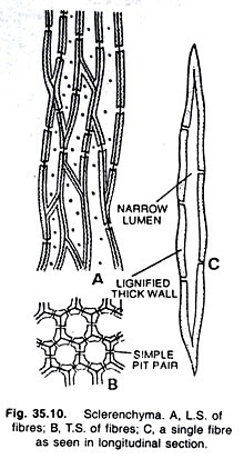
The sclerenchyma (Greek, sclerous, hard; enchyma, an infusion) consists of thick walled cells, often lignified, whose main function is mechanical. This is a supporting tissue that withstands various strains which result from stretching and bending of plant organs without any damage to the thin-walled softer cells.
The individual cells of sclerenchyma are termed sclerenchyma cells. Collectively sclerenchyma cells make sclerenchyma tissue. Sclerenchyma cells do not possess living protoplasts at maturity. The walls of these cells are uniformly and strongly thickened. Most commonly, the sclerenchyma cells are grouped into fibres and sclereids.
Fibres:
The fibres are elongate sclerenchyma cells, usually with pointed ends. The walls of fibres are usually lignified. Sometimes, their walls are so much thickened that the lumen or cell cavity is reduced very much or altogether obliterated. The pits of fibres are always small, round or slit-like and often oblique.
The pits on the walls may be numerous or few in number. The middle lamella is conspicuous in the fibres. In most kinds of fibres, however, on maturation of cells the protoplast disappears and the permanent cell becomes dead and empty. Very rarely the fibres retain protoplasts in them.
The fibres are abundantly found in many plants. They may occur in patches, in continuous bands and sometimes singly among other cells. As already mentioned, they are dead and purely mechanical in function. They provide strength and rigidity to the various organs of the plants to enable them to withstand various strains caused by outer agencies. The average length of fibres is 1 to 3 mm. in angiosperms, but exceptions are there.
In Linum usitatissimum (flax), Cannabis sativa (hemp), Corchorus capsularis (jute), and Boehmeria nivea (ramie), the fibres are of excessive lengths ranging from 20 mm. to 550 mm. Such long, thick-walled and rigid cells constitute exceptionally good fibres of commercial importance.
In addition to these plants common long fibre yielding plants are Hibiscus cannabinus (Madras hemp), Agave sisalana (sisal hemp), Sansevieria and many others.
The fibres are divided into two large groups xylem fibres and extraxylary fibres. The xylem fibres develop from the same meristematic tissues as the other xylem cells and constitute an integral part of xylem. On the other hand, some of the extraxylary fibres are related to the phloem.
The fibres that form continuous cylinders in monocotyledonous stems arise in the ground tissue under the epidermis at variable distances. They are known as cortical fibres. The fibres forming sheaths around the vascular bundles in the monocotyledonous stems arise partly from the same procambium as the vascular cells, partly from the ground tissue.
The fibres present in the peripheral region of the vascular cylinder, often close to the phloem are known as pericyclic fibres. The extraxylary fibres are sometimes combined into a group termed bast fibres.
Generally the term extraxylary fibres is used for bast fibres, which are classified as follows — phloem fibres, fibres originating in primary or secondary phloem; cortical fibres, fibres originating in the cortex; perivascular fibres (Van Fleet, 1948), fibres found in the peripheral region of the vascular cylinder inside the innermost cortical layer but not originating in the phloem.
The extraxylary fibres may vary in length, and their ends are sometimes blunt, rather than tapering, and may be branched. The longest fibres (primary phloem fibres) measured Boehmeria nivea (ramie). The cell walls of extraxylary fibres are very thick. The pits are simple or slightly bordered. Some possess lignified walls, others non-lignified.
The fibres of Linuin usitatissimum are non-lignified, and their secondary walls consist of pure cellulose. On the other hand, the extraxylary fibres of the monocotyledons are strongly lignified. Concentric lamellations are found in extraxylary fibres. In the fibres of Linum usitatissimum the individual lamellae vary in thickness from 0.1µ to 0.2µ.
Xylem fibres typically possess lignified secondary walls. They vary in size, shape, thickness of wall, and structure and abundance of pits.
Sclereids:
The sclereids are widely distributed in the plant body. They are usually not much longer than they are broad, occurring singly or in groups. Usually these cells are isodiametric but some are elongated too. They are commonly found in the cortex and pith of gymnosperms and dicotyledons, arranged singly or in groups. In many species of plants, the sclereids occur in the leaves.
The leaf sclereids may be few to abundant. In some leaves the mesophyll is completely permeated by sclereids. Sclereids are also common in fruits and seeds. In fruits they are disposed in the pulp singly or in groups (e.g., Pyrus). The hardness and strength of the seed coat is due to the presence of abundant sclereids.
The secondary walls of the sclereids are typically lignified and vary in thickness. In many sclereids the lumina are almost filled with massive wall deposits and the secondary wall shows prominent pits. Commonly the pits are simple and rarely bordered pits may also occur.
The sclereids are grouped into four categories (Foster, 1949).
They are as follows:
1. Brachysclereids:
These stone cells or sclereids are short and more or less isodiametric. They are commonly distributed in cortex, phloem and pith of stem and in the pulp of fruits.
2. Macrosclereids:
They are more or less rod-like cells forming palisade-like epidermal layer of many seeds (of Leguminosae) and fruits and frequently found in xerophytic leaves and stem cortices.
3. Osteosclereids:
They are bone-shaped sclereids, i.e., columnar cells are enlarged at their ends. Such sclereids are commonly found in the hypodermal layers of many seeds and fruits. They are also found in xerophytic leaves.
4. Astrosclereids:
They are star-shaped sclereids; such sclereids with lobes projecting, like hairs are commonly found in the intercellular spaces of the leaves and stems of hydrophytes.
Complex Tissues:
Here the vascular tissues have been treated as complex tissues. The most important complex tissues are- xylem and Phloem.
Xylem:
Xylem is a conducting tissue, which conducts water and mineral nutrients upward from the root to the leaves. The xylem is composed of different kinds of elements.
They are:
(a) Tracheids,
(b) Fibres and fibre-tracheids,
(c) Vessels or tracheae and
(d) Wood parenchyma.
The xylem is also meant for mechanical support to the plant body.
(a) Tracheids:
The tracheid is a fundamental cell type in xylem. It is an elongate tube like cell having tapering, rounded or oval ends and hard and lignified walls. The walls are not much thickened. It is without protoplast and non-living on maturity. In transverse section the tracheid is typically angular, though more or less rounded forms occur.
The tracheids of secondary xylem have fewer sides and are more sharply angular than the tracheids of primary xylem. The end of a tracheid of secondary xylem is somewhat chisel-like. They are dead empty cells. Their walls are provided with abundant, bordered pits arranged in rows or in other patterns.
The cell cavity or lumen of a tracheid is large and without any contents. The tracheids possess various kinds of thickenings in them and they may be distinguished as annular, spiral, scalariform, reticulate or pitted tracheids. Tracheids alone make the xylem of ferns and gymnosperms, while in the xylem of angiosperms they occur associated with the vessels and other xylary elements.
The tracheids are specially adapted to function of conduction. The thick and rigid walls of tracheids also aid in support and where there are no fibres or other supporting ceils, the tracheids play a prominent part in the support of an organ.
(b) Fibres and Fibre-tracheids:
In the phylogenetic development of the fibre, the thickness of the wall increases while the diameter of the lumen decreases. In most types the length of the cell also decreases and the number and size of the pits found on the walls also decrease. Sometimes the lumen of the cell becomes too much narrow or altogether obliterated and simultaneously pits become quite small in size.
At this stage it is assumed that either there is very little conduction of water or no conduction through such type of cells, typical fibres are formed. Between such cells (i.e., fibres) and normal tracheids there are many transitional forms which are neither typical fibres nor typical tracheids. These transitional types are designated as fibre-tracheids.
The pits of fibre- tracheids are smaller than those of vessels and typical tracheids. However, a line of demarcation cannot be drawn in between tracheids and fibre- tracheids and between fibre-tracheids and fibres. When the fibres possess very thick walls and reduced simple pits, they are known as libriform wood fibres because of their similarity to phloem fibres (liber = phloem fibres).
The libriform wood fibres chiefly occur in woody dicotyledons (e.g., in Leguminosae). The walls of fibre tracheids and fibres of many genera of different families possess gelatinous layers. The cells possessing such layers are known as gelatinous tracheids, fibre-tracheids and fibres. In certain fibre-tracheids the protoplast persists after the secondary wall is mature and may divide to produce two or more protoplasts.
These protoplasts are separated by thin transverse partition walls and remain enclosed within the original wall. Such fibre tracheids are called septate fibre-tracheids. In fact, they are not individual cells but rows of cells. Here, the transverse partitions are true walls, and each chamber has a protoplast with nucleus.
(c) Vessels:
In the phylogenetic development of the tracheid the diameter of the cell has increased and the wall has become perforated by large openings. Due to these adaptations and specializations water can move from cell to cell without any resistance.
In the more primitive types of vessels, the general form of the tracheid is retained and increase in diameter is not much. In the most advanced types, increase in diameter is much and the cell becomes drum-shaped (e.g., Quercus alba).
The tracheid is sufficiently longer than the cambium cell from which it is derived. The primitive vessel is slightly longer than the cambium cell. The most advanced type of vessel retains the length of cambium cell or is somewhat shorter, with a diameter greater than its length (drum- shaped vessel).
The ends of the cells change in shape in the series from least to highest specialization. The angle formed by the tapering end wall becomes greater and greater until the end wall is at right angles to the side walls (as in drum-shaped vessel in Quercus alba). Some intermediate forms possess tail-like lips beyond the end wall.
Usually the diameter of vessels is much greater than that of tracheids and because of the presence of perforations in the partition walls they form long tubes through which water is being conducted from root to leaf.
The pits are often more numerous and smaller in size than are those of tracheids and cover the wall closely. When found in abundance they are either scattered or arranged in definite patterns on the walls of the vessels.
The openings in vessel-element walls are known as perforations. These openings are restricted to the end walls except in certain slender, tapering types. The area in which the perforations occur is known as perforation plate.
Commonly this is an end wall. The stripes of cell wall between scalariform perforations are the perforation bars. The perforation plate when bears single opening is described as having simple perforation. If there are two or more openings, they are known as multiple perforations.
The secondary walls of vessel-elements develop in a wide variety of patterns. Generally, in the first-formed part of the primary xylem a more limited area of the primary wall is covered by secondary wall layers than in the later-formed primary xylem and in the secondary xylem.
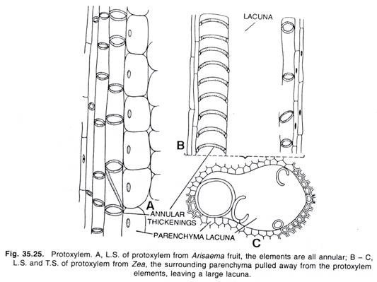
The secondary thickenings are deposited in the vessels as rings, continuous spirals or helices, with the individual coils of a helix here and there interconnected with each other, giving the wall a ladder-like appearance.
Such secondary thickenings are called — annular, spiral or helical and scalariform respectively. In a still later ontogenetic type of vessel elements, the reticulate vessel elements, the secondary wall appears like a reticulum.
When the meshes of the reticulum are transversely elongated, the thickening is called scalariform-reticulate. The pitted elements are characteristic of the latest primary xylem and of the secondary xylem.
Vessels are characteristic of the angiosperms. However, certain angiospermic families lack the vessels — the Winteraceae, Trochodendraceae and Tetracentraceae. In many monocotyledons (e.g., Yucca, Dracaena) they are absent from the stems and leaves.
They are found in some species of Selaginella, in two species of Pteridium among the pteridophytes; among the gymnosperms, in the Gnetales (Ephedra, Welwitschia and Gnetum).
Ontogeny of the Vessel:
The vessels are formed from pro-cambium cells or derivatives of cambium by the fusion of the cells end to end during the last stages of development. During this fusion the end walls are lost and the lumina of the series of the cells are freely open into one another, forming a long tube. From the meristematic stage the vessel elements increase greatly in diameter.
The vessels with scalariform perforations and the elongate, simply perforate types may increase in length to some extent, the tips forming tails which penetrate between surrounding cells. The vessels developing from stratified cambium cells do not elongate and sometimes even become shorter.
During the rapid growth in cell size, the primary cell wall remains constant in thickness except in those areas which later disintegrate to form the perforations. These areas become thicker and limited in their margins. In the sectional view they are lens-shaped or plate like and can be seen to be three layered, composing of the primary walls of the two adjacent cells and the middle lamella.
When the cell reaches its maturity, the cytoplasm of the cell begins to disintegrate. In certain woody plants the nucleus becomes quite small and flat and lies in scant cytoplasm against the wall where perforation is about to occur. As soon as the primary wall becomes mature, the perforation of the end wall and loss of the protoplast begin.
The wall in the perforation area becomes thinner and thinner and ultimately disintegrates. The maturation in all members of a vessel series takes place from one end to the other and not simultaneously (see Fig. 35.22).
(d) Wood Parenchyma:
The parenchyma cells which frequently occur in the xylem of most plants. In secondary xylem such cells occur vertically more or less elongated and placed end to end, known as wood or xylem parenchyma. The radial transverse series of the cells form the wood rays and are known as wood or xylem ray parenchyma.
The xylem parenchyma cells may be as long as the fusiform initials or they may be several times shorter, if a fusiform derivative divides transversely before differentiation into parenchyma (wood parenchyma). The shorter type of xylem parenchyma cells is the more common.
The ray and the xylem parenchyma cells of the secondary xylem may or may not have secondary walls. If a secondary wall is present, the pit pairs between the parenchyma cells and the tracheary elements may be simple, half-bordered or bordered. In between, parenchyma cells only simple pit pairs occur.
The xylem parenchyma cells are noted for storage of food in the form of starch or fat. Tannins, crystals and various other substances also occur in xylem parenchyma cells. These cells assist directly or indirectly in the conduction of water upward through the vessels and tracheids.
Phloem:
The xylem and phloem have evolved along more or less on similar lines. In xylem a series of tracheids, structurally and functionally united, has become a vessel whereas in phloem a series of cells similarly united, forms a sieve tube.
The fundamental cell type of xylem is tracheid, whereas in phloem the basic cell type is the sieve element. There are two forms of sieve element the more primitive form is the sieve cell of gymnosperms and lower forms where series of united cells do not exist, the unit of a series, the sieve tube element.
Phloem like xylem, is a complex tissue, and consists of the following elements:
(a) Sieve elements,
(b) Companion cells,
(c) Phloem fibres and
(d) Phloem parenchyma.
In the pteridophytes and gymnosperms only sieve cells and phloem parenchyma are present. In some gymnosperms, sieve cells, phloem parenchyma and phloem fibres are present. In angiosperms, sieve tubes, companion cells, phloem parenchyma, phloem fibres, sclereids and secretory cells are present.
(a) Sieve Elements:
The conducting elements of the phloem are collectively known as sieve elements. They may be segregated into the less specialized sieve cells and the more specialized sieve tubes or sieve tube elements. The morphologic specialization of sieve elements is expressed in the development of sieve areas on their walls and in the peculiar modifications of their protoplasts.
The sieve areas are depressed wall areas with clusters of perforations, through which the protoplasts of the adjacent sieve elements are interconnected by connecting strands. In a sieve area each connecting strand remains encased in a cylinder of substance called callose.
The wall in a sieve area is a double structure consisting of two layers of primary wall, one belonging to one cell and the other to another, cemented together by intercellular substance.
Like the pits in the tracheary elements, the sieve areas occur in various numbers and are variously distributed in sieve elements of different plants. The wall parts bearing the highly specialized sieve areas are called sieve plates (Esau, 1950). If a sieve plate consists of a single sieve area, it is a simple sieve plate.
Many sieve areas, arranged in scalariform, reticulate, or any other manner, constitute a compound sieve plate. However, just as vessels may have perforation plates in their side walls, sieve tube elements may have sieve plates in their lateral walls.
The two types of sieve elements, the sieve cells and the sieve-tube elements differ in the degree of differentiation of their sieve areas and in the distribution of these areas on the walls. Sieve cells are commonly long and slender, and they are tapering at their ends. In the tissue they overlap each other, and the sieve areas are usually numerous on these ends.
In sieve-tube elements, the sieve areas are more highly specialized than others and are localized in the form of sieve plates. The sieve plates occur mainly on end walls. Sieve-tube elements are usually disposed end to end in long series, the common wall parts bearing the sieve plates. These series of sieve-tube elements are sieve-tubes.
The lower vascular plants and the gymnosperms generally have sieve cells, whereas most angiosperms have sieve-tube elements.
The sieve-tube elements show a progressive localization of highly specialized sieve areas on the end walls; a gradual change in the orientation of these end walls from very oblique to transverse; a gradual change from compound to simple sieve plates; and a progressive decrease in conspicuousness of the sieve areas on the side walls.
The sieve elements generally possess primary walls, chiefly of cellulose. The characteristic of the primary walls of sieve elements is their relative thickness (Esau, 1950). The thickening of the wall generally becomes evident during the late stages of differentiation of the element. In some plants this wall is exceptionally thick. The thick sieve element is usually called the nacre wall.
The most important characteristic feature of the sieve-element protoplast is that it lacks a nucleus when the cell completes its development and becomes functional. The loss of the nucleus occurs during the differentiation of the element.
In the meristematic state the sieve element resembles other pro-cambial or cambial cells in having a more or less vacuolated protoplast with a conspicuous nucleus. Later the nucleus disorganizes and disappears.
The important property of the sieve-element protoplast of dicotyledons is the presence of variable amounts of a relatively viscous substance, the slime. The slime is proteinaceous in nature. The slime appears to be located mainly in the cell-sap together with various organic and inorganic ingredients.
The slime originates in the cytoplasm in the form of discrete bodies, the slime-bodies. They may be spherical, or spindle shaped, or variously twisted and coiled. They occur singly or in multiples in one element.
(b) Companion Cells:
The companion cell is a specialized type of parenchyma cell which is closely associated in origin, position and function with sieve-tube elements. When seen in transverse section the companion cell is usually a small, triangular, rounded or rectangular cell beside a sieve-tube element.
These cells are living having abundant granular cytoplasm and a prominent elongated nucleus which is retained throughout the life of the cell. Usually the nuclei of the companion cells serve for the nuclei of sieve tubes as they lack them. The companion cells do not contain starch.
They live only so long as the sieve-tube element with which they are associated and they are crushed with those cells. The companion cells are formed by longitudinal division of the mother cell of the sieve tube element before specialization of this cell begins. One daughter cell becomes a companion cell and other a sieve tube element.
The companion cell initial may divide transversely several times producing a row of companion cells so that one to several companion cells may accompany each sieve-tube element. A companion cell or a row of companion cells formed by the transverse division of a single companion- cell initial may extend the full length of the sieve-tube element.
The number of companion cells accompanying a sieve-tube element is fairly constant for a particular species. The solitary and long companion cells occur in primary phloem of herbaceous plants whereas numerous companion cells occur in the secondary phloem of woody plants.
The companion cells occur only in angiosperms where they accompany most sieve-tube elements. In the phloem of many monocotyledons, they are abundant, together with sieve tubes making up the entire tissue. The sieve cells of the gymnosperms and vascular cryptogams have no companion cells.
(c) Phloem Fibres:
In many flowering plants, fibres form a prominent part of both primary and secondary phloem. The phloem fibres are rarely found or absent in phloem of living pteridophytes. They are also not found in some gymnosperms and angiosperms. Only simple pits are found on the walls of phloem fibres. The walls may be lignified or non-lignified.
The Cannabis (hemp) fibres are lignifed, whereas fibres of Linum (flax) are of cellulose and without lignin. Because of the strength of strands of phloem fibres, they have been used for a long time in the manufacture of cords, ropes, mats and cloth. The fibre used in this way has been known since early times as bast or bass, and this way the phloem fibres are also known as bast fibres.
The sclereids are occasionally found in the primary phloem. The older secondary phloem of many trees also contains the sclereids. These cells develop from phloem parenchyma as the tissue ages and the sieve tubes cease to function.
(d) Phloem Parenchyma:
The phloem contains parenchyma cells that are concerned with many activities characteristic of living parenchyma cells, such as storage of starch, fat and other organic substances. The tannins and resins are also found in these cells. The parenchyma cells of primary phloem are elongated and are oriented, like the sieve elements. There are two systems of parenchyma found in the secondary phloem.
These systems are — vertical and the horizontal. The parenchyma of the vertical system is known as phloem parenchyma. The horizontal parenchyma is composed of phloem rays. In the active phloem, the phloem parenchyma and the ray cells have only primary un-lignified walls.
The walls of both kinds of parenchyma cells have numerous primary pit fields. The phloem parenchyma is not found in many or most of monocotyledons.
Plant Tissue: Group # 3. Secretory Tissue:
The tissues that are concerned with the secretion of gums, resins, volatile oils, nectar, latex and other substances are called secretary tissues.
These are further subdivided into two groups:
(a) Laticiferous tissue and
(b) Glandular tissue.
(a) Laticiferous Tissue:
Usually latex is present in the families of many flowering plants. This substance may be white, yellow or pinkish in colour. This is a viscous fluid and established to be colloidal in nature. Many substances like sugars, proteins, gums, alkaloids, enzymes, rubber, etc., remain suspended in a matrix of watery fluid.
Starch grains may be abundantly present. The latex of some plants is of great importance, especially as a source of rubber (e.g., Haevea, Ficus, etc.), chicle (Achras), and papain (Carica). The laticiferous ducts, in which latex is found may be of two types — latex cells or non-articulate latex ducts and latex vessels or articulate latex ducts.
The functions and the contents of the two are same but they differ in their nature and morphology. They contain numerous nuclei in the thin layer of cytoplasm along the cell wall. The function of these tissues is not yet clearly understood. They may act as food storage organs or as reservoirs of waste products.
Nonarticulate latex ducts or latex cells:
These ducts are independent units which extend as branched structures for long distances in the plant body. They originate as minute structures, elongate quickly and ramify in all directions of the plant body by repeated branching, but they do not fuse together, thus no netted structures are formed as they are formed in articulate ducts.
The walls of the ducts are soft and very often thick. Such ducts are commonly found in Calotropis, Euphorbia, Nerium, Vinca, etc.
Branched nonarticulated laticifers commonly occur in leaves. Here they travel through the vascular bundles, ramify in the mesophyll, and often reach the epidermis.
The unbranched nonarticulated laticifers of Vinca and Cannabis occur in the primary phloem but are apparently absent in the secondary tissues.
Articulate latex ducts or latex vessels:
These ducts or vessels are the result of anastamosing of many cells together. They originate in the meristems from rows of cells by the abortion, of the separating walls early in the ontogeny of the cells. They grow more or less as parallel ducts which by means of branching and frequent anastamoses form a complex network.
A duct of this type resembles with the xylem vessel only in the respect that it is made up of a series of cells united to form a tube otherwise the latex tube is living and coenocytic. Such latex vessels are commonly found in angiospermic families — Papaveraceae, Compositae, Euphorbiaceae, Moraceae, etc.
Articulated laticifes show various arrangements and a frequent association with the phloem. In Carica papaya the laticifers apparently occur not only in the phloem but also in the xylem. The laticiferous system that makes Hevea brasiliensis (Euphorbiaceae) such an outstanding rubber producer is the secondary system that develops in the secondary phloem.
(b) Glandular Tissue:
This tissue consists of special structures, the glands. These glands contain some secretory or excretory products. The glands may consist of isolated cells or small groups of cells with or without central cavity. They are of various kinds, and more common types are those which secrete digestive enzymes, called digestive glands and those which secrete nectar, known as nectaries.
They may be internal or external. The common internal glands which are usually lying embedded in the interior tissues of plant body are oil glands, mucilage secreting glands secreting gums, resin and tannin, etc., digestive glands secreting enzymes, water secreting glands also known as hydathodes. The common external glands which occur on the epidermis may be glandular epidermal hairs, nectaries, etc.
Oil glands:
These are internal glands which frequently contain essential oils in them. These oils are volatile and odoriferous. These glands originate due to split of certain cells, but they are formed in abundance by the breaking down of cells containing the volatile oil.
On the disintegration of the cells the oil stores up in the large cavities of glands. These cavities are lysigenous in nature. Such oil glands are commonly found in Citrus, Eucalyptus and other plants.
Glands secreting resins, gums, etc.:
In the gymnosperms and. in many angiospermic families, resins, gums, oils and many other substances are secreted and conducted in ducts. In many gymnosperms (e.g., in Pinus) these ducts or canals form extensive systems extending both vertically and horizontally. In other plants (e.g., in Umbelliferae) the ducts are local in occurrence and limited in extent.
In Pinus and many other gymnosperms, the resin ducts are schizogenous in nature, and when mature, have the structure of a tube with an epithelial lining. These glands are internal.
Digestive glands:
In certain insectivorous plants there are special glands which secrete protein-digesting enzymes. These enzymes act upon insects so that the products of digestion can be absorbed by the plant. In Drosera, the secretory tissue is at the tips of the leaf tentacles. Here in addition to the digestive enzymes, there are secreted viscid substances which hold the insects. These are internal glands.
Hydathodes (water secreting glands):
Many plants possess special structures which exudate water under conditions of low transpiration and abundant soil moisture. These modified special structures are known as hydathodes or water stomata. The water stoma resembles an ordinary stoma, and morphologically it is considered to be enlarged stoma which has lost the power of movement and serves for water secretion.
Commonly, the hydathodes occur at the tips of the leaves of Pistia and other aroids, water hyacinth, grasses, garden nasturtium and many other plants of humid climate. The hydathodes are internal glands.
Nectaries:
Many insect-pollinated plants produce nectar which attracts insects. This substance is secreted by special cellular structures, the nectaries. Definite and elaborate nectaries occur in certain families (e.g., Euphorbiaceae). In the less specialized nectaries the secreting cells are superficial upon the floral parts. Here, the epidermal cells do not possess cuticle.
The nectar is exuded through the wall and exposed upon the outer surface. The septal nectaries are commonly found in many monocotyledonous flowers where they make pockets in the septal walls of syncarpous ovaries where the carpel walls are fused incompletely and the epidermal cells are glandular. These glands are external in nature.


