Meristematic Tissue of Plants: Introduction, Types and Theories (With Diagram)!
Introduction:
Meristematic tissue, commonly called meristem, is composed of cells which are immature, not fully differentiated ones, and which possess the power of cell division.
The vascular plants exhibit an ‘open system’ of growth involving formation of new tissues and organs throughout its life. It is possible due to presence of certain organised regions where the cells are in perpetually immature condition and can form new cells throughout.
These organised regions are the meristems. Primitive plants have cells all similar, carrying on different functions. With advance in the line of evolution growth has been restricted to certain parts of the plant body, the function of cell division being confined to the meristematic cells.
The open system of growth in plants is in marked contrast to the development of an animal body.
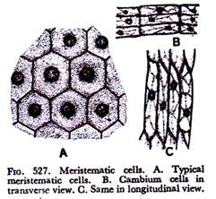
The meristematic cells have generally some distinctive features (Fig. 527). The cells are usually isodiametric in shape; they are compactly set without evident intercellular spaces; have dense cytoplasm with small vacuoles and large prominent nuclei; ergastic matters are lacking; the plastids are in proplastid stages; cell wall, made of celluose, is thin and homogeneous.
Though in general, meristematic cells possess the characters stated above, but departures cannot be ruled out. The meristematic cells of vascular cambium are fusiform in shape (Fig. 527 B & C) often with conspicuous vacuoles.
Comparatively thick walls with primary pit fields are found in some meristematic cells. Ergastic matters like starch and tannins may also be present. Considering these departures some authors are in favour of using the term eumeristem or true meristem for those having the general characters stated above.
It has been stated that meristems are the formative regions where new cells are added to the body. Besides, the meristems perpetuate themselves.
The cells which remain meristematic and thus continue cell division are called initiating cells; whereas the cells formed by them, the so-called derivatives, gradually change their shape, enlarge, lose the power of cell division and ultimately become mature cells with some definite characters and functions.
These changes involving enlargement and specialisation represent the process which may be referred to as differentiation. The differentiation of a particular tissue has been characterised as “a progressive loss of embryonic features of meristematic cells and the progressive attainment of the state of maturity” by a well-known authority.
The derivatives of the initiating cells gradually differentiate into mature cells and lose the power of cell division, at least temporarily. These completely differentiated cells are called permanent ones. Some permanent cells may get back the meristematic nature, a phenomenon which has been referred to as dedifferentiation by some workers. That shows that though cells have attained permanent form, but they have retained the power of cell division.
The living parenchyma cells and the epidermal cells are common reversible permanent cells. In fact, all living cells retain the potentialities of division and growth, though they have become permanent. Strictly speaking, permanent cells are those which have completely lost the capacity of division, as for example, irreversibly specialised cells like the sieve tube elements and the dead elements like tracheids and cork cells.
Various systems of classification of meristems have been proposed, based on characters like origin and nature of initiating cells, stages of development, topography and function. No system is exclusive and rigid.
Types:
The common important types of meristems according to their origin and development are the following:
Promeristem or Primordial Meristem:
Promeristem is the very foundation stage, the region of formation of new organs and tissues. It may be called the earliest embryonic condition consisting of young initials and their derivatives.
All the cells possess the characters of true meristematic cells, viz., diameters alike, dense cytoplasm, large nuclei, proplastids, absence of ergastic matters and thin walls. Promeristem is definitely of limited extent.
As soon as the cells begin to show tendency of differentiation, they have passed the earliest promeristematic condition.
Primary Meristem:
Primary meristems are composed of cells which are direct descendants of embryonic cells and which have all through retained meristematic nature. So primary meristem may be called a later developmental stage.
Chief primary meristems are those located at the tips of stem, root, and appendages. Primary meristems build up the primary body of the plants.
Secondary Meristem:
Primary meristems gradually differentiate into permanent tissues. Some living permanent cells may regain the power of cell division. They constitute the secondary meristem, as they originate from permanent cells.
The cork cambium or phellogen, arising from epidermis, cortical and other cells during secondary increase in thickness, is an example of secondary meristem.
It is to be noted that primary meristems are responsible for the building up of the primary body of the plant; and the secondary meristems, formed later, add new cells to the primary body with definite purposes like effective protection and repair.
The definitions given above are not always accurate. In case of adventitious organs the apical meristem develops within permanent tissues secondarily though they are primary in structure and function.
According to their position in the plant body meristems are put into three groups, viz., apical, intercalary and lateral (Fig. 528).
Apical Meristem:
Apical meristems occur at the apices of the stems, roots, main and lateral, of the vascular plants. Growth in length of the axis is entirely due to their activities; so they are also called growing points. The initiating cells may be solitary or in groups.
Solitary initiating cells go by the name apical cells, whereas those occurring in groups are called apical initials. Solitary apical cells (Figs. 530 & 533A) occur in many pteridophytes like ferns and horse-tails.
Other vascular plants possess apical initials which may be terminal or terminal and sub terminal (Figs. 532 & 533). The apical initials may occur in one or more tiers.
In case of one tier all the cells of the plant body derive their origin from it; whereas if there is more than one tier different parts of the plant body originate from different tiers of initials.
The derivatives of the apical meristem differentiate in course of time into permanent tissues which together constitute the primary body of the plant.
Intercalary Meristem:
These are the portions of apical meristems which are separated from the apex during the growth of the axis and remain intercalated between permanent cells.
The position is such that apical meristems go ahead during development and a portion of it is left behind which is inserted between permanent cells.
Intercalary meristems are found in the stem and leaf sheaths of many monocotyledons, particularly grasses, and in the horse-tails. Here nodal regions are usually composed of permanent cells and so intercalary meristems are internodal. These meristems are short-lived, they very soon become permanent and merge with the tissues surrounding them.
Lateral Meristem:
These meristems occur laterally in the axis, parallel to the sides of the organs in dicotyledons and gymnosperms. They are composed of initials which divide periclinally. The derivatives gradually differentiate into permanent tissues called secondary tissues.
These tissues are added to the existing ones and are responsible for increase in thickness. The growth in thickness thus secured due to addition of secondary tissues is referred to as secondary growth.
The cambium of the vascular bundles and the phellogen or cork cambium are lateral meristems. It should be noted, however, that cambium is a primary meristems, whereas phellogen or cork cambium is secondary in origin.
A classification of meristems chiefly based on function was followed in physiological anatomy. Haberlandt in 1890 suggested that primary meristem at the apex of the stem and root is segregated into three tissues, viz., protoderm, procambium and ground meristem (Fig. 529). The protoderm develops into epidermis, the procambium into primary vascular tissues and the ground meristem into fundamental or ground tissues.
This classification is helpful in tracing the three tissue systems of mature regions—epidermal, ground or fundamental, and vascular, consisting of epidermis, ground tissues and vascular tissues respectively. The advantage of this set of terminology from topographical point of view is undeniable, though the interpretation of development of shoot and root from these zones is a quite complex problem.
Meristems on the basis of planes of divisions are of three types, viz., mass meristem rib meristem, and plate meristem. Mass meristems grow by dividing in all planes, so that the bodies formed are either isodiametric or have no definite shape.
This pattern of growth is noticed in endosperm, young embryo and also in the formation of spores and sperms. The rib meristems, also called file meristems, divide anticlinally to the long axis and give rise to longitudinal files or rows of cells.
This growth pattern is clear in the development of cortex and pith. The plate meristems divide chiefly anticlinally in two planes, so that new cells are formed but number of layers does not increase.
The uniseriate epidermis and multiseriate flat blade of the leaf are illustrations of growth forms by plate meristems.
Theories of Structural Development and Differentiation:
The classical concept of the apices of shoot and root had been that they represent the earliest self-perpetuating promeristems consisting of homogeneous eumeristematic or truly meristematic cells.
Modern workers are of opinion that a distinct zonation is exhibited by the immediate derivatives of the promeristem. By zonation is meant the existence of distinct regions differing from one another by characters like nature of cells, plane of cell division, position of the initiating cells and rate of maturation of cells.
The growth and development of the apical meristems of shoot, root, and flower and their differentiation have been most notable problems of plant anatomy.
Intensive studies on those problems have been going on since the nineteenth century and a few theories have been proposed from time to time. A brief review of those theories is being given here.
Apical Cell Theory:
The solitary apical cells (Figs. 530 & 533A) were discovered in the cryptogams—in algae, bryophytes and pteridophytes. This led early workers to believe that a solitary apical cell was a constant unit of apical meristem governing the whole process of growth.
They assumed that the same condition prevails in all higher plants. On that assumption apical cell theory was advanced by Hofmeister in 1857 and supported by Nageli (1878) in the nineteenth century.
Subsequent works revealed that complex apices of gymnosperms could not be interpreted in the light of this theory. So it was not applicable to seed plants.
Histogen Theory:
The older theory was replaced by histogen theory as an attempt towards interpretation of the growing points of seed plants. Hanstein proposed
this theory in the nineteenth century and received support from Strasburger (1868).
According to this theory plant body develops from groups of initials forming a mass of meristem of considerable depth, as opposed to superficial apical cells of former theory, and that three distinct zones or strata can be recognised at the growing points of stem and root.
Every zone consisting of a group of initials was called a histogen or tissue builder. The histogens arise from separate sets of initials and have different courses of development.
The three histogens (Fig. 531) suggested were (i) dermatogen, the outermost uniseriate layer; (ii) plerome, the massive central core consisting of cells extended in longitudinal direction; and (iii) periblem, the region composed of isodiametric cells lying between the dermatogen and the plerome.
The dermatogen cells divide anticlinally and develop into uniseriate epidermis. The periblem forms the cortex; and the plerome gives rise to the massive central cylinder or stele, consisting of primary vascular tissues and ground tissues like pith, pith rays and pericycle.
Hanstein’s histogen theory as a basis of interpretation of the shoot and root apex held ground for fairly long time. Though it is followed even now in case of root apex, it has been found to be inadequate as regards shoot apex of angiosperms mainly for two reasons, viz. (i) sharp distinction between dermatogen and periblem is absent; and (ii) origin of different regions from the sharply defined histogens cannot be demonstrated.
Moreover in many gymnosperms and angiosperms shoot apices hardly show any distinction between periblem and plerome.
Tunica-corpus Theory:
As an interpretation of the apical growth in the shoot apex the third theory—tunica-corpus theory—was proposed by Schmidt and supported by Foster and others in the early part of the twentieth century.
According to this theory, two tissue zones occur at the apex. They are the tunica or cover, consisting of one or more layers of cells forming the outer enveloping region, and the corpus, the central core, a mass of cells surrounded by tunica (Fig. 532).
The two regions differ in structure and appearance due to varying rates and methods of growth. The cells of tunica are smaller and lie in layers with planes of cell division predominantly anticlinal.
So growth here is primarily in area. The corpus cells are larger and they divide in various planes, so that a mass of irregularly arranged cells is formed. Increase of the mass here is thus in volume.
The tunica may be one-layered or many-layered with massive or slender corpus. The number of initials is also variable. In fact, in lower vascular plants like pteridophytes and even in some gymnosperms a sharp distinction between tunica and corpus cannot be traced.
In angiosperms, however, two sets of initials may be definitely located, the two sets giving rise to tunica and corpus. Thus the two have independent origin.
In spite of the fact that many variations and fluctuations occur, the tunica-corpus theory has given a renewed impetus in the interpretation of the apical growth in the stems of angiosperms.
As a result, the stages in the development of the primary body from the initiating cells are better understood.
Growth Patterns:
Differentiation involves some striking changes in the characteristics of the individual cells, viz., enlargement accompanied by increase of vacuolar sap, collection of ergastic matters, development of plastids with colours from the proplastids, disappearance of protoplasts in extreme cases, and growth and chemical changes of the cell wall.
The increase in the size of the cell may be more or less uniform in all directions or due to unequal rate of enlargement in different directions the cells may appear distinctly different from the meristematic cells from which they have derived their origin.
The cellular arrangement of a tissue is really determined by the growth pattern of its meristem. The increase and changes in the shape of the cells in a differentiating tissue bring about some adjustments in their relation on each other. The formation of intercellular spaces, either small ones at the comers or fairly large ones changing the very appearance of the tissue is quite common.
In expanding organs mutual adjustment occurs due to growth of the entire walls of the cells in a co-ordinated manner without involving separation of the walls. This type, called symplastic growth, is noticed in the formation of the derivatives of the apical meristem.
A second type, called intrusive growth, involving intrusion of cells among others is common in case of formation of fibres, tracheids and laticifers.
Here the elongating cells grow at the apex, normally at both ends, resulting in the separation of the primary walls of the adjacent cells. Early workers mentioned gliding or sliding growth as basis of these changes.
Gliding growth used to mean the slipping of the wall of a cell over that of a contiguous one during growth, so that new contacts were made. This has been supplanted by symplastic and intrusive growth mentioned above.


