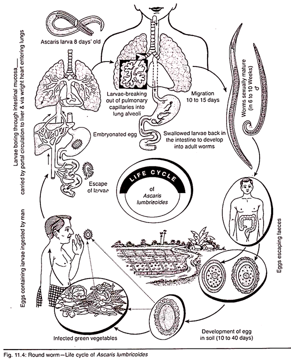
In this article we will discuss about the various stages involved in the life cycle of roundworm which is otherwise known as Ascaris lumbricoides (explained with diagram).
Ascaris lumbricoides is one of the most familiar endoparasites of man. It has also been reported from sheep, pigs, cattle etc. It inhabits the small intestine, more frequently of children than of adults, where it is supposed to feed on the semidigested food of the host.
Ascaris is monogenetic i.e., it requires only one host to complete its life cycle and no intermediate host is required. Comprehensive reviews of the life cycle have been given by Crompton and Pawlowski (1985) and Crompton (1989). Man is the only known definitive host of Ascaris lumbricoides.
The various stages in the life cycle are described below:
Stage 1. Copulation and fertilization:
Copulation occurs in the small intestine of host (man) where the adult worm lives. During copulation the male orients its body at right angle to that of the female in such a way that its cloacal aperture apposes the vulva of the female and the sperms are easily transferred into the vagina from where they ascend up in the uterus and fertilizes the eggs in the oviduct.
Stage 2. Eggs in faeces and structure of eggs:
The eggs are laid in the host’s intestine which are deposited outside along with faeces of host. A female Ascaris produces roughly about 2,00,000 eggs daily. The egg production is astounding. Cram (1925) estimated the number of eggs contained in a mature female worm to be as high as 2,70,00,000 and the eggs per gram of faeces for each female worm may be in excess of 2000.
When the eggs are passed in faeces, their further development is largely dependent on oxygen tension, moisture content and temperature of their environment. Due to high temperature inadequate moisture and oxygen supply in the host’s intestine, the fertilized eggs do not start their further development.
The fertilized eggs are round or oval in shape. They usually measure about 52-84 μm by 45-67 μm. The zygote has a thick, clear inner shell covered over by a warty, albuminous coat which is always bile-stained and brownish (golden-brown) in colour. The egg contains a very large conspicuous, unsegmented ovum (the nucleus is concealed by a large amount of coarse yolk granules). There is a clear crescentic area at each pole of the zygote (Fig. 11.4).
Stage 3. Cleavage (Segmentation of fertilised egg) and early development:
Cleavage of fertilised egg is of spiral and determinate type. The first division is transverse which results in a dorsal cell and a ventral cell. The dorsal cell divides vertically into an anterior and a posterior cell, while the ventral cell divides horizontally into an upper and a lower cell. The four celled embryo, thus formed, is first T-shaped in appearance.
In the next cleavages, the 4-celled embryo becomes the 16-celled embryo and attains the form of a hollow ball. It is termed as blastula containing the blastocoel. The blastula undergoes the process of invagination and becomes the gastrula. The juvenile is formed within 10-14 days from the onset of cleavage. It has an alimentary canal, a nerve-ring and a larval excretory system.
For its close resemblance with Rhabditis (a nematode found in the soil and human faeces), the juvenile is also termed as Rhabditiform larva. This larva of the first stage is not infective. In another week’s time it undergoes moulting within the egg-shell and becomes the second stage of Rhabditoid which is capable of infecting the host. Under suitable conditions of moisture, oxygen and temperature, the infective eggs are known to remain viable for about six years.
Stage 4. Infection of new host (man):
Man acquires infection when the egg containing Rhabditoid larva is swallowed by the host along with raw vegetables, improperly cooked vegetables or with the drinking water. Infection is brought about by ingestion of viable eggs, which are triggered to hatch under the influence of the intestinal conditions especially PCO2; hatching largely occurs in the duodenum but some takes place in the stomach.
Stage 5. Migration through the lungs:
In the small intestine by the action of host’s digestive juice the egg-shells dissolve and the juveniles hatch out. It performs active thrashing movements and bores through the intestinal epithelium to enter in the hepatic circulation which carries it to the liver.
According to Douvres et al (1969), on hatching the larvae burrow into the intestinal mucosa, penetrate blood vessels and appear as second stage larvae in the liver within six hours of post-infection. Some larvae penetrate lymphatics but apparently become inhibited and it is doubtful if these larvae develop further. They remain in the liver for a few days and develop to the early third stage larva.
From the liver it finally reaches the heart through the post caval vein. Larvae are then carried to the lungs via pulmonary arteries. The larva generally remains in the lung for few days and gradually increases in size. Then it ruptures out of blood capillary and finally bores its way into the lung alveolus.
Stage 6. Re-entry into the stomach and the small intestine:
After about six days stay there, the larva moults there for the second time. Then they pass through the trachea with cough and when the cough is swallowed, pass to the oesophagus, stomach and finally to the intestine. The larva here undergoes moulting for two times and becomes adult. The period of migration from the time of infection to that of reaching the intestine is said to be about 10 days.
Stage 7. Sexual maturity and egg liberation:
The larvae on reaching their habitat grow into adult worms and become sexually matured in about 6-10 week’s-time. The gravid female begins to discharge eggs in the stool of host (man) within about two months from the time of infection. The cycle of Ascaris lumbricoids is again repeated.
Within the intestine, the larvae begin the third moult on the ninth day and are in the fourth stage by the tenth day. So in the life cycle of A. lumbricoides there are four moultings or ecdysis—one outside, while within the egg shell, one in the lung and two in the intestine.

