Here is a list of viruses that are found in animals: 1. Papovaviruses 2. Simian Virus-40 3. Adenoviruses 4. Herpesviruses 5. Pox Viruses 6. Picornavirus 7. Togaviruses 8. Rabies Viruses 9. Influenza Viruses 10. Reoviruses.
1. Papovaviruses:
Papovaviruses are one of the four important dsDNA viruses (e.g. papovaviruses, adenoviruses, herpes viruses and pox viruses) which produce tumour in many animals.
The term papova is derived from the first two letters of the three prototypes, papilloma virus, polyoma virus and simian vacuolating virus-40 (SV40). The other important viruses of this group are JC virus (associated with neurological degeneration), BX virus (which suppresses immune system of humans), K virus of mice, etc.
Capsid is of 45-55 nm, naked, icosahedral; virion consists of dsDNA and protein. Capsid is made up of 72 capsomers which are built by 420 subunits. Capsid contains one major polypeptide (VP1) and two identical minor polypeptide (VP2 and VP3). Virus enters the cell and migrates to the nucleus where it replicates. The dsDNA encodes the early proteins and capsid proteins.
2. Simian Virus-40 (SV40):
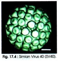
S V40 is an oncogenic virus. It is naked and icosahedral in morphology with a diameter of 45 nm. (Fig. 17.4). Capsid consists of 72 capsomers. SV40 is similar to polyoma virus in size and structure. Polyoma is associated with tumour in mice.
The dsDNA in its native form is supercoiled (i.e. covalently closed circle) helix having the sedimentation coefficient of 21S. Total G+C content of nucleic acid is 41 %. After breaking the phosphodiester bond, single stranded DNA helix is converted into a relaxed circular form. This form has the sedimentation coefficient of 16S. A linear form (of 14S) is formed after double stranded break in the supercoil.
(i) Replication:
Virus enters the cell and directly migrates to the nucleus. Replication of the viral RNA takes place inside the nucleus. Before the replication begins, early proteins are synthesized in the nucleus of the infected cells.
The mechanism of DNA replication can be divided into the following four stages:
(a) Initiation:
DNA replication begins at a site known as origin of replication as the ori genes are present at this site. Initiation requires a gene product A which is a globular protein. The ori region is rich in adenine and thymine.
(b) Elongation:
Replication in two direction starts from the point of ori region. The RNA polymerase acts at this region and an RNA polymer of about 10 nucleotide in length is formed. Using (+) DNA as template a complementary (-) DNA strand develops on the RNA primer.
The chain elongates discontinuously on both the strands and form short fragements of DNA which is known as Okazaki fragements. In turn the Okazaki fragements are covalently sealed to form a continuous strand. DNA polymerase and DNA ligase are required for the complementary chain.
(c) Segregation of complementary DNA:
Until the two complementary strands reach the termination, chain elongation continues. Both the strands are terminated at about 180° from the ori region. Each duplex contains an original strand and a linear strand.
(d) Maturation:
During maturation the two ends of the linear strand is sealed by the ligase and two complete circular DNA molecules are formed. The histone proteins get attached to DNA and results in super coiled form through winding of the DNA strands.
(ii) Protein Synthesis:
Within 12h of infection and before start of DNA replication, there begins early protein synthesis. The synthesis of antigen (i.e. tumour antigen) occurs by viral DNA which results in increased DNA metabolism in the infected host cell. Late proteins are synthesized when DNA replication is over. Polyadenylation (addition of poly A) takes place at 3′ end of mRNA which is not coded by the mRNAs.
3. Adenoviruses:
Adenoviruses were first isolated in human adenoids (tonsils) from which its name is derived. The adenoviruses are common pathogens of humans and animals. More than 100 serologically distinct types of adenovirus have been identified including 49 types that infect humans. Moreover, several strains have been the subject of intensive research and are used as tools in mammalian molecular biology.
Several adenoviruses cause respiratory and conjunctival diseases such as pneumonia, acute follicular conjunctivitis, epidemic keratoconjunctivitis, cystitis and gastroenteritis. In infants, pharyngitis and pharyngeal-conjunctival fever are common. In addition, a few types of human adenoviruses induce undifferentiated sarcomas in newborn hamsters and other rodents and can transform certain rodent and human cell cultures.
Adenoviruses are unusually stable to chemical or physical agents and adverse pH conditions. This ability helps in its prolonged survival outside of the body and water. Adenoviruses are primarily spread via respiratory droplets; however, they can also be spread by fecal routes as well.
Adenoviruses are classified as group I under the Baltimore classification scheme. Adenoviruses are put iii the family Adenoviridae which is divided into two genera: mastadenoviruses (the mammalian adenoviruses) and aviadenoviruses (the avian adenoviruses). However, more than 100 antigenic types of adenoviruses e.g. mastadenoviruses and aviadenoviruses have been identified that infect mammals and birds.
Since adenoviruses readily infect human and other mammalian cells, their genomes have been developed into vectors in experimental therapy. Vector genomes carry deletions in the E1 and E3 regions; the gaps in the genome are used to take up foreign genes, e.g. the gene for the cystic fibrosis trans-membrane conductance regulator (CFTR).
Deletions in E1 minimize the potential of these vector genomes to elicit an infection cycle in human cells. The first clinical applications in patients suffering from the genetic disease cystic fibrosis have been reported but problems with adenovirus toxicity remain.
4. Herpesviruses:
The name ‘herpes’ comes from the Greek word herpein which means ‘to creep’. These viruses cause chronic/latent/recurrent infections. Epidemiology of the common herpesvirus infections puzzled clinicians for many years. In 1950, Burnet and Buddingh showed that herpes simplex virus (HSV) could become latent after a primary infection, becoming reactivated after later provocation.
In 1954, Weller isolated varicella zoster VZV (HHV-3) from chicken pox and zoster, indicating the same causal agent. So far, about 100 herpesviruses have been isolated from many animal species.
Herpesviruses belong to the family Herpesviridae (viruses with double stranded DNA genomes) (Class 1), which have envelope with spikes on icosahedral virion. To date, there are eight known human herpesviruses; some of them are oncogenic such as Simplex virus (herpes simples virus, HSV), Varicellovirus (caricella Zoster virus, CZV), Lymphocryptovirus (Epstein-Barr virus).
5. Pox Viruses:
The family Poxviridae is a legacy of the original grouping of viruses associated with diseases that produced poxs in the skin. Modem viral classification is based on the shape and molecular features of viruses and the smallpox virus remains as the most notable member of the family. It has two sub-families: Chordopoxvirinae and Entomopoxvirinae.
Some of the important genera are:
Orthopoxvirus (type species: Vaccinia virus; diseases-cowpox, vaccinia, smallpox), Para poxvirus, Avipoxvirus, Capri poxvirus, Leporipoxvirus, Suipoxvirus, Swinepox virus, Molluscipoxvirus (type species: Molluscum contagiosum virus),Yatapoxvirus, Entomopoxvirus A, Entomopoxvirus B, Entomopoxvirus C. Poxviruses can infect both vertebrate and invertebrate animals.
There are four genera of poxviruses that may infect humans e.g. orthopox (variola virus, vaccinia virus, cowpox virus, monkeypox virus, smallpox), Parapox (orf virus, pseudo cowpox, bovine papular stomatitis vims), yatapox (tanapox virus, yaba monkey tumor virus), and molluscipox contagiosum virus (MCV).
The most common viruses are vaccinia (found in Indian subcontinent) and molluscum contagiousum but monkeypox infections are gradually increasing in west and central African rainforest countries.
An example of such a group and the problems of complexity are shown by the members of the poxvirus family. These viruses have oval or brick-shaped 200-400 nm long particles. These particles are so large that they were first observed using high resolution optical microscopes in 1886. At that time they were thought to be ‘the spores of micrococci’.
6. Picornavirus:
Picornaviruses are among the most diverse (more than 200 serotypes) and ‘oldest’ known viruses. A temple record of from Egypt (1400 B.C.) shows a picture of poliomyelitis in a Priest, Ruma. In 1898, Loeffler and Frosch first recognized foot and mouth disease virus (FMDV).
Picornaviruses belong to the family Picornaviridae which is one of the largest of the viral families. Under Baltimore’s viral classification system picornaviruses are classified as Group IV Viruses because they contain a single stranded, positive sense RNA genome of 7.2 – 9.0 Kb in length.
As the term denotes (pico=small, rna=RNA) picorna viruses are the smallest in size (18-30 nm). They are icosaherdal and contain a (+) ssRNA because it acts as mRNA.
There are five groups of picorna viruses:
(i) Human enterovirus which are found in alimentary canal e.g. poliovirus, ECHO (enteric cytoplasmic human orphan) virus causing paralysis, diarrhoea,
(ii) Cardio-viruses of rodent e.g. encephalomyocarditis virus,
(iii) Rhinovirus which causes respiratory infection like common cold, bronchitis and foot and mouth disease virus e.g. FMD virus in catties,
(iv) Aptho-viruses, and
(v) Hepato-viruses (cause of hepatitis A).
The viruses that generally replicate in the intestine are called ‘enterovirus’. The most important pathogens from the genus entero-viruses include: poliovirus and Coxsackie A and B viruses.
7. Togaviruses:
Togaviruses belong to the family Togaviridae, which falls into the group IV of the Baltimore classification of viruses. Some examples Alphavirus (type species- Sindbis virus, eastern equine encephalitis virus, western equine encephalitis virus, Venezuelan equine encephalitis virus, Ross River virus) and Rubivirus (type species Rubella virus). Only Alphaviruses are arthropod-borne. Rubella virus has one species, which is quite distinct from Alphaviruses.
Togaviridae is classified as in Table 17.6:
Rubella was first recognized as a distinct disease in 1814. During 1938, Venezuelan Equine Encephalitis was isolated. Rubella vaccine was licensed in 1969. Large epidemic of the chikungunya virus was reported on the island of La Reunion and the surrounding islands in the Indian Ocean. During 2005-2006 in India, the major epidemic of the chikungunya virus was reported in over 1.5 million cases.
It grows in both mammalian and insect cell lines. Transmission of virus takes place from salivary glands of the mosquito to the bloodstream of the vertebrate host. Thereafter, virus particles travel to the skin and reticuloendothelial system (spleen and lymph nodes), where the primary infection occurs.
8. Rabies Viruses:
Rabies (Latin: rabies, madness, rage, fury; also called ‘hydrophobia’) is a viral zoonotic neuro-invasive disease that causes acute encephalitis (inflammation of the brain) in mammals (Fig. 17.31). It is most commonly caused by a bite from an infected animal or by other contact. Rabies has been known for more than 20,000 years.
The first description dates from the 23rd century BC in the Mesopotamia. During 1880s, Pasteur carried out the serial passage of Rabies virus in rabbits, and eventually succeeded in isolating an attenuated preparation which was used to treat patients bitten by mad dogs. There are over 200 Rhabdo-viruses known, which infect man, other mammals, fish, insects and plants.
The family Rhabdoviridae includes the genera Lyssavirus, Ephemerovirus and Vesiculo-virus. The rabies virus is a member of the genus lyssavirus. It is classified under Group V of Baltimore’s classification. Genetically, these viruses have non-segmented (-) sense RNA genome reminiscent of Paramyxoviruses. The family includes six genera.
9. Influenza Viruses:
In a phylogenetic-based taxonomy the RNA viruses includes the negative-sense ssRNA viruses which includes the Order Mononegavirales, and the family Orthomyxoviridae (Greek; orthos – straight; myxa = mucus). The family Orthomyxoviridae includes five genera: Influenza virus A, Influenza virus B, Influenza virus C, Thogotovirus and Isavirus.
The first three genera contain viruses that cause influenza in vertebrates, including birds, humans, and other mammals. Isaviruses infect salmon; thogotoviruses infect vertebrates and invertebrates (e.g. mosquitoes and sea lice).
Orthomyxoviridae consists of 7 to 8 segments of linear negative-sense single stranded RNA. The total length of the genome is 12,000-15,000 nucleotides (nt). The sequence of genome has terminal repeats which are repeated at both ends. At 5′-end the terminal repeats are 12-13 nucleotides long, whereas nucleotide sequences of 3′-terminus are identical.
In most on all RNA species, the terminal repeats at 3′-end, are 9-11 nucleotides long. The 5′ and 3′ terminal sequences of all the genome segments are highly conserved. The nucleic acid is completely genomic in nature. However, each virion may contain defective interfering copies as well.
10. Reoviruses:
The family reoviridae falls under Group III (ds RNA) of Baltimore classification. It is a family of viruses that can affect the gastrointestinal system (such as Rotavirus) and respiratory tract. Viruses of this family have genome consisting of segmented dsRNA. The name Reoviridae is derived from respiratory, enteric and orphan viruses. The orphan virus are either non-Pathogenic or of low virulence.
The virus can be readily detected in feces, and may also be recovered from pharyngeal or nasal secretions, urine, cerebrospinal fluid, and blood. So far, the role of Reovirus in human disease or treatment is not clear.
There are more than 150 species in the family Reoviridae. Examples of reoviruses are: Aquareovirus, Coltivirus, Cypovirus, Fijivirus, Idnoreovirus, Mycoreovirus, Orbivirus, Orthoreovirus, Oryzavirus, Phytoreovirus, Rotavirus, and Seadornavirus.
Some genera and species of reoviridae are given in Table 17.10:


