After reading this article we will learn about:- 1. Sporophyte of Azolla 2. Gametophyte of Azolla 3. Phylogeny.
Sporophyte of Azolla:
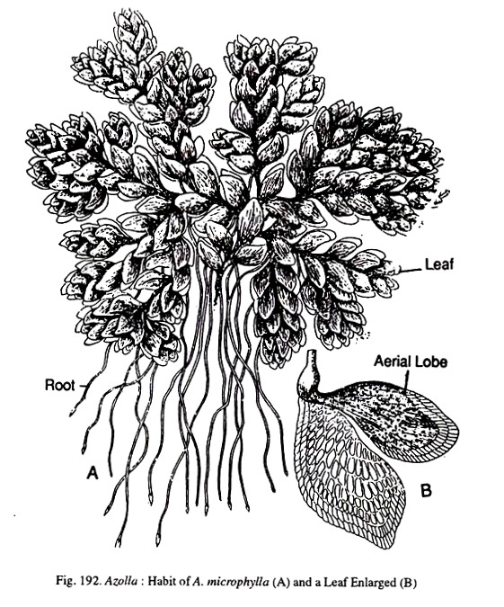
The sporophyte is extremely small when compared with Marsilea and Salvinia. It is distinguishable into stem, leaves and roots. The stem is often called the rhizome. It is profusely branched and on its upper surface is covered with dense leaves. The leaves are alternate and are arranged in two rows. Each leaf has two lobes, the upper lobe being aerial and green in color.
The lower lobe is thin and colourless, and is completely submerged in water. The dorsal lobe encloses large mugilage filled cavities. Inhabiting these mucilage cavities is found a Cyanophycean alga-Anabaena azollae.
According to Oes (1913), the relationship between alga and Azolla is symbiotic. While the alga provides nitrogen to the plant the latter gives it shelter. The same species of Anabaena occurs in Azolla all over the world. The rhizome on its lower surface produces simple roots either singly or in clusters. These roots help in stabilising the plants in water.
Anatomy:
The rhizome, anatomically, resembles other ferns. A cross section shows an epidermis, a middle cortex and a central stele. Epidermis is single layered and it encloses a cortex which is 4-8 cells broad. There are no air cavities in the cortex as in Salvinia. The stele is surrounded by an indistinct endodermis. Internal to the endodermis is the pericycle.
The vascular elements are greatly reduced. There are a few tracheids surrounded by phloem elements. It is difficult to determine the stelar type. While Smith (1955) considers it protostelic, Eames (1936) says it is apparently siphonostelic.
Anatomically, the dorsal lobe of leaf shows two epidermal layers enclosing a thin mesophyll. Stomata are found on both the epidermal layers. The upper epidermis has a number of one to two celled layers. The mesophyll is made up of loosely arranged cells.
Within the mesophyll is found a mucilage filled cavity containing Anabaena. It has been suggested that the hormogones of Anabaena enter into the cavity through a small pore and get themselves established. A cross section of the root shows a thin walled epidermis enclosing a two layered cortex. Next to the cortex is the endodermis made up of six tracheids surrounded by four phloem elements.
Reproduction:
The main method of reproduction in Azolla seems to be vegetative fragmentation. The lateral branches get separated and develop into new individuals.
Spore Production:
The spores in Azolla are produced in sporangia which in turn are enclosed in sporocarps as in Marsilea and Salvinia. The sporocarps are usually borne on the first leaf of the lateral branch. In fertile leaves, the submerged lobe is usually divided only twice, and on each a sporocarp is produced terminally. The upper lobe of the leaf forms a marginal flap covering the sporocarp.
The sporocarps are mono-sporangiate. They have either microsporangia or mega-sporangia. There is a size difference also between mega-sporocarps and micro-sporocarps. The former are small and have only one mega-sporangium, while the latter are large and have a number of microsporangia.
 The wall of sporocarp is two layered. In a mega-sporocarp, the mega-sporangium arises on a small receptacle at the base. The sporangium is covered by a two layered inducium. In a microsporocarp there is a central cushion like receptacle which gives rise to a number of microsporangia.
The development of the sporangium is of the leptosporangiate type. As the sporangia begin to emerge, a ring of meristematic tissue surrounds the sporangium and forms the sporocarp wall. The wall ultimately becomes two layered thick. In some cases the filaments of Anabaena which are commonly present above the stem apex may get enclosed in the top of the sporocarp cavity.
In a megasporagium there are usually eight mega-sporocytes surrounded by a layer of tapetum. The tapetal cells break down and form a Plasmodium within which are enclosed the sporocytes.
The mega-sporocytes undergo reduction division and produce 32 spores of which all but one degenerate. The dis-organising tapetal cells by now form four massulae. Of these one contains the functional megaspore while the other three hold together the remaining 31 abortive spores.
At maturity the wall of the sporocarp as well as of the sporangium break open helping in the further development. The development of the microsporangium is similar to that of mega-sporangium until the sporocyte stage. In the microsporangium all the 32 spores are functional. These spores get enclosed by the tapetal Plasmodium. Here also the Plasmodium forms four massulae, each containing more than one microspore.
In Azolla filiculoides and A. caroliniana, many hooked processes arise from the massulae. These are called ‘Glochidia’. Soon after the maturation, the sporangial wall dehisces and massulae with microspores lie freely in the cavity of the sporocarp. Subsequently, when the sporocarp wall ruptures the massulae with the spores come out. The glochidia help in the attachment of the microspore massulae to the megaspore massulae.
Gametophyte of Azolla:
The mature sporocarps usually sink to the bottom of the pond where the release of the massulae from the sporocarp takes place.
Development of Male Gametophyte:
The microspore germinates within the massula. The spore wall breaks open and a small projection comes out. This projection is cut off by a cross wall at its base. The large cell filling the spore cavity cuts off a small lenticular basal call.
The outer cell divides into three, by cross walls. Of these, the outer and inner cells do not divide and they develop into the cap and basal cells of the antheridium. In the central two cells, a periclinal divisions take place forming a central cell and two jacket cells.
A division in one of the outer cells ultimately results in a total of five jacket cells surrounding a central cell. By further divisions the central cell produces eight spermatocytes. The spermatocytes metamorphose into spermatozoids.
Development of Female Gemetophyte:
Germination takes place in situ. The gametophyte never comes out of the confines of the spore. In the early stages of development Azolla resembles Salvinia. The first division forms a large basal cell and a terminal lenticular cell.
The lenticular cell by further divisions forms an apical cushion from which an archegonium is formed. At this stage, the spore wall breaks open and the gametophyte bulges out a little. The archegonium in its structure and development resembles that of Salvinia. The lower large cell undergoes free nuclear divisions and serves as a store house of reserve food material.
Fertilization:
Fertilisation is effected when the sperms released from the micro-gametophyte reach the archegoniuim.
Embryogeny:
The first division of zygote is transverse. Subsequent divisions form the quadrant, from which develop the four primary organs of the plant namely, foot, root, stem, and leaf. The lower quadrant forms the root and foot while the upper quadrant forms the leaf and stem.
The foot is cylindrical. It does not have further growth. The other three organs grow by means of an apical cell. The first leaf is like a funnel and it surrounds the stem apex. The development of the root is very slow. As the embryo continues its growth the upper portions of the sporocarp and massulae are thrown off. The embryo rises to the surface of water when air chambers develop within the first leaf.
Phylogeny of Azolla:
The two genera, Salvinia and Azolla share many characters and also have many differences. The similarity in reproductive details are very striking though there is difference in the morphology of the plant body between the two genera. It is clear that Azolla has a natural affinity with Savinia, though the same cannot be said with Marsilea.
According to Eames (1964), the Salviniaceae includes Azolla also, because the similarities between the two are so close as to be placed in a single family. He also opines that the Salviniaceae represents a highly specialized offshoot arising from a primitive Leptosporangiate group.


