The following points highlight the top eight types of stoma in the epidermis. The types are: 1. Anomocytic 2. Anisocytic 3. Paracytic 4. Diacytic 5. Actinocytic 6. Gramineous 7. Hemiparacytic 8. Hexacytic.
Type # 1. Anomocytic (also called ranunculaceous or irregular-celled type):
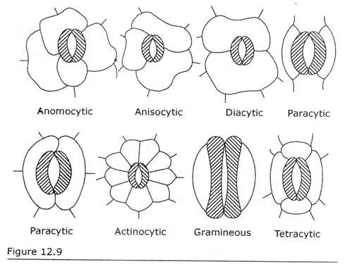
Anomocytic stoma remains surrounded by cells that are not different from other epidermal cells so far as size and shape are concerned. There exist no definite number and arrangement of cells that surround a stoma. A stoma appears to be embedded in epidermal cells. Ex. Apocynaceae, Boraginaceae, Chenopodiaceae, Cucurbita etc. Metcalfe and Chalk mentioned 142 families where anomocytic stoma is present.
Type # 2. Anisocytic (also called Cruciferous or unequal-celled type):
Anisocytic stoma remains surrounded by three unequally sized subsidiary cells, among which one is distinctly smaller in size than the other two. Ex. Cruciferae, Solanum, Petunia, Sedum and Nicotiana etc. Metcalfe and Chalk mentioned 37 families where anisocytic stoma occurs.
Type # 3. Paracytic (also called rubiaceous or parallel-celled type):
Paracytic stoma is accompanied on either side by one or more subsidiary cells. The longitudinal axes of subsidiary cells lie parallel to that of aperture and guard cells. The subsidiary cells may or may not meet over the poles.
In the family Rubiaceae the subsidiary cells usually meet over the poles. The subsidiary cells of Drvnys and Linum etc. usually fall short of the poles. Ex. Rubiaceae, Convolvulaceae, Phaseolus, Arachis and Psoralea etc. Metcalfe and Chalk cited 105 families where paracytic stoma is found.
Type # 4. Diacytic (also called caryophyllaceous or cross-walled type):
Diacytic stoma remains surrounded by a pair of subsidiary cells. The common wall of the subsidiary cells is at right angles to guard cells. Ex. Caryophyllaceae, Acanthaceae, Hygrophila, Dianthus etc. Metcalfe and Chalk mentioned eleven families to have diacytic stoma.
Type # 5. Actinocytic:
Actinocytic stoma remains surrounded by a circle of radiating cells. Ex. Ancistrocladus and Euclea pseudebenus (Ebenaceae). It is to note that Metcalfe and Chalk in the original definition of actinocytic did not state the number of subsidiary cells that enclose a stoma.
Van Cotthem described actinocytic stoma where at least five subsidiary cells surround a stoma (actinocytic = star-celled). Stace described the subsidiary cells of actinocytic stoma as somewhat ‘radially elongated’ cells.
Type # 6. Gramineous:
Gramineous stoma possesses two guard cells that are shaped like dumb-bells. Each guard cell has a narrow middle portion and two bulbous ends. The narrow middle portion is strongly thickened. The subsidiary cells occur parallel to the long axis of pore. Ex. Gramineae and Cyperaceae.
Metcalfe and Chalk in the Introduction of their book illustrated anomocytic, anisocytic, paracytic and diacytic as Type A, Type B, Type C and Type D respectively. They thought that more terms would be necessary and so added another term —actinocytic.
The terms ranunculaceous, cruciferous, rubiaceous and labiatous or caryophyllaceous stoma were coined by Vesque in 1889. Later studies revealed that these types are not confined to respective families only. Identical types occur in very distantly related families. Therefore Metcalfe and Chalk proposed new terms respectively anomocytic, anisocytic, paracytic and diacytic instead of ranunculaceous, cruciferous, rubiaceous and labiatous or caryophyllaceous.
They added the fifth type —actinocytic to the four ‘classic’ ones of Vesque. They did not employ the new terms in the text and commented that ‘the older and more familiar terms have been mentioned in this book to allow time for the new ones to become assimilated’.
In addition to gramineous stoma Metcalfe (1961) described a new stomatal type in monocotyledons. It is termed tetracytic where the guard cells are surrounded by four subsidiary cells —two laterals and two polar, each being present on the four sides. The two laterals lie parallel to guard cell. The two polar subsidiary cells are often smaller.
Tetracytic stoma characterizes numerous monocotyledonous families. It is also reported from dicotyledons, e.g. Tilia and some members of Asclepiadaceae. The seven types of stoma (five from dicotyledons and two from monocotyledons) according to Metcalfe and Chalk and Metcalfe are shown in Fig. 12.9.
 Diagrammatic representation of different types of stoma in dicotyledons and monocotyledons. Guard cells are hatched. The other are subsidiary cells/epidermal cells.
In addition to the above five types of stoma in dicotyledons Van Cotthem illustrated two more types of stoma, which are as follows (Fig. 12.10):
Type # 7. Hemiparacytic:
The stoma is accompanied by a single subsidiary cell, which is placed parallel to the long axis of the pore and this cell may be long or short in length in contrast to the guard cells. Example: Glinus latioides and Trianthema lancastrum etc.
Type # 8. Hexacytic:
The stoma is surrounded by six subsidiary cells among which two are situated on the two polar sides and rest two pairs occur on the two lateral sides being parallel to the long axis of the guard cells.
The size of subsidiary cells may be of two different types:
(a) The two polar cells may be as broad as the stomatal complex (e.g. Geogenanthus), and
(b) The length of the outer most lateral subsidiary cells may as long as stomatal complex (e.g. Commelina).
Stace (1965) illustrated the cyclocytic (Fig. 12.10) stoma in dicotyledons. The stoma is surrounded by four or more subsidiary cells, which form one or two narrow rings around the guard cells.
Example:
Lumnitzera, Laguncularia etc.
The above discussed stomatal types are tabulated in table 12.1:
Stebbins and Khush (1961) recognized four categories of stomatal complex in monocotyledons. The basis of classification is the presence or absence of subsidiary cells and when present the number, shape and arrangement in relation to guard cells form the basis.
Stebbins and Khush did not propose any terminology to aid their concepts and distinguished them as First type, Second type, Third type and Fourth type which are as follows (Fig. 12.11):  First type:
The two reniform guard cells of stoma remain surrounded by subsidiary cells that are 4-6 in number. In Rhoeo, Tradescantia and Zebrina of Commelinaceae there are four subsidiary cells. Each subsidiary cell occurs on each of the four sides of paired guard cells. As a result the stomatal complex has square appearance in surface view.
Commelina of the same family has six subsidiary cells. The additional subsidiary cells occur on the lateral sides of stoma, each being present on each of the two lateral sides. In contrast to Zebrina, Commelina has four subsidiary cells on lateral sides, two being present on each of the two lateral sides of paired guard cells.
Second type:
The two reniform guard cells of stoma remain surrounded by subsidiary cells that are 4-6 in number. Two subsidiary cells are smaller and roundish than the rest. The roundish cells are present at the ends of paired guard cells —each being present on each end. In Pandanus haerbachii (Pandanaceae) there are four subsidiary cells among which two are smaller and roundish than the other two.
The roundish cells occur at the ends and other two elongated subsidiary cells are present on the lateral sides of the paired guard cells. In Phytelephas microcarpa there are well defined six subsidiary cells.
The roundish cells are situated at the ends of paired guard cells, each being on each end. The rest occurs on lateral sides, two being on each side. Such type of stomatal complex with four subsidiary cells occurs in Calycanthaceae.
Third type:
The two guard cells of stoma remain surrounded by two subsidiary cells each being present on each lateral side of guard cells. This is the most common and predominant type of stomatal complex and spreads over 24 monocot families so far investigated. Ex Juncales, Graminales, Cyperales, Typhales etc.
Fourth type:
The two guard cells of a stoma are without any subsidiary cells. A stoma appears to be embedded in the epidermal cells. Ex. Amaryllidales, Iridales, Orchidales etc.
Paliwal (1969) distinguished morphologically five main types of stoma in monocotyledons (Fig. 12.12) on the basis of number and arrangement of subsidiary cells and suggested special terminology for them. The terms were derived from the word ‘sahkoshik’ which in the language of Sanskrit means subsidiary cells.
The stomatal complexes are as follows:
1. Asahkoshik (Sans: ‘A’= without + Sahkoshik):
There are no subsidiary cells in this type of stoma and the stomatal complex consists of two guard cells only. Ex. Members of Liliaceae, Orchidaceae etc.
2. Dwisahkoshik (Sans: ‘Dwi’ = two + Sahkoshik):
In this type of stoma there are two subsidiary cells each of which is situated laterally on either side of the pair of guard cells. Ex. Cyperaceae, Palmae etc.
3. Chatushsahkoshik (Sans: ‘Chatus’ = four + Sahkoshik):
In this type of stoma there are four subsidiary cells, which are arranged in two different ways:
(a) A pair of subsidiary cell is placed laterally on two sides of the guard cells. Ex. Members of Zingiberaceae and
(b) Each of the four subsidiary cells is situated on the four sides of guard cells, i.e. two polar and two laterals. Ex. Rhoeo.
4. Shatsahkoshik (Sans: ‘Shat’ = six + Sahkoshik):
In this type of stoma there are six subsidiary cells of which two are placed on the two polar sides and a pair on two lateral sides of the guard cells. Ex. Members of Commelinaceae, Palmae, Musaceae etc.
5. Bahusahkoshik (Sans: ‘Bahu’ = many + Sahkoshik):
In this type of stoma there are more than six subsidiary cells, which are either arranged irregularly or form a ring around the guard cells. Ex. Agavaceae, Araceae etc.


