In this article we will discuss about the stoma in a plant. This will also help you to draw the structure and diagram of stoma in plant.
A stoma consists of two guard cells that surround an aperture. In the extinct Devonian Pteridophyte Zosterophyllum myretonianum and Z. ilanoveranum had one guard cell with an aperture in its centre that simulates the stoma of moss sporophyte. In the extant Pteridophyte Azolla pinnnta the stoma consists of a unicelled guard cell with a pore.
The guard cell is binucleate and there is no wall separating the nuclei. The aperture leads to a large intercellular space below. This space is termed as sub-stomatal chamber and it is continuous with the intercellular spaces of the mesophyll. Differential expansion between guard cell mother cell and the developing underlying mesophyll cells result in the formation of sub-stomatal cavity or chamber.
There are certain epidermal cells that adjoin the guard cells and differ from the rest of the epidermal cells in size, arrangement and content. Such cells are termed as subsidiary cells.
Conventionally a stoma means a pair of guard cells and the pore. A stoma along with neighbouring subsidiary cells is referred to as stomatal complex/stomatal apparatus (Fig. 12.21). The term stomatal pore indicates the intercellular space only.
 Diagram illustrating stomatal apparatus in transverse section.
i. Guard Cell:
There are two basic shapes of guard cells. One is crescent-shaped with blunt ends as seen in surface view; a pair of such cells is attached to each other by their concave ends thus enclosing a pore. A pair of guard cells and the pore together constitute an elliptical stoma that appears in surface view. Such type of stoma is the characteristic of most dicotyledons (Fig. 12.5 B).
The other is dumb-bell shaped. The two ends of such cells are bulbous and they are continuous with each other through a narrow middle section. Two such guard cells are attached to each other at the bulbous ends.
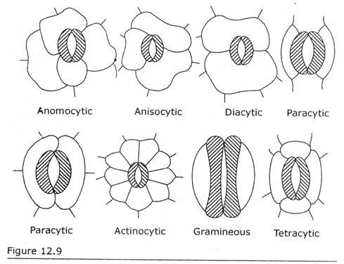
An aperture is formed at the narrow middle portion of the two dumb-bell-shaped guard cells (Fig. 12.9 Gramineous). Such type of stoma is the characteristic of Gramineae and also reported to be present in Cyperaceae, Flagellariaceae, Marantaceae, Anarthriaceae and Rapateaceae etc.
Diagrammatic representation of different types of stoma in dicotyledons and monocotyledons. Guard cells are hatched. the others are subsidiary cells/epidermal cells.
There exist considerable size variations in a guard cell. Depending on the species the length of a guard cell ranges from 10 µ to 80 µ. The width may vary from a few microns to 50 microns. The variations of width are dependent on the condition of a stomatal aperture. .
Conventionally, anatomists call the four sides of a guard cell (Fig. 12.5A) as ventral wall (= wall that occurs towards pore), dorsal wall (= wall that occurs opposite to ventral wall and juxtaposed to neighbouring epidermal cells), outer lateral wall (= wall that occurs parallel to leaf surface toward the atmosphere) and inner lateral wall (=wall that occurs toward the sub-stomatal chamber).
There exist variations in the guard-cell-wall thickenings. In kidney-shaped guard cells (=also called reniform guard cells) the dorsal wall is thin. The ventral wall is heavily thickened and may be ornamented. Localized ledges or projections of thick wall or cuticle often occur on the upper and lower edges of ventral wall of guard cells.
The ledges appear as horns in cross-sectional view. Ledges may be absent, occur only on upper side or occur both on upper and lower sides. When present on both sides, the upper ledge delimits the front cavity above the stomatal pore; the lower ledge is situated below the back cavity of stomatal pore (Fig. 12.6) and above the sub-stomatal chamber.
In dumb-bell-shaped guard cells the bulbous ends are thin walled. The narrow middle section that connects the bulbous ends is thick walled. The middle portion has very thick upper and lower lateral walls and thin ventral and dorsal walls.
Electron microscopic study indicates that the bulbous ends of guard cells of Zea mays have thick walls. The guard cells are attached to subsidiary cells or other epidermal cells by their dorsal wall only.
Figure 12.6 Diagram illustrating ledge of a stoma. Ex. Pastinaca. Guard cells are in cross-sectional view.
Guard-cell walls are rich in pectins that impregnate the cellulose microfibrils. Electron microscopic study reveals that in kidney-shaped guard cells the cellulose microfibrils/micellae have radial orientations, i.e. microfibrils radiate out from the pore around the guard cell (Fig. 12.7). In dumb-bell-shaped guard cells the microfibrils radiate out from the pore and they exhibit axial arrangements.
The guard-cell walls contain lignin, e.g. many vascular cryptogams, gymnosperms (ex. Abies pinsapo and Juniperus chinensis) and some angiosperms. In the guard cell wall of Ophioglossum β-1, 3 glucan, a callose, has been detected next to plasmalemma. Silicon, in the form of silicon dioxide is located below the cuticle of guard cells of sugarcane and Equisetum.
It is thought that the thick-walled region of guard cells and the radial micellation (= radial orientation of microfibrils) play a major role in the mechanism of opening and closing of pore. Stomatal movement is attributed to changes in turgor of guard cells. With the increase of turgor the thin dorsal wall expands more than the thick ventral wall.
As a result the shape of guard cells is deformed in such a way that the pore of stoma opens. There exists a relationship between potassium accumulation in guard cell and opening of stoma. Guard cells have greater concentration of potassium than the neighbouring cells. So it is regarded that greater concentration of potassium in the guard cells leads to the opening of stoma and loss of potassium brings about closure of stoma.
The radial micellation of guard-cell walls allows the expansion of guard cells. With the increase of turgor the thin-walled ends of the dumb-bell-shaped guard cells swell and push the middle portion apart (Fig. 12.8). As a result the pore opens with parallel sides.
All the usual cell organelles are present in the guard cells, e.g. mitochondria, microbodies, peroxisomes, spherosomes (ex. Campanula persicifolia), microtubules, rough endoplasmic reticulum, vesicles, plastid and nucleus etc., among which the last two are in brief discussed below:
a. Plastids:
All functional stomata have chloroplasts in the guard cells. There is report that the functional guard cells lack chloroplasts, e.g. Paphiopedilum and Pelargonium zonale.
The number of chloroplasts varies depending on the species. Anthoceros has two chloroplasts per guard cell, Selaginella has three to six chloroplasts per guard cell and Polypodium vulgare exhibits as many as 100 chloroplasts per guard cell.
The size of chloroplast varies. In some species of Allium it is too small to be observed under light microscope. It is large in ferns. In contrast to the mesophyll chloroplasts, guard-cell-chloroplasts are poorly developed. Guard-cell- chloroplasts have little thylakoid structure and scant granal stacking.
A common feature of guard cell chloroplasts is the abundance of starch. Exceptions are noted in the families Iridaceae, Amaryllidaceae and Liliaceae.
b. Nucleus:
The kidney-shaped-guard cells have prominent nucleus similar to other cells of leaf. The nucleus is usually centrally located close to ventral wall of guard cell. The nucleus exhibits change in shape during stomatal closure and opening. In Vicia faba the nucleus is oval when stomata are closed and it becomes rounded when stomata are open.
In dumb-bell-shaped guard cells a mass of nuclear material is present instead of nucleus. The mass of nuclear material is located at each bulbous end of a guard cell. A strand of nucleoplasm that occurs at the narrow middle portion of guard cell connects the masses of nuclear materials present at the two bulbous ends.
Two dumb-bell-shaped guard cells are attached to each other by their bulbous ends thus forming a stoma. In contrast to crescent-shaped-guard cell the bulbous ends are fused. In the common walls of guard cell pair large pores of about one micron or more in diameter exist. Through the pores exchange of organelles between each guard cell pair occurs. So the guard-cell-pair is regarded as a single binucleate unit.
The guard cells are distinct within the epidermal layers both anatomically and biochemically. They have adapted a specialized set of metabolic pathways that cause rapid change in osmotic potential. Minor changes in the external environment stimulate the metabolic pathways. As for example, a change in carbon dioxide concentration in atmosphere will stimulate a series of reactions that result in opening and closing of stoma.
The stomatal movement is associated with rapid fluxes of K+ and H+ between guard cells and neighbouring cells. Guard cells possess high metabolic activity due to the presence of abundant mitochondria and protein synthesizing machinery. Calvin cycle is not detected in guard cells. It is to note that functional plasmodesmata do not exist between guard cells and neighbouring cells though plasmodesma exists in developing stoma.
ii. Subsidiary Cell:
Subsidiary cell (also called accessory cell) can be defined as specialized epidermal cell that differs in size, arrangement from the rest of epidermal cells and lies close to guard cells. In Pinus and Equisetum subsidiary cell envelops the guard cells. They are smaller than the epidermal cells.
They contain dense cytoplasmic content and greater frequency of cell organelles. Normally subsidiary cells are devoid of chloroplasts, anthocyanins and crystalline inclusions. Subsidiary cell plays a major role in the movement of stoma composed of dumb-bell-shaped guard cells.


