In this article we will discuss about the signalling mechanisms, both in eukaryotes and prokaryotes.
1. Eukaryotic Cell-to-Cell Signaling:
The integrative nature of biological systems could be understood after the pioneering work of Claude Bernard (1813-1878) of France. He gave the concept of the miliew interieur and suggested the system of ductless gland (i.e. endocrine glands) for integrating function and maintaining homeostasis.
In 1902, Bayliss and Staling demonstrated a marked flow of pancreatic juice in dogs after injecting an acid extract of duodenum. Starling coined the term ‘hormone’ (Greek, I excite) for such intercellular messenger molecules. An American physiologist Water Cannon coined the term ‘homeostasis’ (a condition that may vary but is relatively constant).
There are three types of signalling systems in multicellular organisms like mammals: neuronal, endocrine and cytokine signalling (Fig. 27.3). Neuronal signalling occurs over very long distance i.e. brain to toe. Synaptic junctions communicate rapidly. Many chemicals are involved in signalling at junctions and associated with inflammation.
Endocrine signalling involves the release of a hormone from its gland and its transport to blood to a limited number of cells in the target tissue. It occurs at a long distance and limited by the rate of blood flow and diffusion from blood to tissues.
Most of the intracellular signalling is that of the cytokines. Much of this signalling occurs through paracrine signalling (over short distance cell to nearby cell) or by auto-signalling (stimulation of the cell producing the cytokines).
Certain bacterial endotoxins target neuronal signalling; hence these are called neurotoxins as produced by Clostridium tetani and CI. botulinum. These have metalloproteinase activity and cleave specific intracellular proteins. Thus they prevent the neurotransmitters. A recent discovery also points to the interaction of a bacterial toxin with neuroendocrine signalling and synapsis of cytokines.
(i) Endocrine Hormone Signalling:
Endocrine hormones are mostly produced by specific glands (such as pituitary, hypothalamus, and parathyroid glands) and glandular tissues (such as pancreas and intestine).
There are three major groups of hormones: peptide hormones (produced especially by the intestine which has neurotransmitter-like activity), steroid hormones (produced by adrenal cortex, gonads and skin), and thyrosine derivatives (e.g. thyroid hormones T3, T4, etc. and catecholamines, noradrenaline, adrenaline and dopamine having neurotransmitter activity).
After secretion from glands, endocrine hormones are circulated as free hormone or bound to carrier proteins, for example a serum protein, albumin. It binds to several circulating hormones and exerts action (only in bound form) to specific cell receptors in target tissues.
The peptide hormones bind to specific membrane receptors which result in specific intracellular signalling pathway. On the other hand the steroid and sterol hormones enter into cells and bind to cytoplasmic receptor proteins and then move to nucleus and act as a factor for transcription.
Endocrine hormones control the energy metabolism through insulin and glycogen, adrenaline and noradrenaline involving production and breakdown of carbohydrate stored as glycogen in the liver and muscle.
The lack of control of this system is visible in diabetes. Bacterial infection results in hormonal imbalance in body. Certain bacteria and viruses affect neural tissue. Mycobacterium leprae and Treponema pallidum have a tropism for nervous tissue.
The tissue of the gastric and intestinal mucosae are highly regulated. They respond and produce several endocrine signals including gastro-intestinal hormones e.g. gastrin, secretin, cholecystokinin and guanylin. E. coli alters fluid imbalance in the intestine and causes diarrhoea. Now seven different strains of E. coli have been reported which induce different pathological symptoms.
The enter toxigenic E. coli strains produce heat-labile toxin (LT) and heat-stable toxin (ST). ST is the first bacterial analogue of an endocrine hormone (guanyl) that activates guanyl cyclase and control fluid release from intestinal cells so that mucin layer could be kept wet (Fig. 27.4). There are other heat-stable guanyl-like toxin of other strains of E. coli and other bacteria.
(ii) Cytokines:
Cytokines are a large group of over 1000 proteins which are involved in cell- to-cell signalling and control the inflammatory response to bacterial infection. These are polypeptide hormones secreted by a cell that affects growth and metabolism of the same cell (autocrine signalling) or another cell (paracrine signalling).
Their over production causes disease. These are found at the site of infection by the agents. These induce lipid mediators (prostaglandins, leukotrienes, lipoxins, platelet-activating factor and the mediators from mast cells (e.g. histamine and enzymes such as tryptase).
(a) Nomenclature of cytokines:
Cytokines are divided into six sub-families (Table 27.7) on the basis of several criteria such as historical types, sequence homology, localisation of chromosomes and biochemical actions. In 1979, the term interleukin (inter: between, leukin: leukocytes) was coined to denote the proteinaceous factors which modulate the function of the other leukocytes.
At present there are over 20 interleukins (IL-1 to IL-18). The endotoxin-injected mice expressed a tumour necrosis factor (TNF) grouped under cytotoxic cytokines. The TNF kill certain tumour cell lines via induction of apoptosis and are potent pro-inflammatory molecules. The TNF receptor family is itself membrane-bounded proteins (e.g. CD27, CD30 and CD40).
Table 27.7 : Cytokines: nomenclature and sub-families.
The interferon’s (IFNs) are such cytokines which were discovered first. These are involved in inhibiting the growth and spread of viruses. They are of three types: INF-a, INF-P, and INF- Y- Interferon’s also act against protozoa, rickettsia and mycobacteria.
The colony-stimulating factors (CSFs) control the growth and differentiation of neutrophils, monocytes and cell populations derived from monocytes in the bone marrow. The monocytes/macrophages are the phagocytic cells which engulf and kill bacteria. Hence, they are also called as antigen-presenting cells and stimulate T and B lymphocytes.
Growth factors include families of proteins such as fibroblast growth factor (FGF) family, platelet-derived growth factors (PDGF), and transforming growth factor-β (TGFβ). The FGF cytokines act on mesenchyme cells and epithelial cells also.
The peptide chemotactic factors are called chemokines which is a large sub-group of cytokines. Chemokines have molecular mass of 8-10 kDalton, with 20-50% sequence homology at protein level and cysteine as conserved residues which form disulphide bonds within the molecules.
On the basis of chromosomal location of genes and protein structure, chemokines are divided into two families: α-chemokine and β-chemokine families. A third family of chemokines discovered in 1994 currently has one member called lympholactin which is a strong attractant of T cells.
(b) Receptors of cytokines:
Cytokine receptors have high affinity for their ligand. The number of individual receptor present on target cell is low. On the basis of sequence homology and structural motifs cytokine receptors are grouped into a small number of families. At present there are nine receptors for CC chemokines (CCR), five receptor for CXC chemokines and CXCR1, one receptor for fractalkine.
The cytokine receptors are shed from cell via proteolytic cleavage. Cell surface metalloproteinases (sheddases) help the release of cytokine receptors. The released receptors bind the soluble cytokines and inhibit their activity or stimulate the cytokine-receptor lacking cells.
(c) Biological action of cytokines:
Cytokines play a role in physiological development. They are found at all developmental stages in mammals. On the other hand, cytokine receptors present on cell membrane also play a physiological role. They act as portals for vital entry into cells. For example HIV enters through binding to cytokine receptors. Similarly, herpes simplex virus enters through binding the TNF receptor family.
After binding receptors induce selective intracellular signalling resulting in switching on or switching off of particular genes and production of cyclooxygenase II, and nitric oxide (NO) is synthesised after induction of nitric oxide synthetase.
Aspirin and ibuprofen are the non-steroid anti-inflammatory drugs which block cyclooxygenase activity. These drugs reduce pain and fever as the prostaglandins and prostacyclin lower threshold in pain nerve resulting in a relief of pain and fever.
Various molecules are produced after binding cytokines to cytokine-receptor which produce pathology [prostaglandins, NO, tissue plasminogen activator (tPA) and plasminogen activator inhibitor and collaginases]. Tissue damage is directly induced by collagenase and tPA.
Besides, cytokines also induce the synthesis of their own and other cytokines which result in a complex network of interactions. Cytokines can also modify the behaviour of cells in many ways. Various actions of cytokines on cells are shown in Fig. 27.5.
2. Prokaryotic Cell-to-Cell Signalling: Quorum Sensing and Bacterial Pheromones:
Until the 1980s, no attention was paid that bacteria could talk to one another. Thereafter, examples were put forth for cell-to-cell signalling in bacteria. Conjugation is one of the methods of DNA transfer between two bacteria. To establish conjugation, both the bacteria must establish cell-to-cell contact. Enterococcus faecalis is a Gram-positive mammalian pathogen.
Its aggregation in controlled by the secretion of small peptide pheromones. Pheromones induce adhesion production; consequently bacteria form cell clumps which facilitate conjugation. Several pheromones have been isolated which are hepta- or octa-peptides found in low concentration (5×10-11 M).
Endospores of Clostridium tetani are regarded as resting forms of bacteria and a part of virulence mechanism. In contrast some bacteria such as a myxobacterium under adverse environmental conditions undergo complex morphological changes. Polyangium vitellinum forms cyst-like structure consisting of an outer covering of polysaccharide to resist from dehydration.
Myxococcus xanthus forms myxospores (fruiting body) and alternate with vegetative cells This programme is triggered by starvation which causes morphological changes within 4 hours. A dense mound-shaped structure is formed when a cell density of bacteria has reached to about 105. After 20 hours of starvation the cells inside this mound differentiate into myxospores.
Myxospores are heat- and starvation-resistant dormant cells. They germinate during favourable conditions and produce vegetative cells. Again myxospores are formed when conditions are unfavourable. This type of cell differentiation is controlled by extracellular signals. Cell-to-cell signalling mechanism is given in Table 27.8.
Quorum Sensing:
The term quorum refers to ‘a fixed number of members of any committee of the society whose presence is mandatory for proper transaction of business’. Quorum sensing in bacteria is a mechanism through which they take a census of their number. After reaching a quorum of cell number they can transact the business of switching on or switching off of specific genes.
The current knowledge of quorum sensing began with the study of luminescence in Vibrio fischeri and V. harveyi. They are marine bacteria forming symbiotic relationship with monocentrid fish and with bobtail squids (e.g. Euprymna scolopes). The bobtail squid consists of very high concentration of V. fischeri. The light organ is supposed to be part of a counter illumination the details of which are not clear.
The newly hatched squids develop symbiotic association with only certain strains of V.fischeri. Within hours after hatching, light organ is colonised by V.fischeri. The light organ positively selects only certain strains of V.fischeri and negatively selects the others to exclude colonisation of other bacteria present in sea water.
It is not known how this selection is made. One of the possible mechanisms may be the expression of specific adhesin for V.fischeri by epithelium of light organ. The epithelium is exposed to trypsin which directly triggers a specific morphogenetic response in the squid. This results in formation of the complex.
(a) Mechanism of quorum sensing:
It is the feedback control system. Bacteria continuously produce a small amount of signal called auto inducer. Most of the Gram-positive bacteria produce auto inducer which are acylhomoserine lactones (AHLs). Staphylococcus aureus and other bacteria produce peptide auto inducers. E. colt and S. typhimurium produce a quorum sensing molecule of 1 kDalton. These extracellular inducers are diffused out.
Besides, bacteria also recognise the presence of auto inducer. The bacterial membrane protein does this function. It acts both as receptor of auto inducer and activator of gene transcription. V. fischeri produces luminescence. V. fischeri system is the best studied quorum sensing system.
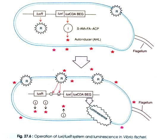
Luminescence is associated with lux operon system which consists of two main regulatory genes luxl and luxR (Fig. 27.6) and other genes (luxCDABEG) which synthesise chemicals to produce light. LuxI encodes a protein which catalyses the synthesis of a wide range of AHLα. Autoinducer of V. fischeri is N-(3-oxo-hexanoyl)-L- homoserine lactone.
LuxR encodes a protein which acts both as a receptor for AHL and as a transducer of the signal that activates the other genes of lux operon. The luxCDABEG genes are expressed after binding AHL to the luxR protein (Fig. 27.6). The luxA and luxB genes synthesise the α- and β- subunits of bacterial luciferase. The other genes encode polypeptides which facilitate the synthesis of the substrate and produces light.
(b) Quorum sensing as a virulence mechanism:
In addition to V. fischeri, there is a large number of Gram-negative bacteria which produce AHLs to quorum sense. These are medically important bacteria, for example Pseudomonas aeruginosa, Proteus mirabilis, Serratia liquefaciens and Yersinia enterocolitica. In these bacteria LuxI/LuxA homologues are involved in quorum sensing system. Ps. aeruginosa utilises two quorum systems, the las and rhl.
The las operon expresses LasR protein which is similar to LuxR and acts as transcriptional activator in the presence of PAI of Pseudomonas. The LasI (the Luxl homologue) produces AHL. The autoinducer of P. aeruginosa at a threshold concentration swich on a group of virulence gene including lasB, lasA apv and toxA.
The rhl system is the second quorum sensing system which involves RhIR (the transcriptional activator protein) along with the autoinducer (N-butyryl-L-homoserine lactone) synthesised by RhIR. This quorum sensing system results in production of extra virulence factor e.g. elastase which cleaves and inhibits the interleukin-2 (the key host defence cytokines). The las system is dominant which is activated before the rhl system.
Many Gram-positive bacteria use oligopeptide as signalling molecules. For example, two different peptides are secreted by Bacillus subtilis. These are necessary for competence (ability for DNA uptake) and sporulation.
In Staphylococcus aureus, a locus agr controls the expression of many virulence factors, namely exotoxins, capsular polysaccharide type 8 and V8 protease. An octapeptide quorum sensing autoinducer is encoded by the agr lucus which induces the agr locus. The quorum sensing autoinducer interacts with host defence system and inhibits the albeit at high concentration (Fig. 27.7).


