In this article we will discuss about Dugesia:- 1. Habit and Habitat of Dugesia 2. Structure 3. Body Wall 4. Locomotion 5. Digestive System 6. Respiratoty and Excretory System 7. Nervous System 8. Sense Organs 9. Reproduction 10. Copulation, Fertilisation and Development 11. Regeneration.
Contents:
- Habit and Habitat of Dugesia Tigrina
- Structure of Dugesia Tigrina
- Body Wall of Dugesia Tigrina
- Locomotion of Dugesia Tigrina
- Digestive System of Dugesia Tigrina
- Respiratoty and Excretory System in Dugesia Tigrina
- Nervous System of Dugesia Tigrina
- Sense Organs of Dugesia Tigrina
- Reproduction in Dugesia Tigrina
- Copulation, Fertilisation and Development of Dugesia Tigrina
- Regeneration of Dugesia Tigrina
1. Habit and Habitat of Dugesia Tigrina:
Dugesia tigrina is gregarious (lives in groups) found on the underside of leaves, logs, debris and rocks submerged in cool, clear and running water of streams, ponds and lakes.
They are not easy to see unless they are moving, for they are small and flat and their dark mottled colour blends perfectly with the rocks or plants to which they cling. They live in damp surroundings as their bodies are not protected against desiccation. Dugesia tigrina is world-wide in distribution.
2. Structure of Dugesia Tigrina:
(i) Shape, Size and Colour:
Dugesia tigrina is about 12 mm long and is dark brownish or blackish in colour.
It is a thin flattened worm with definite sides, there is an anterior end directed forwards in moving, one surface of the body is always uppermost, this is the dorsal surface, while the surface towards the substratum is ventral. The dorsal surface is darker in colour than the ventral surface. It has bilateral symmetry which is in direct correlation with progression towards the anterior end.
(ii) External Morphology:
The anterior end forms an obvious head which is triangular with two laterally projecting head lobes or auricles. On the head are two cup-shaped black eyes.
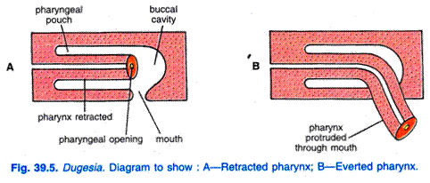
The head is separated from the body by a neck-like constriction. The body is elongated with the dorsal surface slightly arched and the ventral surface flat. Behind the middle of the body on the ventral side is a mouth which leads into a pharyngeal sheath containing a cylindrical pharynx which can be seen through the body wall, the pharynx can be everted through the mouth as a proboscis.
On the ventral surface encircling the margin is an adhesive zone through which an adhesive substance comes out from glands.
The animal clamps itself to the substratum by the adhesive zone. The animal when moving leaves behind a mucus trail, mucus is secreted by mucous glands opening on the ventral surface. Just a little behind the mouth aperture on the ventral surface, there is a genital aperture in sexually mature forms.
3. Body Wall of Dugesia Tigrina:
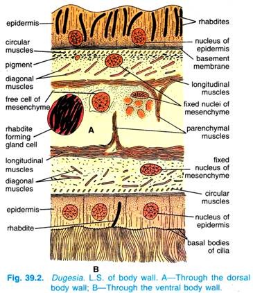
The body wall is made of an outer epidermis and inner muscle layers. Both these layers are separated by a basement membrane. The space between muscle layer and the alimentary canal is filled with a special type of tissue called mesenchyme or parenchyma, therefore, no coelom or body cavity is found in it.
However, a detailed study of the body wall represents the following structures:
(i) Epidermis:
It is single cell-layered thick and made of cuboidal epithelial cells. The epidermis is ciliated all over in most planarians, but in Dugesia tigrina, cilia are found only on the ventral surface where they are absent from the adhesive zone. Between the epidermal cells are sensory cells and mucous gland cells in certain areas. The gland cells provide a mucus coating for the animal and they lay down a slime trail for its locomotion.
In the epidermal cells are characteristic erect hyaline rods called rhabdites, they are more abundant on the dorsal than the ventral side. Rhabdites are secreted by rhabdite gland cells usually located in mesenchyme. After the rhabdites are secreted in the rhabdite gland cells, they migrate to the epidermal cells where they lie. Rhabdites are absent in the adhesive zone.
The function of rhabdites is not known, but they form a slimy substance on discharge to the exterior which may be protective, and help in obtaining living food. Below the epidermis are granules and rods of pigment. The glands are all unicellular, some occur in the epidermis but most of them are in the mesenchyme, they have long necks opening on the surface, they secrete mucus.
(ii) Basement Membrane:
The epidermal cells rest on a structure-less thin basement membrane to which underlying muscle layer is attached. In fact, it marks the boundary between the epidermis and muscle layers and it helps in maintaining general form of the body.
(iii) Muscle Layer:
The basement membrane layer is followed by muscle layer. It contains elongated contractile muscle cells. These cells originate from myoblasts which are mesodermal in origin. The muscle layer is differentiated into an outer layer of circular muscles, middle layer of diagonal muscles and inner layer of longitudinal muscles.
The longitudinal muscle layer is more developed on the ventral side. The dorso-ventral muscles extend across the body between dorsal and ventral surfaces.
(iv) Parenchyma or Mesenchyme:
It is a special type of connective tissue of mesodermal origin. It is filled in the spaces between various internal organs and body wall. It is, in fact, a net-like syncytium containing nuclei, free wandering mesenchyme cells like neoblasts or formative cells and fluid-filled spaces.
The mesenchyme cells serve to transport digested food and excretory wastes; in fact they perform the function of circulatory system of higher animals as this system is wanting in flatworms.
4. Locomotion of Dugesia Tigrina:
Dugesia tigrina is aquatic but it does not swim. Dugesia tigrina moves in two ways. The usual way is gliding. In gliding, head is slightly raised, over a slime tract secreted by its marginal adhesive glands. The beating of the ventral cilia in the slime tract drives the animal along.
Rhythmic waves of movement can be seen passing backward from the head as it glides. A less common method is crawling. In crawling, Dugesia tigrina lengthens, anchors its anterior end with mucus or by its special adhesive organ, and by contracting its longitudinal muscles pulls up the rest of the body. By means of its oblique muscles it can change its direction.
5. Digestive System of Dugesia Tigrina:
It includes the alimentary canal, food, feeding and digestion.
(i) Alimentary Canal:
The flatworms are the first in the animal kingdom to possess the alimentary canal which is incomplete because anus or any exit for defaecation is not found. However, the alimentary canal of Dugesia (Fig. 39.6) consists of mouth, pharynx and intestine.
(ii) Mouth:
It is a small oval or rounded aperture situated on the ventral side behind the middle of the body. It serves both for ingestion and egestion. It leads into a short mouth cavity which joins a cylindrical thick-walled pharynx.
(iii) Pharynx:
The pharynx lies in a pharyngeal cavity or pouch bounded by a muscular sheath called the pharyngeal sheath. The thick-walled, muscular and cylindrical pharynx in the pharyngeal pouch is attached to the anterior end of the pharyngeal sheath. The pharynx can be everted or protruded through the mouth opening as a proboscis which can be extended greatly. The eversible proboscis of Dugesia helps in feeding.
The pharynx in section is circular enclosed in a circular pharyngeal cavity. It consists of following layers from the surface to the lumen, epithelial cells, longitudinal muscle layer, circular muscle layer, outer gland cells, nerve plexus, inner gland cells, longitudinal muscle layer, circular muscle layer and an endodermal epithelial lining.
(iv) Intestine:
The attached end of the pharynx leads into an intestine which divides at once into three branches characteristic of order Tricladida to which Dugesia belongs, one extending forward in the middle line up to the head, and the other two going backwards to the posterior end, one on either side of the pharyngeal cavity.
All the three branches of intestine give off numerous branching diverticula, all ending blindly, there being no anus. The much-branched intestine is a means of increasing the surface area for digestion, absorption and distribution of food. The intestine is made of a single layer of vacuolated columnar cells containing granules, between the columnar cells are some gland cells of triangular shape containing reserve proteins.
(v) Food, Feeding and Digestion:
The animal is carnivorous. The food consists of small living worms, crustaceans and snails, and pieces of larger dead animals.
In feeding, the animal perceives the presence of food at a distance and moves towards it, living food is often entangled in the slimy secretions of mucous glands and rhabdites, then it encloses the food in the everted pharynx and digestive juices are poured into the food, the food is broken up by a pumping action of the pharynx and acted upon by extruded digestive juices for extracellular digestion, after which the food is swallowed.
Digestion is both extracellular and intracellular; the mesenchyme helps to distribute digested food. Undigested food is egested through the mouth. Planarians can live without food for long periods, they obtain nourishment by dissolving their reproductive organs, parenchyma, and muscles, they get smaller in size. The missing parts are regenerated when they feed again.
6. Respiratory and Excretory System of Dugesia Tigrina:
There are no respiratory organs. Exchange of gases takes through the body surface, i.e., respiratory exchange is by diffusion.
Excretory System in Dugesia:
The excretory system of Dugesia tigrina (Fig. 39.7) consists of a system of excretory tubules having a large number of excretory cells called flame cells or protonephridia.
(i) Excretory Tubules:
There is one pair of longitudinal excretory trunks running on each side of the body which open to the dorsal surface by several minute pores called nephridiopores, each pair of trunks is considerably coiled together, and the two pairs are connected together in the head by a transverse vessel.
Each longitudinal trunk divides into a number of branches, the branches divide into extremely fine capillaries which end in flame cells. The capillary is actually a part of the flame cell.
(ii) Flame Cell:
The flame cell is nucleated and has a number of protoplasmic processes reaching into the mesenchyme. The flame cell has an intracellular space which is continued into the capillary. In the space of the flame cell are a number of flagella which vibrate giving the appearance of a flickering candle flame, hence, the name.
The flame cells occur in large numbers placed along the length of the body on each side. The flame cells are excretory units and work like nephridia of annelids. Since, these are very simple in structure and function like nephridia, hence, also referred to as protonephridia.
(iii) Physiology:
Excretory substance is collected from the mesenchyme and is transferred into the cavities of flame cells. The beating of flagella of flame cells causes hydrostatic pressure by which fluid waste passes into longitudinal trunks and goes out of nephridiopores.
The excretory system is spoken of as a protonephridial system. But more important than removal of excretory matter, the system brings about elimination of excess water from the animal, it functions as an osmoregulatory system.
7. Nervous System of Dugesia:
The nervous system of Dugesia tigrina (Fig. 39.8) represents the primitive type of centralised nervous system of higher animals. It consists of the brain, nerve cords and peripheral nerves.
Brain:
In the head is a brain made of bilobed cerebral ganglia in the shape of an inverted V, with limbs near the eyes and the rest lying parallel to the head margin. From the brain numerous nerves extend forward and laterally to the head and auricles.
Nerve Cords and Peripheral Nerves:
The brain is continued into two ventral nerve cords which run to the posterior end, each lying about one-third of the distance from the margin. The nerve cords give off transverse branches on both sides, the two cords are joined by some transverse commissures. The nervous system acts as a coordinating centre for nerve impulses.
In addition to the central nervous system there is a sub-epidermal plexus or nerve net just below the epidermis, and a deeper sub- muscular plexus in the mesenchyme below the muscle layers of the body wall, both are joined to the nerve cords. In fact, the brain and nerve cords constitute the central nervous system, while the various nerves originating from central nervous system constitute the peripheral nerves.
8. Sense Organs of Dugesia Tigrina:
In Dugesia tigrina, sense organs consist of chemoreceptors, auricular organ, tango-receptors, rheoreceptors and eyes or ocelli.
1. Chemoreceptors:
Chemoreceptors are found on the head, they are ciliated pits and grooves in which the epidermis has sunken cells having cilia but no rhabdites, they are supplied by a sensory nerve. They enable the animal to find food by means of a water current which passes over them.
2. Auricular Organ:
On each side of the head is a whitish groove called an auricular organ lying near the base of the auricle. The grooves are ciliated and are provided with nerves, they are organs of chemical sense for smelling and tasting.
3. Tango Receptors:
These are sensory cells for touch stimuli and found distributed at the anterior end most abundantly on the ventral surface around the mouth opening.
4. Rheoreceptors:
These are sensitive to water currents; their sensory processes project much beyond the level of body cilia.
5. Eyes or Ocelli:
Eyes are two round dark spots on the dorsal surface of the head. The eye has a pigment cup with its open mouth facing laterally forward. Projecting into the pigment cup are several retinal cells, they are bipolar nerve cells with expanded inner ends which are striated, and outer ends joined to the brain.
Eyes are capable of a crude discrimination of the direction of light. The pigment cup serves as a shield and light can enter only through its opening to stimulate the photosensitive expanded ends of retinal cells, thus, the animal can detect the direction of light. The animal is negatively phototactic and is most active at night.
9. Reproduction of Dugesia Tigrina:
Dugesia tigrina reproduces both asexually and sexually.
(i) Asexual Reproduction:
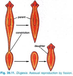
Dugesia tigrina exists in asexual and sexual strains. The asexual form has no reproductive organs, it reproduces by fission. Fission occurs when the animal has attained maximum size; the posterior end adheres firmly, while the anterior region advances forward so that the animal ruptures into two behind the pharynx.
Each new animal regenerates its missing parts. The separated anterior part regenerates the posterior region and the rear end grows into a complete worm. Locomotion and adhesion are essential for fission.
(ii) Sexual Reproduction:
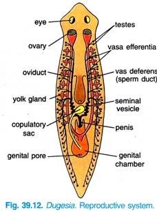
Reproductive organs (Fig. 39.12) are temporary in Dugesia tigrina, they are formed during the breeding season, after which the reproductive organs degenerate and the animal becomes an asexual strain which will reproduce by fission till early summer of the following year. The sexual strain develops hermaphrodite organs and it reproduces sexually every year in early summer.
(i) Male Reproductive Organs:
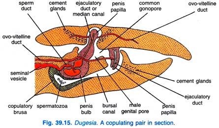
Male reproductive organs (Fig. 39.12) consist of two rows of testes, vasa efferentia, a pair of vas deferens and a penis. The testes are numerous, small and round. They are situated on the right and left borders of the body. Each testis is connected to the vas deferens of its side by a tiny duct, the vas efferens.
Each vas deferens enlarges posteriorly to form a seminal vesicle where the sperms are stored until discharged posteriorly through the muscular penis. The right and left vaas deferens unite at the middle of the body and form a median duct which passes through a muscular penis. The penis opens into a genital atrium, a cavity that terminates in the genital pore through which the penis extends during the copulation.
(ii) Female Reproductive Organs:
There is one pair of small ovaries lying laterally behind the head. From each ovary arises a long oviduct which runs laterally. At the commencement of each oviduct where it arises from the ovary, is a small dilated seminal receptacle. On each side of body there are numerous small yolk glands which join the oviducts, yolk cells from yolk glands pass into oviducts, hence, called ovo-vitelline duct.
Opening into the genital atrium is large club- shaped copulatory sac. Numerous small cement glands open into the genital atrium and oviduct. The genital atrium opens externally by a genital pore situated on the ventral side behind the mouth.
10. Copulation, Fertilisation and Development of Dugesia Tigrina:
During copulation, the two worms come together by their ventral surfaces facing in the same direction. The penis papilla of each emerges by elongation through the genital pore and is inserted into the copulatory sac of the other worm by which mutual insemination occurs.
The sperms are discharged into the copulatory sac where they stay only a short while, they travel up the oviducts to reach the seminal receptacles. As the eggs come out of the ovary they are fertilised, and they pass down the oviducts becoming mingled with yolk cells from yolk glands.
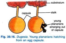
The eggs and yolk cells collect in the genital atrium a capsule or cocoon is formed around from yolk cells. The capsule contains several fertilised eggs, and it is laid through the genital pore under stones.
While passing out the capsule receives the secretion of cement glands, this adhesive secretion forms a stalk on the capsule. The capsules get attached to stones by their stalk. One animal copulates several times during the breeding season and lays a cocoon every few days.
The development is direct, i.e., without any larval form. Each zygote in capsule develops into a young worm in about two weeks, finally the capsule ruptures and the young worms hatch out (Fig. 39.16). These resemble their parents except that they are smaller in size and without reproductive organs which has not yet developed. They feed, grow and develop sex organs to reproduce again.
11. Regeneration of Dugesia Tigrina:
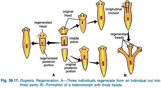
Dugesia tigrina has great powers of regeneration. Regeneration is a process of restitution and involves the development of lost part of the body automatically. If it is cut into two, each part will form the lost portion. A cut piece of moderate size from any part of the body will form a new worm. Some pieces taken from posterior side form animals with reduced heads or no head at all.
The ability of a piece to regenerate into a complete worm depends upon the regeneration of head at the anterior cut surface, because the head controls the morphological pattern.
If a sexually mature planarian is cut into two between the pharynx and the copulatory apparatus, then the reproductive organs degenerate, and each piece will regenerate into an asexual worm. Longitudinal cuts can be made to produce double heads or tails.
It was thought that interstitial cells are responsible for regeneration. But recently it has been shown that on cutting a planarian free cells from the mesenchyme called neo-blasts migrate to the cut surface and give rise to a bud-like structure called blastema which differentiate into the lost part.
In fact, the process of regeneration is completed by two processes occurring together. These processes are epimorphosis, which is related to the formation of lost part, and morphollaxis, which is related to the adjustment and coordination between the old tissue and regenerated tissues.

