In this article we will discuss about Taenia Solium:- 1. Habit and Habitat of Taenia Solium 2. Structure of Taenia Solium 3. Body Wall 4. Digestive System 5. Excretory System 6. Nervous System 7. Reproductive System 8. Life History 9. Pathogenic Effects 10. Prevention and Control Measures of Disease 11. Parasitic Adaptations.
Contents:
- Habit and Habitat of Taenia Solium
- Structure of Taenia Solium
- Body Wall of Taenia Solium
- Digestive System of Taenia Solium
- Excretory System of Taenia Solium
- Nervous System of Taenia Solium
- Reproductive System of Taenia Solium
- Life History of Taenia Solium
- Pathogenic Effects of Taenia Solium
- Prevention and Control Measures of Disease Caused by Taenia Solium
- Parasitic Adaptations of Taenia Solium
1. Habit and Habitat of Taenia Solium:
The name is due to its elongated tape-like appearance. It may attain a length of several metres. It belongs to the class Cestoidea. Sexual forms of Taenia occur as endoparasites in the intestine of man. Asexual forms are encountered in the muscles of pigs or in exceptional cases in the muscle of man.
Man is its primary host and pig is its secondary host. Distribution of Taenia solium is worldwide and is most common in European countries. Nowadays Taenia solium occurs rarely in man. Taenia saginata is more common, because it uses cattle as the secondary or intermediate host.
2. Structure of Taenia Solium:
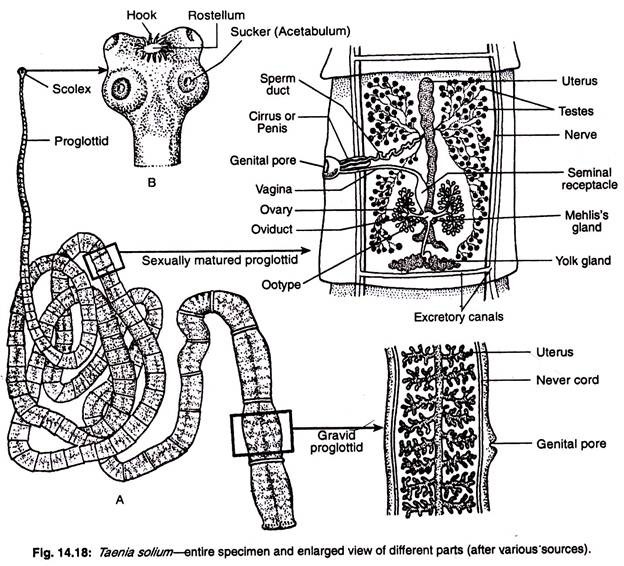
A fully developed Taenia solium may attain a length of 3-5 metres. Antero-posterior ends are clearly distinguishable but it is difficult to differentiate the dorsal from the ventral surface. The body is ribbon-like (Fig. 14.18) and consists of a distinct head or scolex at the anterior region.
The tip of the head bears a conical elevation—the rostellum which can be retracted or extended. The rostellum bears 28 to 33 hooks. The hooks are of two types—larger and smaller, and they alternate with each other. Each hook (Fig. 14.19) has three parts—a base or guard, a conical blade at the tip and a handle projected from the middle. The hooks are arranged in two rows. Such hooks are absent in Taenia saginatus.
When the contractile rostellum is withdrawn the hooks become anteriorly directed and get fixed into the host tissue. The head bears in the middle four cup-like suckers or acetabula (sing, acetabulum). Rostellum and suckers act as organs of attachment to the intestine of the host.
Behind the head there is a narrow and small tubular region—the neck or the zone of proliferation.
The rest of the body or tape is called strobila. The strobila is segmented in appearance, though this segmentation is not similar to the true segmentations of annelids. The chain-like strobila is made up of about 850 segments or sexual units, called proglottids.
The proglottids progressively increase in size and mature towards the posterior extremity. The youngest or newly formed proglottid occupies a position just beneath the neck while the oldest one is at the posterior end. The number of proglottids varies from 800-850 in a full-grown worm.
Structure of a Proglottid:
A proglottid from the middle region of the strobila offers a rectangular outline (Fig. 14.20). The surface is lined by cuticle (recently renamed as epidermis or tegument). It is thick and perforated at intervals by fine canals, at the bottom of which either gland cells or nerve endings are situated. It is followed by longitudinal and circular layers of muscles.
The circular muscles divide the parenchyma cells into an outer cortex and inner medullary region. Towards each lateral margin is found the longitudinal nerve and just median to them lies the longitudinal pair of excretory vessels. A transverse excretory canal is situated at a posterior position of the proglottid.
The anterior-lateral borders of the medulla are housed with testes and posterior lateral borders are with ovary. The male and female genital ducts open to a chamber, called the genital atrium, which is situated in the middle of one of the lateral margins. The atrium opens to the exterior through an aperture, called genital pore.
3. Body Wall of Taenia Solium:
The body is covered by a thick epidermis. The epidermis is many-layered and perforated. It is impregnated with calcium carbonate. The epidermis remains sunk in the parenchyma. Longitudinal muscles run under the epidermis. The parenchyma is divided into an outer cortical zone and inner medullary zone by the circular muscles. Nervous, reproductive and excretory organs are situated within the medullary zone.
For a long time it was regarded that the body of tapeworm is covered by thick cuticle. But recently electron microscopic studies have revealed that the outer layer of the body of all cestodes contains mitochondria and remains continuous with processes emerging from the cytoplasm of some underlying cells. There is recent trend to replace the term cuticle’ by epidermis or tegument.
The tegument is a non-ciliated outer syncytial layer of the body wall of parasitic Platyhelminthes which is thick, tough and resistant to digestive enzymes. The ontogenetic development of the epidermis is not exactly known. The epidermis bears microvilli which increase the absorptive area of the tapeworms where gut is absent. Beneath the epidermis lies the prominent basement membrane.
4. Digestive System of Taenia Solium:
It is completely absent. This absence is due to the parasitic life of the tapeworm. It absorbs nutrient from the intestinal contents of the host.
5. Excretory System of Taenia Solium:
The excretory system of Taenia Solium includes protonephridia. It consists of excretory canals with flame cells.
Excretory Canals:
There are two pairs main longitudinal excretory canals. Of these four longitudinal excretory canals, two are dorsal and other two are ventral. The dorsal longitudinal canals are thin and ventral longitudinal canals are thick. Each pair runs along with the lateral margins of the strobila.
The paired condition of the longitudinal excretory canals is well recognised at the anterior part of the strobila and in the posterior part one from each of the pairs becomes lost. The two pairs of the longitudinal trunks are connected with each other in the head region by a ring-like vessel, called nephridial plexus (Fig. 14.28H).
Similar connections by straight excretory canal are seen in the posterior regions of each proglottid. In the last and penultimate proglottid the longitudinal trunks open into a pulsatile caudal vesicle (Fig. 14.21) which opens to the outside by a single median aperture, called excretory pore.
When the last proglottid is cast off at maturity, the longitudinal trunks open to the exterior separately and independently. The main trunks give off branches from which numerous fine canaliculies each terminating in a flame cell arise. The structure of the flame cell is similar to that of Dugesia and Fasciola.
Flame Cells:
Each flame cell is irregular- shaped, thin-walled parenchymatous cell which bears many pseudoped-like cytoplasmic processes. A large nucleus is situated at the base of the bundle of cilia and the cytoplasm is granular and contains some excretory globules.
The flame cell is hollowed out to form a large central cavity which is continuous with that of the capillary ducts. A bunch of vibratile cilia or flagella arises from the basal granules and hangs down into the lumen of the cell (Fig. 14.27C). It is the flickering movement of these cilia that gives its name flame cell.
Physiology of Excretion:
The flame cells are distributed in the parenchyma tissue. The excretory products in fluid state enter from the parenchyma into flame cells by the flagellar movement.
Due to the absence of contractile blood vessels in platyhelminthes the filtration force from the blood pressure is not created and the flagellar motion of the flame cells creates the pressure difference that drives ultrafiltration. The excretory canal is non-ciliated but the capillaries are ciliated which drives the excretory fluid to the outside of the canal through the nephridiopore.
Many zoologists have mentioned that the flame cells of platyhelminthes can play important roles both in osmoregulation and in the elimination of metabolic wastes such as ammonia, amino acids and urea, but Anderson (1998) has reported that the protonephridia of cestodes is concerned with only excretion, not osmoregulation.
6. Nervous System of Taenia Solium:
The nervous system consists of a pair of ill-defined ganglia situated in the head or scolex. The ganglia are connected to each other by a broad transverse commissure of slender nerve.
Each sucker is provided with a pair of nerves arising from the ganglion. Two longitudinal nerves of considerable thickness arise, one each from the ganglion and run along the lateral margin up to the last proglottid. Each longitudinal nerve gives a pair of accessory nerves.
7. Reproductive System of Taenia Solium:
Taenia is hermaphrodite. Male and female reproductive organs resemble those of the liver fluke. Each proglottid behind the first 200 is equipped with a set of reproductive organs. Male reproductive organs develop first in each proglottid and then female organs make their appearance.
Male Reproductive Organs:
There are numerous rounded testes distributed along the length and breadth of the proglottid. Each testis is provided with a fine efferent duct. The efferent ducts of neighbouring regions join together and form larger ducts. These larger ducts open into the vas deferens or main duct of the testes.
The vas deferens is convoluted, transverse in position and extends towards the lateral margin (left or right) of the proglottid. The tip of the vas deferens is narrow and it pierces a narrow protrusible process the cirrus or penis and opens by male gonopore into a cup-shaped genital atrium.
The base of the cirrus is enclosed in a muscular sac—the cirrus sac.
Female Reproductive Organs:
There are a pair of bilobed ovaries or germ-aria situated in the posterior region of the proglottid. The two lobes are unequal in size and lie one on each side of the median line. The ovaries are made up of numerous branching tubules which converge to form the oviducts. The two oviducts meet and form a common median oviduct.
A single vitelline gland or yolk gland consisting of few lobules is situated at the posterior border of the proglottid and opens into the median oviduct through yolk duct. Numerous round shell glands (also designated as Mehlis’ gland) are situated round the yolk duct and the shell gland ducts open into the oviduct by the side of the yolk duct.
The specialised part of the oviduct where shell gland ducts and yolk ducts are open is called the ootype. The ootype passes anteriorly into a median, elongated and blind uterus. A fertilizing or spermatic duct arises from the ootype. The anterior part of the spermatic duct becomes dialted to form the receptaculum seminalis.
From the receptaculum seminalis arises the vagina which runs forward and lateral and opens into the atrium. In a mature or gravid proglottid the uterus becomes distended and branched and contains full of fertilized eggs. Consequently other structures become reduced and modified.
8. Life History of Taenia Solium:
Taenia solium is a digenetic parasite because its life cycle is completed through two hosts. Man is the primary host and pig is the intermediate host.
Fertilization:
Taenia practices self-fertilization, i.e., eggs are fertilized by sperms from the same proglottid or one proglottid may be inseminated by a proglottid situated anterior to it. This is achieved by the bending of the strobila into folds. The possibility of a cross-fertilization is remote since no host will be in a position to house two large tapeworms at a time. Fertilization occurs inside the ootype.
Formation of Egg Shell:
The fertilized eggs or zygotes become surrounded by mass of yolk cells secreted by the yolk gland. The fertilized ovum and yolk cells are subsequently enclosed in a think egg shell secreted by the shell glands. Finally the eggs pass to the uterus. The first completed eggs are encountered in the uterus of the proglottid ranging between 400-500.
Development:
In the uterus the development of the fertilized egg starts (Fig. 14.22). The first cleavage results a large megamere and a small embryonic cell. Both the megamere and the small embryonic cell divide and the descendants of the megamere ultimately form a chitinous envelope, called embryophore round the embryonic cells or morula.
From the embryonic cells develop the embryo proper. Later on, chitinous six hooks develop in the posterior pole of the embryo and it is now called a hexacanth embryo. The whole structure containing the hexacanth embryo, embryophore and egg shell or capsule is called onchosphere.
Infection to Secondary (intermediate) Host:
Gravid proglottids containing onchosphere break off (apolysis) from the strobila (four or five at a time) and pass out of the body of the host (human) along with the faeces. A newly-shed proglottid wriggles for some time but eventually dies and disintegrates.
The onchospheres are not affected but further development within them do not occur until they enter the body of the secondary host (pig) through its food. In the stomach of the pig, as the egg-shell and embryophore get digested the hexacanth (six- hooked) embryo is released.
The embryo with the help of its hooks bores into the wall of the gut and reaches the blood stream. Then it reaches the voluntary muscles or tongue or any other muscular tissue via heart and becomes encysted.
Cysticercus or Bladder-Worm:
During encystment, the embryo loses its hooks and grows in size by the absorption of nourishment from the host’s tissues. Inside the cyst the embryo increases in size. A large cavity filled with watery fluid is formed inside the cyst and whole structure assumes a bladderlike appearance. The bladder has a thin wall with an outer layer of thick syncytial protoplasmic mass and an inner mesenchymal layer.
At one point in the bladder an invagination occurs and at the bottom of the invagination a small scolex—the proscolex is formed. The embryo at this stage is called cysticercus larva or bladder-worm. The cysticercus of Taenia solium is called cysticercus cellulosa because the wall of cysticercus larva possesses a cellulose layer. It does not develop further.
The cysticercus is oval in shape about 10 mm in length and 6 to 18 mm in diameter with an invaginated scolex bearing suckers, hooks and rostellum. The flesh of pig (pork) containing cysticercus larvae is called “measly perk” which is identified by white dots.
Infection to Primary (final) Host (man):
When man eats imperfectly cooked measly pork, the bladder-worms enter the body of man. The invagination is everted and the scolex attaches itself to the wall of the intestine, develops the neck which buds off proglottids and develops into an adult tapeworm in the gut.
9. Pathogenic Effects of Taenia Solium:
Taenia may cause:
(i) Cerebral cysticercosis,
(ii) May bring about reduction and complete occlusion of the lumen of intestine,
(iii) Anaemia, and
(iv) Gastric disturbances leading to regurgitation of gravid proglottids.
The infection of adult Taenia may cause taeniasis in human beings.
The injury develops for the attachment to the mucous lining of the alimentary canal of the human beings and bacterial infection to the injury may prevalent to the following symptoms:
(a) Pain in the abdomen,
(b) Chronic indigestion,
(c) Excessive appetite,
(d) Diarrhoea,
(e) Weight loss, and
(f) Eosinophilia.
10. Prevention and Control Measures of Disease Caused by Taenia Solium:
The infection of Taenia solium can be prevented by eating properly cooked pork. The taeniasis can be controlled by the use of antihelminthic drugs like oil of chenopodium, quinacrine hydrochloride and atabrin for the removal of adult Taenia from the human intestine. Yomesan 5-Chloro-N-(2 Chloro-4-nitro phenyl) salicyclamide is most effective to kill the parasites in the intestine.
11. Parasitic Adaptations of Taenia Solium:
Taenia exhibits some adaptive features to lead the parasitic mode of life:
1. Body is dorsoventrally flattened which is related to cling on to the host.
2. Locomotory organs are absent. Instead they have developed some organs of attachment.
Scolex containing suckers and spines helps to cling on to the inner wall of the host’s intestine.
3. The body of Taenia is covered externally by tegument which protects from the action of host’s enzyme.
4. In Taenia there is no gut and produces no digestive enzymes of its own. Instead they rely on host’s enzymes to break down food of low molecular weight. After digestion, the digested food material is absorbed through the integument.
5. Anaerobic type of respiration is prevalent in Taenia because specialized organs are absent.
6. CNS and sensory organs are ill-developed because they develop in sheltered habitat.
7. Highly elaboration of reproductive organs are associated with the production of huge number of eggs due to chance of loss for the transference from one host to another. In Taenia, except head and neck, the rest part of the body is serially repeated gonads.
8. The reproductive potential is again increased by asexual reproduction and hermaphroditism.
9. The protein content within the body tissues is less but glycogen and lipid contents are high.


