Read this article to learn about the history, molecular tools, strategies and guidelines of genetic engineering.
Genetic engineering primarily involves the manipulation of genetic material (DNA) to achieve the desired goal in a pre-determined way.
Some other terms are also in common use to describe genetic engineering. i. Gene manipulation ii. Recombinant DNA (rDNA) technology iii. Gene cloning (molecular cloning) iv. Genetic modifications v. New genetics.
Contents
Brief History of Recombinant DNA Technology:
The present day DNA technology has its roots in the experiments performed by Boyer and Cohen in 1973. In their experiments, they successfully recombined two plasmids (pSC 101 and pSC 102) and cloned the new plasmid in E.coli. Plasmid pSC 101 possesses a gene resistant to antibiotic tetracycline while plasmid pSC 102 contains a gene resistant to another antibiotic kanamycin. The newly developed recombined plasmid when incorporated into the bacteria exhibited resistance to both the antibiotics-tetracycline and kanamycin.
The second set of experiments of Boyer and Cohen were more organized. A gene encoding a protein (required to form rRNA) was isolated from the cells of African clawed frog Xenophs laevis, by use of a restriction endonuclease enzyme (ECoRI). The same enzyme was used to cut open plasmid pSC 101 DNA. Frog DNA fragments and plasmid DNA fragments were mixed, and pairing occurred between the complementary base pairs.
By the addition of the enzyme DNA ligase, a recombined plasmid DNA was developed. These new plasmids, when introduced into E.coli, and grown on a nutrient medium resulted in the production of an extra protein (i.e. the frog protein). Thus, the genes of a frog could be successfully transplanted, and expressed in E.coli. This made the real beginning of modern rDNA technology and laid foundations for the present day molecular biotechnology.
Some biotechnologists who admire Boyer- Cohen experiments divide the subject into two chronological categories:
1. BBC-biotechnology Before Boyer and Cohen.
2. ABC-biotechnology After Boyer and Cohen.
More information on the historical developments of genetic engineering and biotechnology is given under the scope of biotechnology.
An outline of recombinant DNA technology:
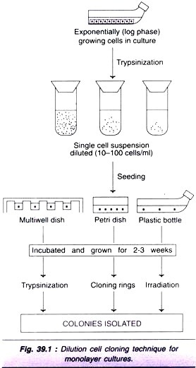
There are many diverse and complex techniques involved in gene manipulation. However, the basic principles of recombinant DNA technology are reasonably simple, and broadly involve the following stages (Fig. 6.1).
1. Generation of DNA fragments and selection of the desired piece of DNA (e.g. a human gene).
2. Insertion of the selected DNA into a cloning vector (e.g. a plasmid) to create a recombinant DNA or chimeric DNA (Chimera is a monster in Greek mythology that has a lion’s head, a goat’s body and a serpent’s tail. This may be comparable to Narasimha in Indian mythology).
3. Introduction of the recombinant vectors into host cells (e.g. bacteria).
4. Multiplication and selection of clones containing the recombinant molecules.
5. Expression of the gene to produce the desired product.
Recombinant DNA technology with special reference to the following aspects is described below:
1. Molecular tools of genetic engineering.
2. Host cells-the factories of cloning.
3. Vectors-the cloning vehicles.
4. Methods of gene transfer.
5. Gene cloning strategies.
6. Genetic engineering guidelines.
7. The future of genetic engineering.
Molecular Tools of Genetic Engineering:
An engineer is a person who designs, constructs (e.g. bridges, canals, and railways) and manipulates according to a set plan. The term genetic engineer may be appropriate for an individual who is involved in genetic manipulations. The genetic engineer’s toolkit or molecular tools namely the enzymes most commonly used in recombinant DNA experiments are briefly described.
Restriction Endonucleases— DNA Cutting Enzymes:
Restriction endonucleases are one of the most important groups of enzymes for the manipulation of DNA. These are the bacterial enzymes that can cut/split DNA (from any source) at specific sites. They were first discovered in E.coli restricting the replication of bacteriophages, by cutting the viral DNA (The host E.coli DNA is protected from cleavage by addition of methyl groups). Thus, the enzymes that restrict the viral replication are known as restriction enzymes or restriction endonucleases.
Hundreds of restriction endonucleases have been isolated from bacteria, and some of them are commercially available. The progress and growth of biotechnology is unimaginable without the availability of restriction enzymes.
Nomenclature:
Restriction endonucleases are named by a standard procedure, with particular reference to the bacteria from which they are isolated. The first letter (in italics) of the enzymes indicates the genus name, followed by the first two letters (also in italics) of the species, then comes the strain of the organism and finally a Roman numeral indicating the order of discovery. A couple of examples are given below.
EcoRI is from Escherichia (E) coli (co), strain Ry13 (R) and first endonuclease (I) to be discovered. Hindlll is from Haemophilus (H) influenza (in), strain Rd (d) and the third endonucleases (III) to be discovered.
Types of endonucleases:
At least 4 different types of restriction endonucleases are known-type 1 (e.g Ecok12), type II (e.g. EcoRI), type III (e.g. EcoPI) and type IIs. Their characteristic features are given in Table 6.1. Among these, type II restriction endonucleases are most commonly used in gene cloning.
Recognition sequences:
Recognition sequence is the site where the DNA is cut by a restriction endonuclease. Restriction endonucleases can specifically recognize DNA with a particular sequence of 4-8 nucleotides and cleave. Each recognition sequence has two fold rotational symmetry i.e. the same nucleotide sequence occurs on both strands of DNA which run in opposite direction (Table 6.2). Such sequences are referred to as palindromes, since they read similar in both directions (forwards and backwards).
Cleavage patterns:
Majority of restriction endonucleases (particularly type II) cut DNA at defined sites within recognition sequence. A selected list of enzymes, recognition sequences, and their products formed is given in Table 6.2. The cut DNA fragments by restriction endonucleases may have mostly sticky ends (cohesive ends) or blunt ends, as given in Table 6.2. DNA fragments with sticky ends are particularly useful for recombinant DNA experiments. This is because the single-stranded sticky DNA ends can easily pair with any other DNA fragment having complementary sticky ends.
DNA Ligases —DNA Joining Enzymes:
The cut DNA fragments are covalently joined together by DNA ligases. These enzymes were originally isolated from viruses. They also occur in E.coli and eukaryotic cells. DNA ligases actively participated in cellular DNA repair process.
The action of DNA ligases is absolutely required to permanently hold DNA pieces. This is so since the hydrogen bonds formed between the complementary bases (of DNA strands) are not strong enough to hold the strands together. DNA ligase joins (seals) the DNA fragments by forming a phosphodiester bond between the phosphate group of 5′-carbon of one deoxyribose with the hydroxyl group 3′-carbon of another deoxyribose (Fig. 6.2).
Phage T4 DNA ligase requires ATP as a cofactor while E.coli DNA ligase is dependent on NAD+. In each case, the cofactor (ATP or NAD+) is split to form an enzyme—AMP complex that brings about the formation of phosphodiester bond. The action of DNA ligase is the ultimate step in the formation of a recombinant DNA molecule.
Homo-polymer tailing:
The complementary DNA strands can be joined together by annealing. This principle is utilized in homo-polymer tailing. The technique involves the addition of oligo (dA) to 3′-ends of some DNA molecules and the addition of oligo (dT) to 3′-ends of other molecules. The homo-polymer extensions (by adding 10-40 residues) can be synthesized by using terminal deoxynucleotidyltransferase (of calf thymus). Homo-polymer tailing, achieved by annealing is illustrated in Fig. 6.3.
Linkers and adaptors:
Linkers and adaptors are chemically synthesized, short, double-stranded DNA molecules. Linkers possess restriction enzyme cleavage sites. They can be ligated to blunt ends of any DNA molecule and cut with specific restriction enzymes to produce DNA fragments with sticky ends (Fig. 6.4). Adaptors contain preformed sticky or cohesive ends. They are useful to be ligated to DNA fragments with blunt ends. The DNA fragments held to linkers or adaptors are finally ligated to vector DNA molecules (Fig 6.4).
Alkaline Phosphatase:
Alkaline phosphatase is an enzyme involved in the removal of phosphate groups. This enzyme is useful to prevent the unwanted ligation of DNA molecules which is a frequent problem encountered in cloning experiments. When the linear vector plasmid DNA is treated with alkaline phosphatase, the 5′-terminal phosphate is removed (Fig 6.5). This prevents both re-circularization and plasmid DNA dimer formation. It is now possible to insert the foreign DNA through the participation of DNA ligase.
DNA Modifying Enzymes:
Some authors prefer to use the broad term DNA modifying enzymes to all the enzymes involved in recombinant DNA technology. These enzymes represent the cutting and joining functions in DNA manipulation. They are broadly categorized as nucleases, polymerases and enzymes modifying ends of DNA molecules, and briefly described below, and illustrated in Fig. 6.6.
Nucleases:
Nucleases are the enzymes that break the phosphodiester bonds (that hold nucleotides together) of DNA. Endonucleases act on the internal phosphodiester bonds while exonucleases degrade DNA from the terminal ends (Fig 6.6A). Restriction endonucleases, described already, are good examples of endonucleases. Some other examples of endo- and exonucleases are listed.
Endonucleases:
i. Nuclease S1 specifically acts on single-stranded DNA or RNA molecules.
ii. Deoxyribonuclease I (DNase I) cuts either single or double-stranded DNA molecules at random sites.
Exonucleases:
i. Exonuclease III cuts DNA and generates molecules with protruding 5′-ends.
ii. Nuclease Bal 31 is a fast acting 3′-exonuclease. Its action is usually coupled with slow acting endonucleases.
A diagrammatic representation of the action of endo-and exonucleases is given in Fig. 6.7.
Besides the DNA cutting enzymes, there are RNA specific nucleases, which are referred to as ribonucleases (RNases).
Polymerases:
The groups of enzymes that catalyse the synthesis of nucleic acid molecules are collectively referred to as polymerases. It is customary to use the name of the nucleic acid template on which the polymerase acts (Fig. 6.6B). The three important polymerases are given below.
a. DNA-dependent DNA polymerase that copies DNA from DNA.
b. RNA-dependent DNA polymerase (reverse transcriptase) that synthesizes DNA from RNA.
c. DNA-dependent RNA polymerase that produces RNA from DNA.
Enzymes modifying the ends of DNA:
There are certain enzymes that act on the terminal ends of DNA and modify these molecules. The important ones are listed.
a. Alkaline phosphatase that removes the terminal phosphate group (see Fig. 6.5).
b. Polynucleotide kinase involved in the addition of phosphate groups.
c. Terminal transferase (also called terminal deoxynucleotidyl transferase) repeatedly adds nucleotides to any available 3′-terminal ends, the most suitable being the protruding 3′-ends. This enzyme is particularly useful to add homo-polymer tails prior to the construction of recombinant DNA molecules.
d. The most commonly used enzymes in recombinant DNA technology/genetic engineering are listed in Table 6.3.
Host Cells —The Factories of Cloning:
The hosts are the living systems or cells in which the carrier of recombinant DNA molecule or vector can be propagated. There are different types of host cells-prokaryotic (bacteria) and eukaryotic (fungi, animals and plants). Some examples of host cells used in genetic engineering are given in Table 6.4.
Host cells, besides effectively incorporating the vector’s genetic material, must be conveniently cultivated in the laboratory to collect the products. In general, microorganisms are preferred as host cells, since they multiply faster compared to cells of higher organism (plants or animals).
Prokaryotic Hosts:
Escherichia coli:
The bacterium, Escherichia coli was the first organism used in the DNA technology experiments and continues to be the host of choice by many workers. Undoubtedly, E.coli, the simplest Gram negative bacterium (a common bacterium of human and animal intestine), has played a key role in the development of present day biotechnology.
Under suitable environment, E.coli can double in number every 20 minutes. Thus, as the bacteria multiply, their plasmids (along with foreign DNA) also multiply to produce millions of copies, referred to as colony or in short clone. The term clone is broadly used to a mass of cells, organisms or genes that are produced by multiplication of a single cell, organism or gene.
Limitations of E. coli:
There are certain limitations in using E.coli as a host. These include- causation of diarrhea by some strains, formation of endotoxins that are toxic, and a low export ability of proteins from the cell. Another major drawback is that E.coli (or even other prokaryotic organisms) cannot perform post-translational modifications.
Bacillus subtilis:
Bacillus subtilis is a rod shaped non-pathogenic bacterium. It has been used as a host in industry for the production of enzymes, antibiotics, insecticides etc. Some workers consider B.subtilis as an alternative to E.coli.
Eukaryotic Hosts:
Eukaryotic organisms are preferred to produce human proteins since these hosts with complex structure (with distinct organelles) are more suitable to synthesize complex proteins. The most commonly used eukaryotic organism is the yeast, Saccharomyces cerevisiae. It is a non-pathogenic organism routinely used in brewing and baking industry. Certain fungi have also been used in gene cloning experiments.
Mammalian cells:
Despite the practical difficulties to work with and high cost factor, mammalian cells (such as mouse cells) are also employed as hosts. The advantage is that certain complex proteins which cannot be synthesized by bacteria can be produced by mammalian cells e.g. tissue plasminogen activator. This is mainly because the mammalian cells possess the machinery to modify the protein to its final form (post-translational modifications).
It may be noted here that the gene manipulation experiments in higher animals and plants are usually carried out to alter the genetic makeup of the organism to create transgenic animals and transgenic plants rather than to isolate genes for producing specific proteins.
Vectors —The Cloning Vehicles:
Vectors are the DNA molecules, which can carry a foreign DNA fragment to be cloned. They are self- replicating in an appropriate host cell. The most important vectors are plasmids, bacteriophages, cosmids and phasmids.
Characteristics of an ideal vector:
An ideal vector should be small in size, with a single restriction endonuclease site, an origin of replication and 1-2 genetic markers (to identify recipient cells carrying vectors). Naturally occurring plasmids rarely possess all these characteristics.
Plasmids:
Plasmids are extra chromosomal, double- stranded, circular, self-replicating DNA molecules. Almost all the bacteria have plasmids containing a low copy number (1-4 per cell) or a high copy number (10-100 per cell). The size of the plasmids varies from 1 to 500 kb. Usually, plasmids contribute to about 0.5 to 5.0% of the total DNA of bacteria (Note: A few bacteria contain linear plasmids e.g. Streptomyces sp, Borella burgdorferi).
Types of plasmids:
There are many ways of grouping plasmids. They are categorized as conjugative if they carry a set of transfer genes (tra genes) that facilitates bacterial conjugation, and non-conjugative, if they do not possess such genes. Another classification is based on the copy number. Stringent plasmids are present in a limited number (1-2 per cell) while relaxed plasmids occur in large number in each cell.
F-plasmids possess genes for their own transfer from one cell to another, while R-plasmids carry genes resistance to antibiotics. In general, the conjugative plasmids are large, show stringent control of DNA replication, and are present in low numbers. On the other hand, non- conjugative plasmids are small, show relaxed control of DNA replication, and are present in high numbers.
Nomenclature of plasmids:
It is a common practice to designate plasmid by a lower case p, followed by the first letter(s) of researcher(s) names and the numerical number given by the workers. Thus, pBR322 is a plasmid discovered by Bolivar and Rodriguez who designated it as 322. Some plasmids are given names of the places where they are discovered e.g. pUC is plasmid from University of California.
pBR322 — the most common plasmid vector:
pBR322 of E.coli is the most popular and widely used plasmid vector, and is appropriately regarded as the parent or grandparent of several other vectors. pBR322 has a DNA sequence of 4,361 bp. It carries genes resistance for ampicillin (Ampr) and tetracycline (TeIr) that serve as markers for the identification of clones carrying plasmids. The plasmid has unique recognition sites for the action of restriction endonucleases such as EcoRI, Hindlll, BamHI, Sail and Pstll (Fig. 6.8).
Other plasmid cloning vectors:
The other plasmids employed as cloning vectors include pUC19 (2,686 bp, with ampicillin resistance gene), and derivatives of pBR322- pBR325, pBR328 and pBR329.
Bacteriophages:
Bacteriophages or simply phages are the viruses that replicate within the bacteria. In case of certain phages, their DNA gets incorporated into the bacterial chromosome and remains there permanently. Phage vectors can accept short fragments of foreign DNA into their genomes. The advantage with phages is that they can take up larger DNA segments than plasmids. Hence phage vectors are preferred for working with genomes of human cells.
Bacteriophage λ:
Bacteriophage lambda (or simply phage λ), a virus of E.coli, has been most thoroughly studied and developed as a vector. In order to understand how bacteriophage functions as a vector, it is desirable to know its structure and life cycle (Fig. 6.9).
Phage λ consists of a head and a tail (both being proteins) and its shape is comparable to a miniature hypodermic syringe. The DNA, located in the head, is a linear molecule of about 50 kb. At each end of the DNA, there are single-stranded extensions of 12 base length each, which have cohesive (cos) ends.
On attachment with tail to E.coli, phage X injects its DNA into the cell. Inside E.coli, the phage linear DNA cyclizes and gets ligated through cos ends to form a circular DNA. The phage DNA has two fates-lytic cycle and lysogenic cycle.
Lytic cycle:
The circular DNA replicates and it also directs the synthesis of many proteins necessary for the head, tail etc., of the phage. The circular DNA is then cleaved (to form cos ends) and packed into the head of the phage. About 100 phage particles are produced within 20 minutes after the entry of phage into E.coli.
The host cell is then subjected to lysis and the phages are released. Each progeny phage particle can infect a bacterial cell, and produce several hundreds of phages. It is estimated that by repeating the lytic cycle four times, a single phage can cause the death of more than one billion bacterial cells.
If a foreign DNA is spliced into phage DNA, without causing harm to phage genes, the phage will reproduce (replicate the foreign DNA) when it infects bacterial cell. This has been exploited in phage vector employed cloning techniques.
Lysogenic cycle:
In this case, the phage DNA (instead of independently replicating) becomes integrated into the E.coli chromosome and replicates along with the host genome. No phage particles are synthesized in this pathway.
Use of phage λ as a vector:
Only about 50% of phage λ DNA is necessary for its multiplication and other functions. Thus, as much as 50% (i.e. up to 25kb) of the phage DNA can be replaced by a donor DNA for use in cloning experiments. However, several restriction sites are present on phage λ which is not by itself a suitable vector. The λ-based phage vectors are modifications of the natural phage with much reduced number of restriction sites.
Insertion vectors:
They have just one unique cleavage site, which can be cleaved, and a foreign DNA ligated in. It is essential that sufficient DNA (about 25%) has to be deleted from the vector to make space for the foreign DNA (about 18kb).
Replacement vectors:
These vectors have a pair of restriction sites to remove the non-essential DNA (stuffer DNA) that will be replaced by a foreign DNA. Replacement vectors can accommodate up to 24kb, and propagate them. Many phage vector derivations (insertion/ replacement) have been produced by researchers for use in recombinant DNA technology.
The main advantage of using phage vectors is that the foreign DNA can be packed into the phage {in vitro packaging), the latter in turn can be injected into the host cell very effectively (Note: No transformation is required).
Phage M13 vectors:
Phage M13 (bacteriophage M13) is a single- stranded DNA phage of E.coli. Inside the host cell, M13 synthesizes the complementary strand to form a double-stranded DNA (replicative form DNA; RF DNA). For use as a vector, RF DNA is isolated and a foreign DNA can be inserted on it. This is then returned to the host cell as a plasmid. Single- stranded DNAs are recovered from the phage particles. Phage M13 is useful for sequencing DNA through Sanger’s method
Cosmids:
Cosmids are the vectors possessing the characteristics of both plasmid and bacteriophage λ. Cosmids can be constructed by adding a fragment of phage λ DNA including cos site, to plasmids. A foreign DNA (about 40 kb) can be inserted into cosmid DNA.
The recombinant DNA so formed can be packed as phages and injected into E.coli (Fig. 6.10). Once inside the host cell, cosmids behave just like plasmids and replicate. The advantage with cosmids is that they can carry larger fragments of foreign DNA compared to plasmids.
Phasid vectors:
Phasids are the combination of plasmid and phage and can function as either one (i.e as plasmid or phage). Phasids possess functional origins of replication of both plasmid and phage λ, and therefore can be propagated (as plasmid or phage) in appropriate E.coli. The vectors phasids may be used in many ways in cloning experiments.
Artificial Chromosome Vectors:
Human artificial chromosome (HAC):
Developed in 1997 (by H. Willard), human artificial chromosome is a synthetically produced vector DNA, possessing the characteristics of human chromosome. HAC may be considered as a self-replicating micro-chromosome with a size ranging from 1/10th to 1/5th of a human chromosome. The advantage with HAC is that it can carry human genes that are too long. Further, HAC can carry genes to be introduced into the cells in gene therapy.
Yeast artificial chromosomes (YACs):
Introduced in 1987 (by M. Olson), yeast artificial chromosome (YAC) is a synthetic DNA that can accept large fragments of foreign DNA (particularly human DNA). It is thus possible to clone large DNA pieces by using YAC. YACs are the most sophisticated yeast vectors, and represent the largest capacity vectors available. They possess centromeric and telomeric regions, and therefore the recombinant DNA can be maintained like a yeast chromosome.
Bacterial artificial chromosomes (BACs):
The construction of BACs is based on one F-plasmid which is larger than the other plasmids used as cloning vectors. BACs can accept DNA inserts of around 300 kb. The advantage with bacterial artificial chromosome is that the instability problems of YACs can be avoided. In fact, a major part of the sequencing of human genome has been accomplished by using a library of BAC recombinant.
Shuttle Vectors:
The plasmid vectors that are specifically designed to replicate in two different hosts (say in E.coli and Streptomyces sp) are referred to as shuttle vectors. The origins of replication for two hosts are combined in one plasmid. Therefore, any foreign DNA fragment introduced into the vector can be expressed in either host. Further, shuttle vectors can be grown in one host and then shifted to another host (hence the name shuttle). A good number of eukaryotic vectors are shuttle vectors.
Choice of a Vector:
Among the several factors, the size of the foreign DNA is very important in the choice of vectors. The efficiency of this process in often crucial for determining the success of cloning. The size of DNA insert that can be accepted by different vectors is shown in Table 6.5.
Gene Cloning Strategies:
A clone refers to a group of organisms, cells, molecules or other objects, arising from a single individual. Clone and colony are almost synonymous. Gene cloning strategies in relation to recombinant DNA technology broadly involve the following aspects (Fig. 6.13).
a. Generation of desired DNA fragments.
b. Insertion of these fragments into a cloning vector.
c. Introduction of the vectors into host cells.
d. Selection or screening of the recipient cells for the recombinant DNA molecules.
Cloning From Genomic DNA or MRNA?
DNA represents the complete genetic material of an organism which is referred to as genome. Theoretically speaking, cloning from genomic DNA is supposed to be ideal. But the DNA contains non- coding sequences (introns), control regions and repetitive sequences. This complicates the cloning strategies; hence DNA as a source material is not preferred, by many workers. However, if the objective of cloning is to elucidate the control of gene expression, then genomic DNA has to be invariably used in cloning.
The use of mRNA in cloning is preferred for the following reasons:
a. mRNA represents the actual genetic information being expressed.
b. Selection and isolation mRNA is easy.
c. As introns are removed during processing, mRNA reflects the coding sequence of the gene.
d. The synthesis of recombinant protein is easy with mRNA cloning.
Besides the direct use of genomic DNA or mRNA, it is possible to synthesize DNA in the laboratory and use it in cloning experiments. This approach is useful if the gene sequence is short and the complete sequence of amino acids is known.
The different strategies for the cloning of genomic DNA and mRNA are described under gene libraries
Genetic Engineering Guidelines:
With the success of Boyer-Cohen experiments (in 1973), it was realised that recombinant DNA technology could be used to create organisms with novel genes. This created worldwide commotion (among scientists, public and government officials) about the safety, ethics and unforeseen consequences of genetic manipulations. Some of the phrases quoted in media in those days are given.
a. Manipulation of life.
b. Playing God.
c. Man-made evolution.
d. The most threatening scientific research.
It was feared that some new organisms, created inadvertently or deliberately for warfare, would cause epidemics and environmental catastrophes. Due to the fears of the dangerous consequences, a cautious approach on recombinant DNA experiments was suggested.
In 1974, a group of ten scientists led by Paul Berg wrote a letter that simultaneously appeared in the prestigious journals-Nature, Science and Proceedings of the National Academy of Sciences.
The dangers of DNA technology were printed out in that letter (highlights given below):
“Recent advances in techniques for isolation and rejoining of segments of DNA now permit construction of biologically active recombinant DNA molecules in vitro. Although such experiments are likely to facilitate the solution of important theoretical and practical biological problems, they would also result in creation of novel types of DNA elements whose biological properties cannot be completely predicted. There is a serious concern that some of these DNA molecules could prove biologically hazardous”.
The letter also appealed to molecular biologists worldwide for a moratorium on many kinds of recombinant DNA research, particularly those involving pathogenic organisms.
Asilomar Recommendations:
In February 1975, a group of 139 scientists from 17 countries held a conference at Asilomar, a conference center in California, USA. They assured the uneasy public that the microorganisms used in DNA experiments were specifically bred and could not survive outside the laboratory. These scientists formulated guidelines and recommendations for conducting experiments in genetic engineering.
NIH Guidelines:
National Institute of Health (NIH), USA constituted the Recombinant DNA Advisory Committee (RAC) which issued a set of stringent guidelines to conduct research on DNA. RAC was in fact overseeing the research projects involving gene splicing and recombinant DNA.
Some of the important original NIH recommendations on recombinant DNA research relate to the following aspects:
a. Physical (laboratory) containment levels for conducting experiments.
b. Biological containment-the host into which foreign DNA is inserted should not proliferate outside the laboratory or transfer its DNA into other organisms.
c. For research on pathogenic organisms, elaborate, controlled and self-contained rooms were recommended.
d. For research on less dangerous organisms, units equipped with high quality filter systems should be used.
e. No deliberate release of any organism containing recombinant DNA into the environment.
It may be noted here that although the NIH guidelines did not have the legal status, most institutions, companies and scientists voluntarily complied.
Relaxation of NIH guidelines:
It was in 1980, the original NIH guidelines were considerably relaxed by NIH-RAC, based on the experience and experimental data obtained from the NIH-sponsored studies on recombinant DNA research. It was almost agreed that the original apprehensions on recombinant DNA research were unfounded.
It is a fact that the genetic engineering research flourished and progressed rapidly after relaxation of NIH guidelines. It may however be noted that NIH- RAC continues to be a watchdog over the DNA technology experiments.
Pharmaceutical products of recombinant DNA:
As the recombinant DNA technology progressed, many pharmaceutical compounds of human health care are being produced through genetic manipulations. Most countries consider that the existing regulations for approval of pharmaceuticals of commercial use are adequate to ensure safety since the process by which the product manufactured is irrelevant. Thus, the recombinant DNA product (protein, vaccine, and drug) is evaluated for its safety and efficacy like any other pharmaceutical product.
Genetically engineered organisms (GEOs):
Recombinant DNA research has resulted in the creation of many genetically engineered organisms. These include microorganisms, animals and plants. The latter two respectively result in transgenic animals and transgenic plants
The Future of Genetic Engineering:
DNA technology has largely helped scientists to understand the structure, function and regulation of genes. The development of new/modern biotechnology is primarily based on the success of DNA technology. Thus, the present biotechnology (more appropriately molecular biotechnology) has its main roots in molecular biology.
Biotechnology is an interdisciplinary approach for applications to human health, agriculture, industry and environment. The major objective of biotechnology is to solve problems associated with human health, food production, energy production and environmental control.
The major contributions of genetic engineering through the new discipline biotechnology are given in this book.
It is an accepted fact that the recombinant DNA technology has entered the main stream of human life and has become one of the most significant applications of scientific research. Biotechnology is regarded as more an art than a science. After the successful sequencing of human genome, many breakthroughs in biotechnology are expected in future.
